
Efficacy and Safety of Optic Nerve Sheath Fenestration for Idiopathic Intracranial Hypertension: A Subgroup-Focused Systematic Review and Meta-Analysis
DOI:
https://doi.org/10.71079/ASIDE.IM.1542545Abstract
Introduction: Optic nerve sheath fenestration (ONSF) is an important surgical management option for patients with idiopathic intracranial hypertension (IIH) who have failed medical treatment. We conducted a systematic review and meta-analysis to evaluate the outcomes of ONSF and identify factors affecting treatment success.
Methods: A literature search was conducted up to December 2024. Primary outcomes included improvement in visual acuity, visual fields, and optic disc swelling resolution. We performed a detailed subgroup analysis based on geographic location, study design, surgical approach, and technical variations.
Results: Nineteen studies with a total of 1,159 patients were included in our study. ONSF significantly improved visual acuity in 34.5% (95% CI: 31.8-37.3%) and visual fields in 69.4% (95% CI: 65.9-72.7%) of cases. A 90.9% improvement rate was observed in reducing optic disc swelling. Significant heterogeneity was noted in visual acuity (I²=92.1%) and visual field improvements (I²=73.8%). The overall complication rate was 9% (95% CI: 5-16%). Centers that included 30 or more patients in their study demonstrated significantly lower postoperative complications.
Conclusions: ONSF demonstrates favorable efficacy in improving visual outcomes with an acceptable safety profile, lower postoperative complications were observed when the procedure was performed in high-volume centers using appropriate surgical techniques. Geographic variations and surgical approaches significantly affected outcomes, highlighting the importance of standardized protocols and adequate surgical experience. Future prospective studies with standardized outcome measures are needed to optimize patient selection and surgical techniques.
Keywords:
Idiopathic Intracranial Hypertension, Optic Nerve Sheath Fenestration, Meta-Analysis, Systematic Review, Visual Outcomes, Benign Intracranial HypertensionFull Text
Introduction
Idiopathic intracranial hypertension (IIH), previously known as pseudotumor cerebri or benign intracranial hypertension, is a neurological condition characterized by elevated intracranial pressure (ICP) without identifiable structural or vascular causes [1]. This disorder mainly affects women of reproductive age and demonstrates a strong association with obesity; despite the current research about IIH, the etiopathogenesis remains not fully understood yet [1]. The hallmark clinical manifestations of the disease include headache, papilledema, and visual disturbances that may progress to blindness if left untreated with proper management [1].
The management of IIH follows a systemic approach, beginning with conservative measures such as weight loss and medical therapy, with acetazolamide, topiramate, and diuretics [2,3,4]. However, around 25% of patients are developing refractory IIH in which they have limited or poor response to the initial medical interventions, necessitating surgical intervention [5]. Among the available surgical options, optic nerve sheath fenestration (ONSF) is considered to be among the surgical interventions for IIH patients to preserve vision in cases of progressive visual deterioration [5,6]. ONSF involves creating an opening in the optic nerve sheath to reduce localized cerebrospinal fluid (CSF) pressure, which, by role, decreases mechanical compression on the optic nerve [7]. While ONSF for IIH has been reported as a successful intervention for IIH in patients with threatened visual loss, previous studies did not have sufficient conclusive evidence about the optimal ONSF approaches and settings regarding its safety and efficacy for IIH patients [5,8,9].
Previous studies have highlighted important considerations in surgical procedures for IIH, but these studies either lacked complete evaluation of visual fields or have become outdated as new surgical techniques and further updated studies have been published [1,5,7,8,9,10,11,12]. The timing of surgical intervention appears critical, with mounting evidence suggesting that delayed intervention may result in irreversible vision loss due to progressive optic nerve damage [13]Also, technical aspects of the procedure, such as the size and location of the fenestration, muscle disinsertion requirements, and the use of minimally invasive approaches, may affect its efficacy and complication rates.
Recent advances in surgical techniques, especially the development of endoscopic and minimally invasive approaches, have renewed interest in evaluating the safety and efficacy of ONSF from different prospections, even in the presence of other minimally invasive interventions such as venous sinus stenting where they may not be suitable for some patients due to several factors and considerations [14]. Additionally, the emergence of large-scale studies with detailed visual outcome data has created an opportunity for a more detailed analysis of factors influencing surgical success with the option of conducting subgroup analysis to explore further more factors affecting ONSF success [15]. Therefore, we aim to evaluate the safety and efficacy of ONSF in preserving vision in patients with IIH based on the most updated evidence in the most detailed manner, given the currently available data. Our study specifically focuses on three key outcomes: improvement in visual acuity, enhancement of visual fields, and resolution of optic disc swelling, in addition to investigating the reported postoperative complications from ONSF. Our study aims to perform detailed subgroup analyses based on geographical location, study design, surgical approach, and technical variations to identify factors that might influence surgical outcomes. Understanding these variables and factors related to ONSF in IIH patients has important considerations for optimizing patient selection and surgical technique, ultimately improving visual outcomes in this challenging patient population.
Methods:
Our systematic review and meta-analysis were conducted with adherence to the Preferred Reporting Items for Systematic Reviews and Meta-Analyses (PRISMA) guidelines. We conducted a literature search across multiple electronic databases, including PubMed/MEDLINE, Embase, Web of Science, Scopus, Google Scholar, and Cochrane Library, from inception through the 22nd of December 2024. The search strategy incorporated Medical Subject Headings (MeSH) terms and keywords related to "optic nerve sheath fenestration", "ONSF" "idiopathic intracranial hypertension”, “IIH”, “benign intracranial hypertension”, and "pseudotumor cerebri". For methodological clarity, we classified all included studies into two categories: observational and interventional. It is important to note that all studies in both categories involved patients who underwent ONSF; our classification refers to study design rather than treatment allocation. Observational studies (n= 16) included retrospective chart reviews, case series, and cohort studies where the studies analyzed and reported the outcomes after ONSF without a predefined intervention protocol. Interventional studies (three studies) were those with prospective enrollment following a standardized surgical protocol and predefined outcome measures, representing a higher level of methodological confidence and validity.
Literature Review and Data Extraction:
Two independent reviewers screened titles and abstracts for eligibility, followed by a full-text review of the possible relevant articles. We included both observational and interventional studies reporting outcomes of ONSF in patients with IIH. Studies were eligible if they reported at least one of our primary outcomes: visual acuity improvement, visual field improvement, or optic disc swelling resolution. We excluded non-English articles, case reports, or case series, which included less than five patients, review articles, systematic reviews and meta-analyses, editorials, and letters. Data extraction was performed independently by two investigators using a standardized form. We collected information on study characteristics (publication year, country, study design), patient demographics, surgical techniques, and clinical outcomes. Complications were categorized into overall complications, diplopia, transient visual loss, worsening of visual functions, and anisocoria.
Risk of Bias Assessment
We assessed the risk of bias using the Risk Of Bias In Non-Randomized Studies—of Interventions (ROBINS-I) tool, which evaluates seven domains: confounding, selection bias, classification of interventions, deviations from intended interventions, missing data, outcome measurement, and selective reporting. The quality of evidence for each outcome was evaluated using the Grading of Recommendations, Assessment, Development, and Evaluations (GRADE) framework, considering the risk of bias, inconsistency, indirectness, imprecision, and publication bias.
Statistical Analysis
For statistical analysis, we performed a single-arm proportion-based meta-analysis using RStudio software with the ’meta’ and ’metafor’ packages built on the R language. We calculated pooled proportions with 95% confidence intervals for each outcome using random-effects models. Heterogeneity was assessed using I² statistics and Cochran’s Q test. We conducted pre-specified subgroup analyses based on country of study, either United States (US) vs. non-US based study, study type (observational vs. interventional), study design (retrospective vs. prospective), sample size (>30 vs. <30 vs. =30), surgical approach (medial transconjunctival vs. other), and surgical technique (with vs. without muscle disinsertion). Publication bias was evaluated using funnel plots, Egger’s test, and the trim-and-fill method was used when appropriate. Statistical significance was set at P<0.05, and all tests were two-sided.
The rationale for our geographic subgroup analysis (US vs. non-US) was based on the differences in practice patterns, patient selection criteria, reporting standards, and healthcare systems between the regions. Previous studies on IIH have demonstrated geographic variations in disease prevalence, management approaches, and outcomes reporting [16,17]. We aim to identify any differences in ONSF outcomes that might inform international standardization efforts and highlight region-specific considerations for optimizing patient selection and surgical technique.
Results
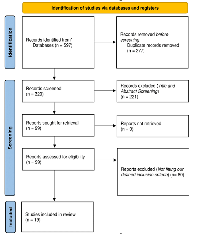
Our systematic search yielded 597 records, with 19 studies meeting the final inclusion criteria after thorough screening Figure 1 [18,19,20,21,22,23,24,25,26,27,28,29,30,31,32,33,34,35,36]. The included studies spanned from 1988 to 2021, comprising 16 observational and three interventional studies, with 14 retrospective and five prospective designs (Supplementary Table 1).
Risk of Bias Assessment:
The ROBINS-I risk of bias assessment highlighted varied methodological quality (Supplementary Figure 1). Three studies (Melson et al., Wadikhaye et al., and Nithyanandam et al.) demonstrated a consistently low risk of bias across all domains (+). Nine studies showed serious risk of bias (X) in their overall assessment, primarily due to confounding (D1) and missing data (D5). Domain D3 (bias in classification of interventions) uniquely showed low risk (+) across all studies. Domain D4 (bias due to deviations from intended interventions) showed mixed results, with eight studies having low risk and 11 showing moderate risk. Seven studies showed consistently poor performance across multiple domains, receiving serious risk ratings in D5, D6, and D7.
GRADE Framework Assessment
Our GRADE framework assessment (Supplementary Table 2) revealed heterogeneous quality across outcomes. Visual acuity improvement evidence (19 studies, n=1,160) received a low-quality rating () due to a serious risk of bias (-1) from lack of standardized visual acuity measurements and high heterogeneity (-1) with I²>50%. Visual field improvement (16 studies, n=719) achieved moderate quality (), downgraded only for serious risk bias (-1) due to varied testing methods, but showed consistent improvement across studies. Optic disc swelling resolution (11 studies, n=351) also received a moderate quality rating (), with consistent resolution rates and low heterogeneity.
Efficacy Outcomes:
Visual acuity improvement results have demonstrated statistically significant differences between non-US and US studies (48.3% vs. 29.8%, p<0.001), prospective versus retrospective designs (80.8% vs. 32.7%, p<0.001), and surgical approaches (medial transconjunctival 30.6% vs. other 48.4%, p-value<0.001). Visual field improvement demonstrated higher rates in US versus non-US studies (72.7% vs. 68.1%, p-value= 0.221) and interventional versus observational studies (83.9% vs. 70.5%, p-value= 0.154). Optic disc swelling resolution showed significantly better outcomes in US studies (97.3% vs. 76.5%, p-value<0.001), interventional studies (98.7% vs 83.6%, p<0.001), and medial transconjunctival approach (98.8% vs 82.8%, p-value<0.001), as listed in Table 1. We have used the single-arm proportion-based meta-analysis as our primary analytical approach due to the nature of available data extracted from included studies. The absence of randomized controlled trials and the limited number of comparative studies precluded traditional two-arm meta-analyses using direct comparison groups and direct comparison metrics, which were unavailable across most included studies.
Safety Outcomes
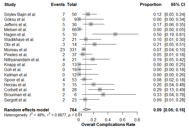
We listed the overall reported complications from our included studies in Table 2. The overall rate was 9% (95% CI: 5-16%, I²=48%) Figure 2. Significant subgroup differences were observed in the country of study (non-US 13.9% vs. US 7.4%, p-value= 0.007), surgical approach (medial transconjunctival 7.7% vs. other 13.2%, p-value= 0.023), and sample size (>30: 6.6% vs <30: 15.2% vs =30: 16.7%, p-value<0.001). Specific complications showed varying rates: worsening of visual functions (8%, I²=51%), diplopia (3%, I²=24%), anisocoria (4%, I²=0%), and transient visual loss (10%, I²=69%). In addition to that, bilateral versus unilateral approaches showed significant differences in transient visual loss (6.9% vs. 15.5%, p-value<0.001) and worsening of visual functions (5.7% vs. 15.5%, p-value=0.018); the forest plots for the efficacy outcomes are shown in (Supplementary Figure 2 – 5), while the subgroup analyses for safety outcomes are listed in (Supplementary Table 3).
Publication Bias
Publication bias assessment (Supplementary Table 4) indicated a moderate risk for visual acuity improvement (Egger’s test p-value= 0.034), with right-skewed funnel plot asymmetry and four potentially missing studies identified through trim-and-fill analysis. Visual field improvement and optic disc resolution showed low publication bias risk (Egger’s test p-value= 0.245 and p-value= 0.789, respectively).
Discussion
In our study, we evaluated the efficacy and safety outcomes of ONSF in IIH patients, focusing on detailed subgroup analyses based on multiple factors. Our findings provide important insights into the factors that may have significant considerations on ONSF outcomes and help identify optimal patient selection criteria.
Our analysis demonstrates that ONSF shows considerable efficacy in improving visual outcomes, with an overall visual acuity improvement rate of 34.5% (95% CI: 31.8-37.3%) and visual field improvement rate of 69.4% (95% CI: 65.9-72.7%). The subgroup analyses revealed several groups for efficacy and treatment success and highlighted several important points that warrant discussion and further investigation. Studies from non-US centers showed significantly higher rates of visual acuity improvement (48.3% vs. 29.8%, p-value<0.001), suggesting potential variations in patient selection criteria or surgical techniques across different geographic regions [3,16,37,38,39,40]. Prospective studies demonstrated markedly better outcomes in visual acuity improvement compared to retrospective designs (80.8% vs 32.7%, p-value<0.001), highlighting the importance of standardized protocols and careful patient monitoring in achieving optimal outcomes, which was previously discussed that we need more optimization and standardization for IIH studies to promote better quality studies and enhance our evidence about the disease [41]. The surgical approach emerged as a crucial factor, with non-medial transconjunctival approaches showing higher success rates (48.4%) compared to medial transconjunctival approaches (30.6%, p-value<0.001). This finding suggests that surgical technique selection may significantly influence visual outcomes. Sample size analysis revealed interesting patterns, with centers performing more than 30 procedures showing more consistent results compared to those with smaller case volumes. Regarding papilledema resolution, US studies demonstrated significantly better outcomes (97.3% vs. 76.5%, p-value<0.001), as did interventional studies compared to observational ones (98.7% vs. 83.6%, p-value<0.001). Also, the medial transconjunctival approach showed superior results in papilledema resolution (98.8% vs 82.8%, p-value<0.001), which is a complex and contradictory finding, especially since the overall success rates were higher in non-medial transconjunctival approach as we mentioned earlier; and based on that further studies with more focused controls and approach-focused outcomes and complications should be conducted to ensure the results about it.
| Outcome/Subgroup Category | Subgroup | Events/Total | Proportion (95% CI) | P-value |
|---|---|---|---|---|
| Visual Acuity Improvement | Overall | 400/1160 | 0.345 (0.318-0.373) | <0.001* |
| Country | Non-USUS | 153/317243/816 | 0.483 (0.428-0.538)0.298 (0.267-0.330) | <0.001* |
| Study Type | ObservationalInterventional | 312/93184/202 | 0.335 (0.306-0.366)0.416 (0.350-0.485) | 0.034* |
| Study Design | RetrospectiveProspective | 354/108142/52 | 0.327 (0.300-0.356)0.808 (0.681-0.892) | <0.001* |
| Surgical Approach | Medial TransconjunctivalOther | 261/854135/279 | 0.306 (0.276-0.337)0.484 (0.426-0.542) | <0.001* |
| Sample Size | >30<30=30 | 242/800128/27726/56 | 0.302 (0.272-0.335)0.462 (0.404-0.521)0.464 (0.340-0.593) | <0.001* |
| Visual Field Improvement | Overall | 499/719 | 0.694 (0.659-0.727) | <0.001* |
| Country | Non-USUS | 169/248323/444 | 0.681 (0.621-0.736)0.727 (0.684-0.767) | 0.221 |
| Study Type | ObservationalInterventional | 466/66126/31 | 0.705 (0.669-0.738)0.839 (0.674-0.929) | 0.154 |
| Study Design | RetrospectiveProspective | 466/66126/31 | 0.705 (0.669-0.738)0.839 (0.674-0.929) | 0.154 |
| Surgical Approach | Medial TransconjunctivalOther | 353/496139/196 | 0.712 (0.670-0.750)0.709 (0.642-0.768) | 0.999 |
| Sample Size | >30<30=30 | 312/436143/20437/52 | 0.716 (0.672-0.756)0.701 (0.635-0.760)0.712 (0.577-0.817) | 0.930 |
| Optic Disc Resolution | Overall | 319/351 | 0.909 (0.874-0.935) | <0.001* |
| Country | USNon-US | 220/22675/98 | 0.973 (0.943-0.988)0.765 (0.672-0.838) | <0.001* |
| Study Type | Interventional | 157/159 | 0.987 (0.955-0.997) | <0.001* |
ONSF, Optic Nerve Sheath Fenestration; CI, Confidence Interval; US, United States; * Denotes Statistical Significance; P-values represent a comparison between subgroups within each category; Overall results for each outcome represent the pooled analysis across all studies
| Study | Diplopia | Transient visual loss | Worsening of visual functions | Anisocoria | Overall complications |
|---|---|---|---|---|---|
| Söylev Bajin et al.[18] | NR | 21/81 | 11/112 | NR | 7/56 |
| Göksu et al. [19] | NR | 0/9 | 0/9 | NR | 0/9 |
| Jefferis et al. [20] | 0/30 | 2/30 | 2/30 | NR | 5/30 |
| Melson et al. [21] | 0/66 | 4/66 | 4/66 | NR | 0/66 |
| Hagen et al. [22] | NR | NR | NR | NR | 5/10 |
| Wadikhaye et al. [23] | NR | 0/21 | 0/21 | 2/21 | 2/21 |
| Obi et al. [24] | 4/14 | 4/14 | 4/14 | NR | 3/14 |
| Moreau et al. [25] | 20/331 | 32/568 | 32/568 | NR | 23/331 |
| Pineles et al. [26] | 2/37 | 9/37 | 9/37 | 2/37 | 2/37 |
| Nithyanandam et al. [27] | NR | 2/21 | 2/41 | NR | 4/21 |
| Knapp et al. [28] | NR | 4/27 | 4/27 | NR | 0/13 |
| Goh et al. [29] | 0/19 | 0/19 | 0/19 | 0/19 | 0/19 |
| Acheson et al. [30] | NR | 3/14 | 3/14 | NR | NR |
| Kelman et al. [31] | 0/12 | 0/12 | 0/12 | 0/12 | 0/12 |
| Spoor et al. [32] | NR | NR | NR | NR | 4/53 |
| Herzau et al. [33] | NR | 3/15 | 3/27 | NR | 3/15 |
| Corbett et al. [34] | NR | 5/40 | 5/NR | NR | 8/28 |
| Brourman et al. [35] | 1/6 | 1/6 | 1/6 | NR | 2/6 |
| Sergott et al. [36] | NR | NR | NR | NR | 2/23 |
NR, Not Reported; ONSF, Optic Nerve Sheath Fenestration
It is important to mention that we had statistically significant heterogeneity in visual acuity (I²=92.1%) and visual field improvements (I²=73.8%), which warrants careful interpretation. This heterogeneity likely originates from multiple sources identified through additional analyses. First, patient-specific factors varied considerably across studies, with mean age ranging from 26.5 to 40.8 years, mean BMI from 26.8 to 39.6 kg/m², and disease duration before ONSF from 1.3 to 36 weeks. Second, follow-up protocols differed significantly, with some studies reporting outcomes at three months and others at up to two years post-procedure, introducing temporal bias in outcome assessment. Third, there was an inconsistency in measurement techniques, varying from Snellen charts to log MAR for visual acuity and from Goldmann to automated perimetry for visual fields, which created methodological variability that contributed to heterogeneous results. Also, the threshold for defining improvement was inconsistently applied across studies, with some requiring one-line improvement in visual acuity while others demanded two or more lines for positive classification.
Our safety analysis revealed an overall complication rate of 9% (95% CI: 5-16%, I²=48%), which is lower than previously reported in some studies from previous literature. The subgroup analyses of complications provided important significant considerations and factors to highlight risks and possible preventive strategies.
Geographic variation was significant, with non-US centers reporting higher complication rates (13.9% vs 7.4%, p=0.007). This difference might reflect variations in surgical expertise, patient selection, guidelines, or reporting practices between the US and other countries [17,38]. The surgical approach significantly affected complication rates, with the medial transconjunctival technique showing lower complications (7.7% vs 13.2%, p=0.023), suggesting it might be the safer approach, although the surgical technique findings warrant further verification as they are contradictory within our analysis findings between different variables. Sample size-based subgroup analysis demonstrated that centers in which they performed more than 30 procedures reported in their research paper had significantly lower complication rates (6.6%) compared to those with fewer cases (15.2% for <30 cases, p<0.001), highlighting the importance of surgical experience and center volume, and may also be considered that there is a possible effect from the influence of sample size power on overall results. Specific complications showed varying patterns, with diplopia being the most common (3%, I²=24%), followed by anisocoria (4%, I²=0%).
Our findings both support and extend the conclusions of the previous meta-analyses. Unlike Kalyvas et al.’s study [8], which focused broadly on various surgical interventions for IIH; our analysis provides a detailed focus on ONSF outcomes only. Compared to Friso et al.’s [42] pediatric-focused review, our study offers a comprehensive analysis across all age groups. When compared to Santos et al. study [43], they included only ten studies, with limited outcomes assessment compared to our defined methodology and results. Also, in comparison with the recent Prokop et al. meta-analysis [44], they had several limitations in their study, which we worked to overcome in our analysis, including more comprehensive subgroup analysis, analysis of postoperative complications in which they did not perform, handling of publication bias using multiple statistical techniques, correction of bias through trim-and-fill technique, performing more detailed risk of bias assessment, in addition to the introduction of a GRADE framework approach to our analysis in which they did not perform.
Despite the strengths and novel points of our analysis compared to previous studies, our study has several limitations that warrant acknowledgment. First, the retrospective nature of most included studies introduces selection and reporting biases. Second, the heterogeneity in outcome reporting and surgical techniques across studies may affect the generalizability of our findings. Third, the lack of standardized visual outcome measurements across studies made some comparisons challenging. Future studies should focus on prospective data collection with standardized outcome measures and longer follow-up periods. Multicenter randomized controlled trials comparing different surgical approaches would be valuable in definitively establishing the optimal technique. Additionally, studies investigating the role of modern surgical adjuncts and their impact on outcomes would be beneficial.
Conclusions
ONSF demonstrated significant efficacy in improving visual outcomes, with promising results in visual field improvement (69.4%) and papilledema resolution (90.9%). The procedure’s effectiveness varied between different settings and approaches, with prospective studies and non-medial transconjunctival approaches showing superior visual acuity improvement rates. Centers performing more than 30 procedures demonstrated better outcomes and lower complication rates, suggesting a volume-outcome relationship in ONSF procedures. The overall safety profile was favorable, with a 9% complication rate, mostly including manageable complications such as diplopia (3%) and anisocoria (4%). The medial transconjunctival approach emerged as the safer technique with significantly lower complication rates (7.7% vs 13.2%), despite the fact that non-medial transconjunctival approaches demonstrated better efficacy outcomes earlier. Geographic variations in both efficacy and safety outcomes address the importance of standardizing surgical techniques and patient selection criteria. US centers showed better papilledema resolution rates and better safety profiles compared to non-US centers, suggesting possible differences in practice patterns that warrant further investigation. These findings support ONSF as a viable surgical option for IIH patients, especially when performed in experienced centers using appropriate surgical techniques. Future studies should focus on prospective studies with standardized outcome measures and surgical protocols to further optimize patient outcomes. Also, developing formal training programs and surgical guidelines could help reduce the observed outcome variations across different centers and regions.
Key finding
ONSF demonstrates favorable efficacy in improving visual outcomes with an acceptable safety profile, lower postoperative complications were observed when the procedure was performed in high-volume centers using appropriate surgical techniques. Geographic variations and surgical approaches significantly affected outcomes, highlighting the importance of standardized protocols and adequate surgical experience. Future prospective studies with standardized outcome measures are needed to optimize patient selection and surgical techniques.
From the authors' abstract.
中文摘要
视神经鞘开窗术在特发性颅内高压中的疗效与安全性:聚焦亚组的系统综述与荟萃分析
背景:视神经鞘开窗术(ONSF)是对已失败药物治疗的特发性颅内高压(IIH)患者的重要外科管理选项。我们开展系统综述与荟萃分析,以评估ONSF的疗效并识别影响治疗成功的因素。
方法:截至2024年12月进行文献检索。主要结局包括视力改善、视野改善以及视盘肿胀消退。我们根据地理位置、研究设计、手术方式和技术变异进行详细亚组分析。
结果:共纳入19项研究,涉及1,159名患者。ONSF显著改善视力的比例为34.5%(95% CI: 31.8-37.3%),视野改善比例为69.4%(95% CI: 65.9-72.7%)。视盘肿胀的改善率为90.9%。视力改善(I²=92.1%)和视野改善(I²=73.8%)存在显著异质性。总体并发症率为9%(95% CI: 5-16%)。研究中包含30例或以上患者的中心显示术后并发症显著降低。
结论:ONSF在改善视力方面表现出良好疗效,且安全性可接受;在高病例量中心采用适当手术技术时,术后并发症更低。地理差异和手术方式显著影响疗效,强调了标准化方案和充分手术经验的重要性。未来需开展前瞻性研究,采用标准化结局指标,以优化患者筛选和手术技术。
本中文摘要为机器辅助翻译,仅供参考;以英文摘要为准。 Machine-assisted Chinese translation of the English abstract; the English abstract is the version of record.
References
1. Colman BD, Boonstra F, Nguyen MN, Raviskanthan S, Sumithran P, White O, Hutton EJ, Fielding J, van der Walt A. Understanding the pathophysiology of idiopathic intracranial hypertension (IIH): a review of recent developments. J Neurol Neurosurg Psychiatry. 2024;95(4):375-83. [PMID: 37798095, https://doi.org/10.1136/jnnp-2023-332222].
2. Shah S, Khan A, Khan M, Lakshmanan R. Paediatric idiopathic intracranial hypertension: Epidemiology, clinical features and treatment outcomes in a tertiary care centre in Western Australia. J Paediatr Child Health. 2024;60(10):499-504. [PMID: 39014968, https://doi.org/10.1111/jpc.16622].
3. Bsteh G, Macher S, Krajnc N, Marik W, Michl M, Muller N, Zaic S, Harreiter J, Novak K, Wober C, Pemp B. An interdisciplinary integrated specialized one-stop outpatient clinic for idiopathic intracranial hypertension-a comprehensive assessment of clinical outcome. Eur J Neurol. 2024;31(10):e16401. [PMID: 39152571, PMCID: PMC11414812, https://doi.org/10.1111/ene.16401].
4. Mollan SP, Davies B, Silver NC, Shaw S, Mallucci CL, Wakerley BR, Krishnan A, Chavda SV, Ramalingam S, Edwards J, Hemmings K, Williamson M, Burdon MA, Hassan-Smith G, Digre K, Liu GT, Jensen RH, Sinclair AJ. Idiopathic intracranial hypertension: consensus guidelines on management. J Neurol Neurosurg Psychiatry. 2018;89(10):1088-100. [PMID: 29903905, PMCID: PMC6166610, https://doi.org/10.1136/jnnp-2017-317440].
5. Kalyvas A, Neromyliotis E, Koutsarnakis C, Komaitis S, Drosos E, Skandalakis GP, Pantazi M, Gobin YP, Stranjalis G, Patsalides A. A systematic review of surgical treatments of idiopathic intracranial hypertension (IIH). Neurosurg Rev. 2021;44(2):773-92. [PMID: 32335853, https://doi.org/10.1007/s10143-020-01288-1].
6. Bouthour W, Bruce BB, Newman NJ, Biousse V. Factors associated with vision loss in idiopathic intracranial hypertension patients with severe papilledema. Eye (Lond). 2025;39(1):185-91. [PMID: 39478195, PMCID: PMC11732981, https://doi.org/10.1038/s41433-024-03408-3].
7. El-Masri S, Wilson M, Goh J, Sanfilippo P, Fok A, Hardy T, Chakrabarti R, Van Der Walt A. A 20-year multicentre retrospective review of optic nerve sheath fenestration outcomes. Ther Adv Neurol Disord. 2023;16:17562864231197994. [PMID: 37719666, PMCID: PMC10501074, https://doi.org/10.1177/17562864231197994].
8. Kalyvas AV, Hughes M, Koutsarnakis C, Moris D, Liakos F, Sakas DE, Stranjalis G, Fouyas I. Efficacy, complications and cost of surgical interventions for idiopathic intracranial hypertension: a systematic review of the literature. Acta Neurochir (Wien). 2017;159(1):33-49. [PMID: 27830325, https://doi.org/10.1007/s00701-016-3010-2].
9. Spitze A, Lam P, Al-Zubidi N, Yalamanchili S, Lee AG. Controversies: Optic nerve sheath fenestration versus shunt placement for the treatment of idiopathic intracranial hypertension. Indian J Ophthalmol. 2014;62(10):1015-21. [PMID: 25449938, PMCID: PMC4278113, https://doi.org/10.4103/0301-4738.146012].
10. Sachdeva V, Singh G, Yadav G. Recent Advances in the Management of Idiopathic Intracranial Hypertension (IIH). Neuro-ophthalmic Disorders. 2020:17-40. [https://doi.org/10.1007/978-981-13-8522-3_2].
11. Subramanian PS. Novel Approaches to the Treatment of Idiopathic Intracranial Hypertension. Curr Neurol Neurosci Rep. 2024;24(8):265-72. [PMID: 38864967, https://doi.org/10.1007/s11910-024-01347-w].
12. Azzam AY, Mortezaei A, Morsy MM, Essibayi MA, Ghozy S, Elamin O, Azab MA, Elswedy A, Altschul D, Kadirvel R, Brinjikji W, Kallmes DF. Venous sinus stenting for idiopathic intracranial hypertension: An updated Meta-analysis. J Neurol Sci. 2024;459:122948. [PMID: 38457956, https://doi.org/10.1016/j.jns.2024.122948].
13. Mollan SP, Grech O, Alimajstorovic Z, Wakerley BR, Sinclair AJ. New horizons for idiopathic intracranial hypertension: advances and challenges. Br Med Bull. 2020;136(1):118-26. [PMID: 33200788, https://doi.org/10.1093/bmb/ldaa034].
14. McCluskey PJ, Lam D, Ang T, Todd MJ, Halmagyi GM. Optic nerve sheath fenestration for treating papilloedema in the era of cerebral venous sinus stenting. Clin Exp Ophthalmol. 2023;51(4):349-58. [PMID: 36754636, https://doi.org/10.1111/ceo.14212].
15. Gupta L, Vora P, Benningfield M, Altman EA, Sudhakar P, Timoney PJ. Optic Nerve Sheath Fenestration: A Retrospective Review of Its Safety, Efficacy, and Impact on Optical Coherence Tomography. Ophthalmic Plast Reconstr Surg. 2025;41(2):213-20. [PMID: 39378203, PMCID: PMC11879765, https://doi.org/10.1097/IOP.0000000000002792].
16. Ghaffari-Rafi A, Mehdizadeh R, Ko AWK, Ghaffari-Rafi S, Leon-Rojas J. Idiopathic Intracranial Hypertension in the United States: Demographic and Socioeconomic Disparities. Front Neurol. 2020;11:869. [PMID: 33013623, PMCID: PMC7506031, https://doi.org/10.3389/fneur.2020.00869].
17. McCluskey G, Doherty-Allan R, McCarron P, Loftus AM, McCarron LV, Mulholland D, McVerry F, McCarron MO. Meta-analysis and systematic review of population-based epidemiological studies in idiopathic intracranial hypertension. Eur J Neurol. 2018;25(10):1218-27. [PMID: 29953685, https://doi.org/10.1111/ene.13739].
18. Soylev Bajin M, Durmaz Engin C, Yaman A, Ayhan Z, Gokcay F, Celebisoy N, Men S, Akdal G, Halmagyi GM. Optic nerve sheath decompression saves sight in severe papilloedema: results from 81 eyes in 56 patients with pseudotumor cerebri. Acta Ophthalmol. 2021;99(7):e991-e998. [PMID: 33377617, https://doi.org/10.1111/aos.14732].
19. Goksu E, Bozkurt B, Ilhan D, Ozak A, Cirak M, Yagmurlu K. Endoscopic Bilateral Optic Nerve Decompression for Treatment of Idiopathic Intracranial Hypertension. Brain Sci. 2021;11(3). [PMID: 33806665, PMCID: PMC7998922, https://doi.org/10.3390/brainsci11030324].
20. Jefferis JM, Littlewood RA, Pepper IM, Hickman SJ, Salvi SM. Optic nerve sheath fenestration via a supero-medial eyelid skin crease approach for the treatment of idiopathic intracranial hypertension in a UK population. Eye (Lond). 2021;35(5):1418-26. [PMID: 32555545, PMCID: PMC8182891, https://doi.org/10.1038/s41433-020-1024-8].
21. Melson AT, Warmath JD, Moreau A, Farris BK. Superonasal Transconjunctival Optic Nerve Sheath Decompression: A Simplified Technique for Safe and Efficient Decompression. J Neuroophthalmol. 2021;41(1):e16-e21. [PMID: 32141981, https://doi.org/10.1097/WNO.0000000000000898].
22. Hagen SM, Wegener M, Toft PB, Fugleholm K, Jensen RH, Hamann S. Unilateral Optic Nerve Sheath Fenestration in Idiopathic Intracranial Hypertension: A 6-Month Follow-Up Study on Visual Outcome and Prognostic Markers. Life (Basel). 2021;11(8). [PMID: 34440522, PMCID: PMC8400184, https://doi.org/10.3390/life11080778].
23. Wadikhaye R, Alugolu R, Mudumba VS. A 270-Degree Decompression of Optic Nerve in Refractory Idiopathic Intracranial Hypertension Using an Ultrasonic Aspirator - A Prospective Institutional Study. Neurol India. 2021;69(1):49-55. [PMID: 33642270, https://doi.org/10.4103/0028-3886.310080].
24. Obi EE, Lakhani BK, Burns J, Sampath R. Optic nerve sheath fenestration for idiopathic intracranial hypertension: a seven year review of visual outcomes in a tertiary centre. Clin Neurol Neurosurg. 2015;137:94-101. [PMID: 26164677, https://doi.org/10.1016/j.clineuro.2015.05.020].
25. Moreau A, Lao KC, Farris BK. Optic nerve sheath decompression: a surgical technique with minimal operative complications. J Neuroophthalmol. 2014;34(1):34-8. [PMID: 24275984, https://doi.org/10.1097/WNO.0000000000000065].
26. Pineles SL, Volpe NJ. Long-Term Results of Optic Nerve Sheath Fenestration for Idiopathic Intracranial Hypertension: Earlier Intervention Favours Improved Outcomes. Neuroophthalmology. 2013;37(1):12-9. [PMID: 28163750, PMCID: PMC5289276, https://doi.org/10.3109/01658107.2012.757787].
27. Nithyanandam S, Manayath GJ, Battu RR. Optic nerve sheath decompression for visual loss in intracranial hypertension: report from a tertiary care center in South India. Indian J Ophthalmol. 2008;56(2):115-20. [PMID: 18292621, PMCID: PMC2636070, https://doi.org/10.4103/0301-4738.39115].
28. Knapp CM, Sampath R. Optic Nerve Sheath Fenestration: A Five Year Audit. Neuro-Ophthalmology. 2009;29(5-6):173-7. [https://doi.org/10.1080/01658100500481438].
29. Goh KY, Schatz NJ, Glaser JS. Optic nerve sheath fenestration for pseudotumor cerebri. J Neuroophthalmol. 1997;17(2):86-91. [PMID: 9176777, https://doi.org/10.1097/00041327-199706000-00003].
30. Acheson JF, Green WT, Sanders MD. Optic nerve sheath decompression for the treatment of visual failure in chronic raised intracranial pressure. J Neurol Neurosurg Psychiatry. 1994;57(11):1426-9. [PMID: 7964827, PMCID: PMC1073203, https://doi.org/10.1136/jnnp.57.11.1426].
31. Kelman SE, Sergott RC, Cioffi GA, Savino PJ, Bosley TM, Elman MJ. Modified optic nerve decompression in patients with functioning lumboperitoneal shunts and progressive visual loss. Ophthalmology. 1991;98(9):1449-53. [PMID: 1945323, https://doi.org/10.1016/s0161-6420(91)32113-4].
32. Spoor TC, Ramocki JM, Madion MP, Wilkinson MJ. Treatment of pseudotumor cerebri by primary and secondary optic nerve sheath decompression. Am J Ophthalmol. 1991;112(2):177-85. [PMID: 1867302, https://doi.org/10.1016/s0002-9394(14)76698-x].
33. Herzau V. Fenestration of optic nerve sheaths in pseudotumor cerebri. Neuro-Ophthalmology. 2009;9(1):65-72. [https://doi.org/10.3109/01658108909019509].
34. Corbett JJ, Nerad JA, Tse DT, Anderson RL. Results of optic nerve sheath fenestration for pseudotumor cerebri. The lateral orbitotomy approach. Arch Ophthalmol. 1988;106(10):1391-7. [PMID: 3273487, https://doi.org/10.1001/archopht.1988.01060140555022].
35. Brourman ND, Spoor TC, Ramocki JM. Optic nerve sheath decompression for pseudotumor cerebri. Arch Ophthalmol. 1988;106(10):1378-83. [PMID: 3178548, https://doi.org/10.1001/archopht.1988.01060140542020].
36. Sergott RC, Savino PJ, Bosley TM. Modified optic nerve sheath decompression provides long-term visual improvement for pseudotumor cerebri. Arch Ophthalmol. 1988;106(10):1384-90. [PMID: 3178549, https://doi.org/10.1001/archopht.1988.01060140548021].
37. Shaia JK, Sharma N, Kumar M, Chu J, Maatouk C, Talcott K, Singh R, Cohen DA. Changes in Prevalence of Idiopathic Intracranial Hypertension in the United States Between 2015 and 2022, Stratified by Sex, Race, and Ethnicity. Neurology. 2024;102(3):e208036. [PMID: 38181397, PMCID: PMC11097766, https://doi.org/10.1212/WNL.0000000000208036].
38. Fraz MA, Kim BM, Chen JJ, Lum F, Chen J, Liu GT, Hamedani AG, SOURCE Consortium. Nationwide Prevalence and Geographic Variation of Idiopathic Intracranial Hypertension among Women in the United States. Ophthalmology. 2025;132(4):476-83. [PMID: 39510331, PMCID: PMC11930622, https://doi.org/10.1016/j.ophtha.2024.10.031].
39. Azzam AY, Nassar M, Morsy MM, Mohamed AA, Wu J, Essibayi MA, Altschul DJ. Epidemiological Patterns, Treatment Response, and Metabolic Correlations of Idiopathic Intracranial Hypertension: A US-Based Study From 1990 to 2024. medRxiv. 2024. [PMID: 39711710, PMCID: PMC11661347, https://doi.org/10.1101/2024.12.08.24318685].
40. Friedman DI. Special Considerations in the Treatment of Idiopathic Intracranial Hypertension. Curr Neurol Neurosci Rep. 2024;25(1):8. [PMID: 39656331, https://doi.org/10.1007/s11910-024-01398-z].
41. Biousse V, Newman NJ. Clinical trials for idiopathic intracranial hypertension: what are we treating? Nat Rev Neurol. 2024;20(1):3-4. [PMID: 37923819, https://doi.org/10.1038/s41582-023-00892-1].
42. Friso S, Giacobbo V, Toscano LM, Baldo B, Guariento C, Lacarra F, Pin JN, Ancona C, Sartori S, Causin F, Toldo I. A systematic review of surgical and interventional radiology procedures for pediatric idiopathic intracranial hypertension. Front Pediatr. 2024;12:1466688. [PMID: 39539766, PMCID: PMC11557315, https://doi.org/10.3389/fped.2024.1466688].
43. Corecha Santos R, Gupta B, Santiago RB, Sabahi M, Kaye B, Dabecco R, Obrzut M, Adada B, Velasquez N, Borghei-Razavi H. Endoscopic endonasal optic nerve sheath decompression (EONSD) for idiopathic intracranial hypertension: Technical details and meta-analysis. Clin Neurol Neurosurg. 2023;229:107750. [PMID: 37146367, https://doi.org/10.1016/j.clineuro.2023.107750].
44. Prokop K, Opechowska A, Sieskiewicz A, Lisowski L, Mariak Z, Lyson T. Effectiveness of optic nerve sheath fenestration in preserving vision in idiopathic intracranial hypertension: an updated meta-analysis and systematic review. Acta Neurochir (Wien). 2024;166(1):476. [PMID: 39585430, PMCID: PMC11588784, https://doi.org/10.1007/s00701-024-06345-y].
Data Availability Statement
This review article does not contain any new primary data. All information discussed is derived from previously published sources and publicly available databases, as cited in the manuscript.
License
Copyright (c) 2025 Feras Almasoud, Abduljabbar Alabduljabbar, Abdulaziz Alotaibi, Abdulbari Hanash, Yazeed Bader Alaql, Abdulwahab Alshehri, Yousef Almohammadi, Mohammad Alenazi, Abdulmajeed Alharbi, Muhammed Amir Essibayi, David J Altschul, Ahmed Y. Azzam

This work is licensed under a Creative Commons Attribution 4.0 International License.
Copyright and Licensing
All articles published in ASIDE Internal Medicine are open access and freely available online immediately upon publication, without embargo.
This journal applies the terms of the Creative Commons Attribution 4.0 International License (CC BY 4.0). Under this license, anyone may copy, redistribute, remix, transform, and build upon the material for any purpose, even commercially, provided that proper credit is given to the original author(s), a link to the license is provided, and any changes made are indicated.
-
License link: https://creativecommons.org/licenses/by/4.0/
-
Machine-readable license metadata is embedded in each article and its DOI record.
Copyright Holder
Copyright in all articles remains with the author(s). ASIDE Internal Medicine is published and owned by the American Society for Inclusion, Diversity, and Equity in Healthcare (ASIDE). By submitting a manuscript, authors agree to grant the publisher a non-exclusive license to publish, distribute, and archive the work under the terms of the CC BY 4.0 license.
Third-party Material
Any material (e.g., images, figures, or tables) not covered by CC BY 4.0 will be clearly indicated in the article. Users must obtain permission directly from the copyright holder for reuse of such third-party material.
Author Rights and Self-Archiving
Authors are permitted to deposit all versions of their work (preprint, author’s accepted manuscript, and version of record) in institutional or subject repositories of their choice immediately upon publication, without embargo, provided that the DOI and license information are included.

Additional Files
Published
How to Cite
Article history
- Received
- 19 Feb 2025
- Received in revised form
- 31 Mar 2025
- Accepted
- 12 Apr 2025
- Published
- 15 Apr 2025