
Concurrent Diabetic Ketoacidosis and Acute Coronary Syndrome: A Systematic Review of Case Reports
DOI:
https://doi.org/10.71079/ASIDE.IM.071925107Abstract
Background: Diabetic ketoacidosis (DKA) and acute coronary syndrome (ACS) represent serious medical emergencies with a complex bidirectional relationship. The clinical presentations and outcomes of these conditions when they co-occur remain incompletely characterized in the literature. We aim to investigate this correlation.
Methods: We systematically searched the PubMed, Scopus, and Web of Science databases, using terms related to acute coronary syndrome (including myocardial infarction, unstable angina, STEMI, and NSTEMI) combined with diabetic ketoacidosis terms, from inception to April 2025, for case reports. The CARE checklist was applied to assess the risk of bias in the included reports.
Results: Twenty-one case reports met inclusion criteria, describing 11 males and 9 females (one unspecified) with a mean age of 51 years. Patients had both type 1 (42.8%) and type 2 (57.1%) diabetes mellitus. Chest pain was the most common presenting symptom (52.3%), but was absent in nearly half of the cases. Six patients (28.5%) on sodium-glucose cotransporter-2 (SGLT2) inhibitors presented with euglycemic DKA. ST-segment elevation was observed in 61.9% of patients, while five patients had normal coronary arteries despite elevated troponin levels. All patients survived after receiving standard DKA management and appropriate cardiac interventions.
Conclusion: This systematic review highlights the importance of maintaining high clinical suspicion for concurrent DKA and ACS, even when typical symptoms such as chest pain or hyperglycemia are absent. We recommend routine cardiac evaluation, including ECG, troponin assessment, and echocardiography, for all DKA patients to ensure early recognition and appropriate management of these potentially life-threatening conditions.
Keywords:
Diabetic Ketoacidosis, Acute Coronary Syndrome, Case Reports, Systematic ReviewFull Text
Introduction
Diabetic ketoacidosis (DKA) is one of the most serious complications of diabetes mellitus (DM), with approximately 220,000 patients with a primary diagnosis of DKA in the United States, representing nearly 60 cases per 100,000 hospital admissions [1]. This high prevalence contributes significantly to the financial burden of DKA, with hospitalization costs for DKA reaching nearly 6.76 billion dollars, with an average length of stay of only three days [2]. DKA results from insulin depletion that often happens in cases of undiagnosed DM, with a triad of hyperglycemia, ketosis, and high anion gap [3,4]. On the other hand, nearly 805,000 acute myocardial infarctions (AMI) occur each year, with approximately 600,00 of them being first-time myocardial infarction (MI) events [5]. Acute coronary syndrome (ACS) refers to conditions caused by a sudden reduction or blockage of blood supply to the heart. It includes unstable angina, non-ST-elevation myocardial infarction (NSTEMI), and ST-elevation myocardial infarction (STEMI) [6].
DKA and ACS are medical emergencies requiring immediate intervention to prevent potentially fatal outcomes. However, the relationship between the two conditions appears bidirectional and clinically significant, as DM is one of the modifiable risk factors for ACS. At the same time, ACS can precipitate DKA by inducing physiological stress responses [7]. Additionally, there is a strong association between DM and cardiovascular diseases, with approximately one-third of diabetes patients reporting serious cardiovascular events [8]. The precise pathophysiological mechanisms connecting DKA and ACS remain incompletely understood, despite the clear epidemiological association between the two conditions, as MI and heart failure account for 28% mortality in DKA cases [9]. The diagnostic challenge is further complicated by euglycemic diabetic ketoacidosis (DKA), particularly in patients using sodium-glucose cotransporter-2 (SGLT2) inhibitors, where normal or mildly elevated glucose levels may delay recognition of ketoacidosis in those presenting with cardiac symptoms. To better understand this relationship, we aim to systematically investigate reported cases of DKA and ACS, describe the clinical presentation and laboratory patterns when both conditions co-occur, and evaluate the effect of this comorbidity on patient outcomes and hospitalization course.
Methods
In this systematic review of case reports, we followed the updated version of the Preferred Reporting Items for Systematic Reviews (PRISMA) [10].
Data Source and Search Terms
We searched PubMed, Scopus, and Web of Science from inception until April 2025 to find relevant case reports on ACS and DKA, with notifications enabled on PubMed, using these search terms ((acute coronary syndrome) OR "ACS" OR (myocardial infarction) OR "MI" OR (unstable angina) OR "UA" OR (non-ST-segment elevation) OR "NSTEMI" OR (ST-segment elevation) OR "STEMI") AND ((diabetic ketoacidosis) OR "DKA" OR (diabetic acidosis)). For detailed search strategy and filters applied in each database, as shown in Supplementary Table 1.
Eligibility Criteria and Study Selection
We included case reports published in English that reported DKA complicated by ACS or vice versa. Other study designs, animal studies, and studies published in languages other than English were excluded. After searching the mentioned databases, we imported the search results into EndNote (Clarivate Analytics, PA, USA) to eliminate duplicates. The remaining unique references were then exported to Rayyan [11]. Four researchers independently screened the titles and abstracts of all identified studies. They then sorted out full-text articles of potentially eligible studies and assessed them for inclusion. Conflicts were resolved by discussion. Finally, a manual review of backwards and forward citations was done for all references cited in the included studies.
Data Extraction
Four independent authors extracted data from each of the final included studies’ papers into Microsoft Excel spreadsheets to ensure the accuracy of our data. Any conflicts were resolved by discussion or by another reviewer. Extracted data included patients’ age, sex, diabetes type, presentation, troponin, BNP, ECG, coronary angiography findings, home medication, blood sugar levels, pH, serum potassium (K), serum bicarbonate , HbA1c level, and the outcome of the presented case.
Risk of Bias and Quality Assessment
The CARE checklist was used to assess the risk of bias between included studies. Eight key domains were evaluated: 1. Demographics Clearly Described 2. History Presented as Timeline 3. Current Clinical Condition Described 4. Diagnostic Tests Described 5. Treatment Procedure Described 6. Post-Intervention Condition Described 7. Adverse Events Described 8. Takeaway Lessons Provided. Each domain was assessed as yes, no, unclear, or not applicable for each study. Two independent researchers evaluated each study, and any disagreements were resolved by consensus.
Statistical Analysis
Due to the heterogeneity in case report methodology and the descriptive nature of the data, meta-analysis was not feasible. Data synthesis was performed using descriptive statistics in Microsoft Excel 365. Categorical variables are expressed as frequencies and percentages, with denominators specified for each variable based on available data. Missing data were handled by calculating percentages from the total number of cases with available information for each specific parameter.
Results
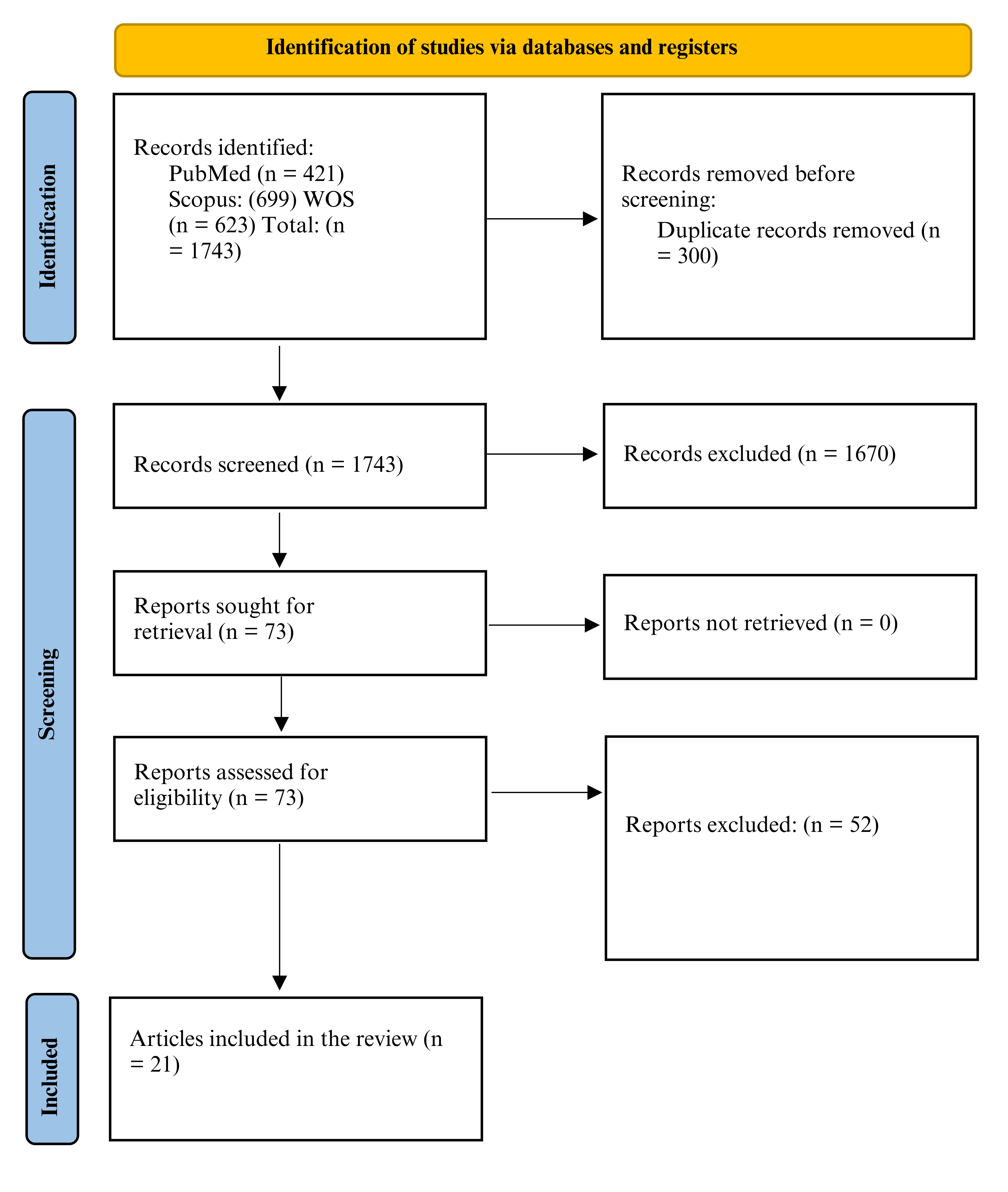
In our systematic review, 1,743 records were initially identified from the searched databases, of which only 1,278 were retrieved after removing 465 duplicate articles. A total of 1212 records were excluded following title and abstract screening, resulting in 66 records for full-text screening. Of these, 45 records did not meet our eligibility criteria. Finally, we included 21 case reports [12,13,14,15,16,17,18,19,20,21,22,23,24,25,26,27,28,29,30,31,32], as shown in Figure 1.
Patients’ Characteristics
Out of review included 21 patients, with a male predominance of 11 males (52.3%) and 9 females (42.8%). The patients’ ages ranged from 18 to 77, with a mean age of 51. The majority of patients were between 50 and 60 years old. Out of these 21 patients, 12 (57.1%) were diagnosed with type 2 diabetes mellitus (T2DM) and 9 (42.8%) with type 1 diabetes mellitus (T1DM). Additionally,
| Author | Age, years | Sex | Diabetes type | Presentation | Troponin, ng/ml | Type of ACS |
|---|---|---|---|---|---|---|
| Baral 2021 [12] | 77 | M | T2DM | SOB, cough, fever for 3 days, oxygen saturation 80% on 10 liters nasal cannula (improved to 95% on a non-rebreather mask with 15L/min). | Initial: 0.923, Peaked at 23.684 | STEMI |
| Baytu0̆11fan 2023 [13] | 61 | M | T2DM | CP, generalized weakness, polyuria, nausea & vomiting for several days. | 161.7 | STEMI |
| Bouzhir 2024 [14] | 76 | F | T2DM | Severe CP at rest, palpitations post-cataract surgery, persistent symptoms after 30 hours, & a dry cough for 10 days. | 3,859 | STEMI |
| Briggs 2022 [15] | 56 | F | T2DM | 48 hours of nausea, vomiting, generalized abdominal pain, and diarrhea. No CP, SOB, or fever. | 212 | STEMI |
| Çakır 2012 [16] | 18 | F | T1DM | Fatigue, weakness, nausea, vomiting for two days, and abdominal pain. No CP. | WNL | Non-STEMI |
| Doherty 2022 [17] | 24 | M | T2DM | CP radiating to the left arm, sweating, tachycardia, 24-h history of vomiting & diarrhoea after alcohol consumption. | 1212 | STEMI |
| Dorcely 2021 [18] | 61 | M | T2DM | Nausea and CP | Negative | Inferior Q waves, unchanged from prior ECG. |
| Eliades 2014 [19] | 71 | F | T2DM | Abdominal pain, vomiting, confusion, & altered mental status. | 5.67 | STEMI |
| Fronczyk 2016 [20] | 20 | M | T1DM (diagnosed 10 years prior) | Weakness, low-grade fever, epigastric and abdominal pain, nausea, vomiting, burning retrosternal pain, and high blood glucose for 2 days. | WNL | STEMI |
| Gerede 2016 [21] | 58 | F | T1DM (diagnosed 33 years prior) | Confusion, nausea, vomiting, cough, sore throat, fever, dehydration, and an inclination to sleep. | 5.43 | Non-STEMI |
| Goto 2021 [22] | 52 | F | T2DM | Tachypnea, vomiting, decreased blood pressure, metabolic acidosis (euglycemic DKA), and myocardial ischemia. | 324.5 | Non-STEMI |
| Kaefer 2019 [23] | 49 | M | T1DM | DKA, nausea, and vomiting after inappropriate insulin therapy, troponin elevated to 142ng/l after 14 h post-admission and peaked at 142 ng/l at 38 h post-admission, no CP but ST elevation less than 2mm. | 33, 14 h post-admission, peaked at 142, 38 h post-admission | STEMI |
| Landa 2021 [24] | 48 | M | T1DM | Diffuse pain and fatigue. He began feeling tired three days prior to the presentation with associated nausea and vomiting, but denied any fever, chills, or CP. | 0.04 | STEMI |
| Lee 2014 [25] | 60 | M | T2DM (New diagnosis) | Intermittent central CP radiated to left shoulder & neck, not affected by posture or exertion, with polydipsia, polyuria, weight loss, fever, sore throat. | 0.77 μg/L | STEMI |
| Mhanna 2020 [26] | 50 | F | T1DM | Lethargy and low Blood pressure. | On admission: 0.21; second day: 20 | STEMI |
| Odubanjo 2017 [27] | Middle age | NA | T1DM | Nausea & vomiting for 1 day. | On admission: 0.012, day 2: 7.3 | STEMI |
| Oriot 2023 [28] | 77 | F | T2DM | CP, later respiratory distress & atrial fibrillation. | 143 | STEMI |
| Petersen 2023 [29] | 28 | M | T2DM | CP, diagnosed as NSTEMI after PCI. | NA | Non-STEMI |
| Umadat 2022 [30] | 61 | M | T1DM | Epigastric abdominal pain, nausea, & vomiting. | 336 | Nonspecific T-wave abnormalities |
| Wray 2020 [31] | 19 | M | T1DM | One day of nausea, vomiting, diffuse back & abdominal pain. Tender abdomen & back without trauma. | Undetectable | STEMI |
| Zughaib 2023 [32] | 54 | F | T2DM | Several days of typical CP, intractable nausea & vomiting. Repeated ED visits. | 0.13 | STEMI |
ACS, Acute coronary syndrome; AFib, Atrial fibrillation; CP, Chest pain; DKA, Diabetic ketoacidosis; ED, Emergency department; F, Female; M, Male; NA, Not available; NSTEMI, Non-ST-elevation myocardial infarction; PCI, Percutaneous coronary intervention; SOB, Shortness of breath; STEMI, ST-elevation myocardial infarction; T1DM, Type 1 diabetes mellitus; T2DM, Type 2 diabetes mellitus; WNL, Within normal limits; ng/mL, Nanograms per milliliter; g/L, micrograms per liter.
| Author | EF% | Coronary Angiography | Home Medication | RBS (mg/dL) | pH | K (mmol/L) | HCO 3 (mEq/L) | HbA1c | Outcome |
|---|---|---|---|---|---|---|---|---|---|
| Baral 2021 [12] | 15–20% | 95% occlusion RCA at ostium, hazy filling defect in stent, patent LAD stent | Aspirin, clopidogrel, atorvastatin, metoprolol | 541 | 7.36 | 5.8 | 16 | NA | DES in RCA. Discharged after 32 days with EF 25–30%. |
| Baytu0̆11fan 2023 [13] | 50% | Total occlusion of proximal LAD | Amlodipine/perindopril, metformin, dapagliflozin | 171 | 7.03 | 5.03 | 5.6 | 9.60% | Successful PCI, insulin and bicarbonate, discharged day 6. |
| Bouzhir 2024 [14] | 43% | No significant stenosis | NA | NA | NA | NA | NA | NA | Full LV recovery, EF 62% post-event. |
| Briggs 2022 [15] | 40–45% | 100% occlusion mid-circumflex, LAD 50%, RCA 60% | Rivaroxaban | 392 | 7.2 | NA | NA | NA | Successful PCI with stent, discharged post-op day 2. |
| Çakır 2012 [16] | NA | Not performed | None | 476 | 7.07 | 2.1 | NA | 11.07% | Resolved with insulin, saline. Discharged stable. |
| Doherty 2022 [17] | 49% | Occlusion of distal PLV branch, RCA thrombus | Olanzapine, fluoxetine | 20.4 mmol/L | metabolic acidosis | 3.2 | 15 | 64 mmol/mol | PCI and insulin therapy. Discharged day 4. |
| Dorcely 2021 [18] | Stress-induced abnormalities | Total occlusion mid-RCA, non-obstructive elsewhere | Empagliflozin, metformin, liraglutide, rosuvastatin, ezetimibe, omeprazole | 84 | metabolic acidosis | NA | 17 | 8.30% | CP and DKA resolved. SGLT2 discontinued. |
| Eliades 2014 [19] | 30% | Normal coronaries | Insulin, metformin | NA | NA | NA | NA | NA | EF improved to 45–50% in 10 days. |
| Fronczyk 2016 [20] | No contractility abnormalities | Normal coronaries | Insulin pump | 33.1 mmol/L | 7.18 | Normal | 5.6 | 10.37% | Pain and ECG normalized. Discharged day 11. |
| Gerede 2016 [21] | No wall motion abnormality | Non-obstructive lesions | Insulin, ramipril | 522 | 7.159 | 6.3–6.4 | 5.8 | NA | ECG/biochem normalized. Discharged day 8. |
| Goto 2021 [22] | NA | Coronary stenting | Empagliflozin, sitagliptin, ezetimibe, rosuvastatin, clopidogrel | 178 | 6.84 | - | 2.1 | NA | ICU, VA-ECMO, CRRT. Discharged ICU day 15. |
| Kaefer 2019 [23] | Apical dyskinesia | No occlusion | - | 38.9 mmol/L | 6.93 | - | 2.7 | 10.4 | Angioplasty, stenting. |
| Landa 2021 [24] | Fair LV function | NA | Insulin only | 952 | 6.94 | 7.6 | <5 | 10.4 | ICU insulin drip. |
| Author | EF% | Coronary Angiography | Home Medication | RBS (mg/dL) | pH | K (mmol/L) | HCO 3 (mEq/L) | HbA1c | Outcome |
|---|---|---|---|---|---|---|---|---|---|
| Lee 2014 [25] | Normal | 50% LCX obstruction | NA | 35.6 mmol/L | 7.25 | NA | 13 | 17% | ICU admission, abscess drainage, 10-week stay. |
| Mhanna 2020 [26] | 60–65% to 25% | Normal coronaries | NA | 1637 | 6.8 | 7.6 | 2.7 | NA | Extubated, Life Vest, outpatient insulin. |
| Odubanjo 2017 [27] | Normal | Non-obstructive | NA | 565 | 6.99 | 5.8 | <5 | NA | DKA resolved. Treated myocarditis. Discharged day 4. |
| Oriot 2023 [28] | 41% | Triple-vessel disease, LAD stented | Metformin, nebivolol, statin, aspirin, spironolactone, citalopram | 247 | 7.2 | NA | 6.1 | 7.80% | Discharged day 22, basal insulin. |
| Petersen 2023 [29] | NA | LAD NSTEMI | Full diabetes and cardiac regimen | 154 | 7.048 | 5.8 | <7 | 11.1 | ICU stay, DKA resolved in 5 days, outpatient follow-up. |
| Umadat 2022 [30] | NA | 99% LAD, 90% diagonal | Aspirin, statin, insulin pump | 326 | 7.39 | 5 | 14 | 8% | Post-DKA PCI with stenting. Discharged on pump. |
| Wray 2020[31] | NA | Normal coronaries | NA | 700 | 7.09 | 5.7 | 9.07 | NA | PCI showed normal coronaries. Discharged day 3. |
| Zughaib 2023 [32] | NA | Ulcerated RCA plaque, mild LAD/LCX disease | Metformin, Empagliflozin | 135 | NA | 3.2 | NA | NA | PCI and stent. Resolved CP and DKA. Discharged. |
DES, drug-eluting stent; EF, ejection fraction; RBS, random blood sugar; RCA, right coronary artery; LAD, left anterior descending artery; LCX, left circumflex artery; PLV, posterior left ventricular branch; NSTEMI, non–ST-elevation myocardial
infarction; PCI, percutaneous coronary intervention; DKA, diabetic ketoacidosis; VA-ECMO, veno-arterial extracorporeal membrane oxygenation; SGLT2, sodium–glucose cotransporter-2 inhibitor; T1DM, type 1 diabetes mellitus; HbA1c,
hemoglobin A1c; NA, not available; mg/dL, milligrams per deciliter; mmol/L, millimoles per liter; mEq/L, milliequivalents per liter; pH, potential of hydrogen; HCO3, bicarbonate; SC, subcutaneous; BID, twice daily; TIMI, Thrombolysis In Myocardial Infarction flow grade; GRACE, Global Registry of Acute Coronary Events; CP, chest pain; AFib, atrial fibrillation; ICU, intensive care unit.
six patients (28.5%) were reported to be on SGLT2 inhibitors, as shown in Table 1.
Clinical Presentation
The most common presenting symptoms included chest pain (n = 11, 52.3%), nausea and vomiting (n = 5, 23.8%), and fatigue or weakness (n = 4, 19.0%). Other reported symptoms included abdominal pain, shortness of breath, and altered mental status, with a duration of symptoms before presentation ranging from several hours to several days, as shown in Table 1.
Vital Signs
Initial vital signs showed variable presentations. Blood pressure measurements ranged from hypotensive (lowest recorded 74/64 mmHg) to hypertensive (highest recorded 193/83 mmHg). Heart rate was frequently elevated, ranging from 69 to 155 beats per minute. Respiratory rates were notably high in several patients, ranging from 15 to 40 breaths per minute, with many patients presenting with Kussmaul breathing, a characteristic of metabolic acidosis. The body temperature was generally within the normal range; however, some patients presented with fever.
Laboratory Findings
Blood glucose levels varied widely, ranging from 84 to 1637 mg/dL, with a notable finding that five patients presented with euglycemic DKA (glucose <250 mg/dL) while on SGLT2 inhibitors. Arterial pH was significantly reduced in most cases, ranging from 6.80 to 7.39, indicating severe acidosis in many patients. Serum bicarbonate levels were markedly decreased, with values as low as <5 mEq/L, confirming metabolic acidosis. Additionally, serum potassium levels varied from hypokalemic (2.1 mmol/L) to hyperkalemic (7.6 mmol/L), as shown in Table 2.
Cardiac Markers
Troponin levels were elevated in most patients, ranging from slightly elevated to markedly increased (maximum recorded 23.684 ng/mL). Several patients showed progressive elevation of troponin levels during hospitalization, peaking hours after admission, as shown in Table 1.
Glycemic Control
Glycated hemoglobin (HbA1c) values were reported in 11 patients, ranging from 7.8% to 17%, indicating poor long-term glycemic control in most cases, with a mean HbA1c of 10.4% Table 2.
Electrocardiographic Findings
ECG abnormalities were observed in almost all patients. ST-segment elevation was the most common finding (n=13, 61.9%), predominantly in the inferior (leads II, III, aVF) or anterolateral leads (V1-V6). ST-segment depression was noted in 9 patients (42.8%). Other ECG findings included T-wave inversions, bundle branch blocks, and atrial fibrillation, as shown in Table 1.
DKA management
All patients received standard DKA management, including intravenous insulin infusion and fluid resuscitation. Electrolyte replacement, particularly potassium, was administered as needed. In one case of severe acidosis, sodium bicarbonate was used, as shown in Table 2.
Coronary Interventions
Coronary angiography was performed in 18 patients (85.7%), and percutaneous coronary intervention (PCI) with stenting was performed in 4 patients (19%). Notably, five patients had normal coronary arteries on angiography despite presenting with clinical and ECG findings suggestive of ACS, as shown in Table 2.
Risk of Bias and Quality Assessment
Quality assessment using the CARE checklist revealed that studies demonstrated high compliance for most reporting domains: demographics (21/21, 100%), current clinical condition (21/21, 100%), diagnostic tests (21/21, 100%), and treatment procedures (21/21, 100%) were adequately described in all studies, while post-intervention conditions (20/21, 95%) and timeline presentation of history (17/21, 81%) were well-documented in the majority of cases. However, adverse event reporting was suboptimal, with only 8 out of 21 studies (38%) providing adequate documentation, and takeaway lessons were present in 12 out of 21 studies (57%).
Discussion
The relationship between DKA and ACS is bidirectional, with each condition capable of precipitating and influencing the pathogenesis of the other. ACS can precipitate DKA through a stress response, that occurs due to the release of significant amount of counter-regulatory hormones, such as epinephrine, glucagon and cortisol [23]. This hormonal surge raises blood glucose level, antagonizes the action of insulin, promotes fat breakdown, facilitates the production of ketones and eventually triggers the development of DKA [33]. On the other hand, DKA can lead to the occurrence of myocardial ischemic damage by a multitude of mechanisms, including free fatty acids accumulation, metabolic acidosis and counter-regulatory hormones [34].
When acidosis of the blood occurs, it leads to the acceleration of intracellular ionic movement of calcium , resulting in the accumulation of inside the myocardial cells. This excess intracellular facilitates the process of protein breakdown, known as proteolysis [35]. This process is accompanied by a reduced ability of calcium to bind to the contractile proteins in the heart muscle—an effect caused by acidosis—which ultimately leads to impaired pump function, a phenomenon known as myocardial stunning. Ultimately, the combined effect of proteolysis and myocardial stunning results in myocardial injury leading to an increase in the serum cardiac troponin level [36]. Moreover, acute decompensation of diabetes is associated with a sudden increase in the levels of counter-regulating hormones, such as epinephrine, cortisol, and glucagon, to try to increase the blood glucose level and provide energy during exacerbation [37,36]. However, these hormones can significantly increase the workload and metabolic demand of the cardiac muscle. In patients who already have coronary artery disease (CAD), this increase in the myocardial oxygen demand can induce the occurrence of ACS [37]. Additionally, acute diabetic decompensation such as DKA is associated with a significant increase in the level of circulating free fatty acids (FFA). The excess FFA can be incorporated into the myocytes of the heart muscle, forming micelle-like formations that weaken and destabilize the cell membrane [38]. Additionally, the severe lack of insulin, associated with increased ketone bodies and FFA, results in dysfunctional glucose uptake by the cardiac muscles and promotes the alternative uptake of FFA as the main energy source [38,37].
Generally, the interpretation of cardiac biomarkers, such as troponin, in DKA patients can be challenging, as ACS may either initiate the onset of DKA or arise as a consequence of it [31]. Additionally, the increase in troponin associated with DKA patients could be due to either the presence of underlying CAD disease, which is exacerbated by metabolic stress and acidosis, or to the infiltration of myocytes by the excess FFA associated with the severe insulin deficiency [39,40]. This could explain the absence of coronary abnormalities on some patients despite a rise in troponin. Moreover, our analysis revealed that both type 1 and type 2 diabetes mellitus (T1DM and T2DM) were present almost equally (42.8% vs. 57.1%), underscoring that neither diabetes phenotype is exempt from the risk of ACS during DKA. This demographic pattern aligns with established epidemiological data for acute coronary syndrome, where middle-aged males represent the highest-risk population, suggesting that traditional ACS risk factors may contribute to the concurrent presentation. T1DM patients are at risk of acquiring DKA due to their absolute insulin deficiency. In contrast, T2DM patients suffer from relative insulin deficiency, which is exacerbated by the rise in counter-regulatory hormones in cases of severe events, such as infection or stress [41,42].
Clinical presentation of patients included a wide variety of symptoms and signs, such as chest pain, abdominal pain, nausea, vomiting, fatigue, altered mental status, hypertension, hypotension and tachypnea. The most common symptom presented was chest pain, however it should be noted that it was absent in nearly half of the patients. Our analysis demonstrated that some patients-mainly old patients- did not report chest pain, despite having high troponin and coronary artery abnormalities. This could be attributed to the sensory neuropathy associated with DM, which can decrease the pain perception of these patients, preventing the sensation of chest pain during the anginal attack [43]. The absence of chest pain in these patients poses a critical diagnostic challenge, potentially leading to delayed recognition of ACS. This atypical presentation, combined with the metabolic focus required for DKA management, creates a scenario where cardiac evaluation may be deprioritized. Healthcare providers managing DKA patients should maintain heightened awareness for silent myocardial ischemia, particularly in patients with cardiovascular risk factors, unexplained hemodynamic instability, or ECG changes that cannot be attributed solely to electrolyte abnormalities [44].
This fact complicates the process of ischemic heart disease diagnosis in DM patients, as in these patients the diagnosis is mainly based on the presence of non-specific angina-equivalent symptoms such as dyspnea and dizziness along with abnormal glucose level [45].
In the majority of cases, DKA was associated with an increased blood glucose level; however, in six cases of individuals receiving SGLT2i drugs at home, euglycemic DKA with a blood glucose level below 250 mg/dL was a prominent feature. Notably, euglycemic DKA occurred in 6 patients (28.6%), which contrasts sharply with the general DKA population, where euglycemic presentations account for only 2.6-3.2% of cases [48). This atypical presentation of DKA can delay the diagnosis and obscure the coexisting myocardial damage [46]. SGLT2i drugs can cause euglycemic DKA by enhancing urinary glucose excretion, thereby lowering blood glucose levels, which in turn can lead to a reduction in insulin secretion or dosing. Additionally, SGLT2i can also stimulate glucagon release, which, when accompanied by a diminished insulin level, promotes a shift in metabolism towards lipolysis, leading to increased ketone production [47]. Thus, the reliance on high blood glucose level alone to diagnose DKA is not a proper clinical practice, especially in patients who are receiving oral anti-diabetic drugs [48]. Additionally, this also underscores the importance of heightened monitoring of DKA patients who are on SGLT2i drugs and the need for immediate discontinuation of the drug in cases of DKA suspicion [49]. The association between SGLT2 inhibitors and euglycemic DKA in our cohort raises important ethical considerations regarding informed consent and risk-benefit communication. Clinicians prescribing SGLT2 inhibitors must ensure that patients understand the risk of DKA, particularly those with additional risk factors, such as insulin deficiency, acute illness, or planned procedures. The cardiovascular benefits of SGLT2 inhibitors are well-established, but the potential for life-threatening ketoacidosis, especially with atypical presentations, necessitates careful patient selection, education, and monitoring protocols [50].
Regarding the analysis of the treatments provided, all of the included cases received the standard DKA therapy, which included aggressive fluid resuscitation, electrolyte correction and IV insulin infusion [51]. Only one patient who was associated with severe metabolic acidosis (PH>7, HCO3=5.6) was indicated to receive intravenous bicarbonate administration. This aligns with current recommendations which deter from routine use of bicarbonate in the treatment of DKA, due to the risk of cerebral edema [52]. Hence, the use of bicarbonate should be solely exclusive in cases with refractory metabolic acidosis that failed to be corrected with standard DKA therapy [53]. Furthermore, 18 cases underwent PCI for the treatment of ACS, of which five patients had normal coronaries suggesting either demand ischemia or coronary vasospasm.
In our review, despite the severity of the presentation of some cases, the majority of the patients survived the acute phase and no mortality was reported. However, some cases, particularly patients with severe metabolic acidosis and elevated cardiac biomarkers, required prolonged hospitalization after undergoing the PCI procedure or after admission to the ICU. Patients with euglycemic DKA on SGLT2i drugs suffered from delayed diagnosis which might have influenced their clinical course and prolonged their hospital stay. Furthermore, whereas no mortality was reported in the selected cohort, the lack of long-term follow up for potential complications limit our understanding of the true long-term prognosis of those patients.
Another important observation is the significantly high HbA1c level in the reported cases, indicating in the process significantly poorly controlled diabetic status. In literature, patients with HbA1c levels above 9% are significantly more likely to develop DKA compared to those with lower levels with one study reporting that this risk could be five times more likely compared to patients with HbA1c levels between 7% and 8% [54]. Moreover, the level of HbA1c could correlate to the severity level of DKA as well as the severity level of the associated metabolic acidosis [55]. Furthermore, elevated HbA1c level alone is an independent risk factor for ACS and is associated with significantly worse outcomes and prognosis [56]. This highlights the importance of good monitoring of diabetic patients, especially patients who are at high risk for CAD [57]. Proper control of blood glucose level over the long-term can be beneficial in preventing the exacerbating events of DKA and ACS [58]. Our findings advance clinical understanding in several key areas: First, the prevalence of euglycemic DKA in concurrent presentations is substantially higher than in general DKA populations, suggesting unique pathophysiological or pharmacological mechanisms. Second, the high frequency of atypical presentations without chest pain necessitates revised diagnostic protocols that include routine cardiac evaluation in DKA patients with cardiovascular risk factors. Third, the prominence of SGLT2 inhibitor-associated cases establishes this drug class as a key risk factor requiring enhanced monitoring and patient education. These insights should inform updated clinical guidelines for both emergency department protocols and SGLT2 inhibitor prescribing practices.
This systematic review aggregated all the known case reports on this subject in the literature, in order to provide a comprehensive, up-to-date assessment of the relationship between DKA and ACS. However, this study still has some limitations that should be considered, including the inherent biases of the study design. A case report was the best available option to address this issue, given the rarity of concurrent DKA and ACS, higher-level evidence, such as cohort studies or RCTs, is not feasible. Additional limitations included heterogeneity in the diagnostic criteria and management protocols, publication bias, a small sample size, and a lack of control groups and long-term follow-up. Thus, larger-scale observational prospective or retrospective studies are required to address these limitations.
Conclusion
In conclusion, our review highlights the bidirectional association between DKA and ACS. Clinicians should maintain a low threshold for suspicion of concurrent DKA in patients presenting with ACS, particularly when traditional DKA symptoms (polyuria, polydipsia, nausea) are absent. Routine ECG, troponin assessment and echocardiography are essential to differentiate between true ischemia and metabolic mimicry. Ultimately, early recognition, standard treatment and use of coronary angiography are pivotal to optimizing outcomes.
Conflicts of Interest
The authors declare no competing interests that could have influenced the objectivity or outcome of this research.
Funding Source
This research did not receive any specific grant from funding agencies in the public, commercial, or not-for-profit sectors
Acknowledgments
None
Institutional Review Board (IRB)
This study is a systematic review of previously published case reports. No human subjects research was conducted and no institutional review board approval was required.
Large Language Model
None
Authors Contribution
AE handled conceptualization, data curation, validation, methodology, reference management, and writing of the original draft, while AFG provided resources, data curation, validation, methodology, and writing of the original draft. ON was responsible for data curation, validation, and writing of the original draft, and LMA managed data curation, methodology, and writing of the original draft. MYE, MM, and AAA oversaw data curation, validation, methodology, and preparation of tables. AH supervised the work and contributed to writing, review, and editing.
Data Availability
All studies used in the research are available in various databases.
References
- Ramphul K., Joynauth J.. An Update on the Incidence and Burden of Diabetic Ketoacidosis in the U.S. Diabetes Care. 2020;43(12):e196-e197. doi:10.2337/dc20-1258 PMID: 33051332
- Desai D., Mehta D., Mathias P., Menon G., Schubart U. K.. Health Care Utilization and Burden of Diabetic Ketoacidosis in the U.S. Over the Past Decade: A Nationwide Analysis. Diabetes Care. 2018;41(8):1631-1638. doi:10.2337/dc17-1379 PMID: 29773640
- Delaney M. F., Zisman A., Kettyle W. M.. Diabetic ketoacidosis and hyperglycemic hyperosmolar nonketotic syndrome. Endocrinol Metab Clin North Am. 2000;29(4):683-705, V. doi:10.1016/s0889-8529(05)70159-6 PMID: 11149157
- Kitabchi A. E., Wall B. M.. Diabetic ketoacidosis. Med Clin North Am. 1995;79(1):9-37. doi:10.1016/s0025-7125(16)30082-7 PMID: 7808097
- Rao S. V., O'Donoghue M. L., Ruel M., Rab T., Tamis-Holland J. E., Alexander J. H., Baber U., Baker H., Cohen M. G., Cruz-Ruiz M., Davis L. L., de Lemos J. A., DeWald T. A., Elgendy I. Y., Feldman D. N., Goyal A., Isiadinso I., Menon V., Morrow D. A., Mukherjee D., Platz E., Promes S. B., Sandner S., Sandoval Y., Schunder R., Shah B., Stopyra J. P., Talbot A. W., Taub P. R., Williams M. S.. Correction to: 2025 ACC/AHA/ACEP/NAEMSP/SCAI Guideline for the Management of Patients With Acute Coronary Syndromes: A Report of the American College of Cardiology/American Heart Association Joint Committee on Clinical Practice Guidelines. Circulation. 2025;151(13):e865. doi:10.1161/CIR.0000000000001328 PMID: 40163565
- Sarkees M. L., Bavry A. A.. Acute coronary syndrome (unstable angina and non-ST elevation MI). BMJ Clin Evid. 2009;2009:0209. PMID: 19445778 PMCID: PMC2907796
- Yusuf S., Hawken S., Ounpuu S., Dans T., Avezum A., Lanas F., McQueen M., Budaj A., Pais P., Varigos J., Lisheng L., INTERHEART Study Investigators. Effect of potentially modifiable risk factors associated with myocardial infarction in 52 countries (the INTERHEART study): case-control study. Lancet. 2004;364(9438):937-52. doi:10.1016/S0140-6736(04)17018-9 PMID: 15364185
- Einarson T. R., Acs A., Ludwig C., Panton U. H.. Prevalence of cardiovascular disease in type 2 diabetes: a systematic literature review of scientific evidence from across the world in 2007-2017. Cardiovasc Diabetol. 2018;17(1):83. doi:10.1186/s12933-018-0728-6 PMID: 29884191 PMCID: PMC5994068
- Al-Mallah M., Zuberi O., Arida M., Kim H. E.. Positive troponin in diabetic ketoacidosis without evident acute coronary syndrome predicts adverse cardiac events. Clin Cardiol. 2008;31(2):67-71. doi:10.1002/clc.20167 PMID: 18257021 PMCID: PMC6653175
- Page M. J., McKenzie J. E., Bossuyt P. M., Boutron I., Hoffmann T. C., Mulrow C. D., Shamseer L., Tetzlaff J. M., Akl E. A., Brennan S. E., Chou R., Glanville J., Grimshaw J. M., Hrobjartsson A., Lalu M. M., Li T., Loder E. W., Mayo-Wilson E., McDonald S., McGuinness L. A., Stewart L. A., Thomas J., Tricco A. C., Welch V. A., Whiting P., Moher D.. The PRISMA 2020 statement: an updated guideline for reporting systematic reviews. BMJ. 2021;372:n71. doi:10.1136/bmj.n71 PMID: 33782057 PMCID: PMC8005924
- Ouzzani M., Hammady H., Fedorowicz Z., Elmagarmid A.. Rayyan-a web and mobile app for systematic reviews. Syst Rev. 2016;5(1):210. doi:10.1186/s13643-016-0384-4 PMID: 27919275 PMCID: PMC5139140
- Baral N., Montalbano A., Khan A., Qureshi M., Luitel P.. Silent Myocardial Infarction and Acute Multiorgan Failure in a COVID-19 Patient: A Case Report. JNMA J Nepal Med Assoc. 2021;59(242):1048-1051. doi:10.31729/jnma.6933 PMID: 35199691 PMCID: PMC9107826
- Baytugan N. Z., Celik A. I., Bezgin T., Cagdas M.. Euglycemic diabetic ketoacidosis associated with ST segment elevation myocardial infarction following SGLT-2 inhibitor therapy. Am J Emerg Med. 2023;71:250 e1-250 e3. doi:10.1016/j.ajem.2023.07.007 PMID: 37451968
- Bouzhir A., Taoufik Benchekroun R., Doghmi N., Zarzur J., Cherti M.. Unmasking the Intricate Association Between Takotsubo Syndrome, Atrial Fibrillation, and Diabetes Mellitus: A Case Report and Literature Review. Cureus. 2024;16(8):e66516. doi:10.7759/cureus.66516 PMID: 39252720 PMCID: PMC11381128
- Briggs B., Zitelny E., Zitelny T.. Abdominal and Pelvic Computed Tomography for the Diagnosis of ST-Elevation Myocardial Infarction: The Challenges of Acute Coronary Syndrome in Diabetic Patients. Cureus. 2022;14(10):e30816. doi:10.7759/cureus.30816 PMID: 36451633 PMCID: PMC9701564
- Cakir Evrim, Ozbek Mustafa, Colak Nujen, Cakal Erman, Delibasi Tuncay. Hypokalemia with ECG Abnormalities Mimicking Acute Coronary Syndrome in a Diabetic Ketoacidotic Patient. Journal of Academic Emergency Medicine Case Reports. 2012;3(1):18-20. doi:10.5505/jaemcr.2011.78942
- Doherty D. J., Sykes R., Connelly P., Hood S.. ST-elevation myocardial infarction due to coronary thrombus in a young patient with diabetic ketoacidosis and a new diagnosis of type 2 diabetes. BMJ Case Rep. 2022;15(3). doi:10.1136/bcr-2021-247090 PMID: 35236686 PMCID: PMC8895887
- Dorcely B., Nitis J., Schwartzbard A., Newman J. D., Goldberg I. J., Sum M.. A Case Report: Euglycemic Diabetic Ketoacidosis Presenting as Chest Pain in a Patient on a Low Carbohydrate Diet. Curr Diabetes Rev. 2021;17(2):243-246. doi:10.2174/1573399816666200316112709 PMID: 32178617 PMCID: PMC8020366
- Eliades M., El-Maouche D., Choudhary C., Zinsmeister B., Burman K. D.. Takotsubo cardiomyopathy associated with thyrotoxicosis: a case report and review of the literature. Thyroid. 2014;24(2):383-9. doi:10.1089/thy.2012.0384 PMID: 23560557 PMCID: PMC3926154
- Fronczyk Aneta, Molęda Piotr, Majkowska Liliana. Clinical and ECG patterns of pseudoinfarction in a young man with type 1 diabetes, diabetic ketoacidosis and normokalaemia. Clinical Diabetology. 2016;5(2):73-76. doi:10.5603/dk.2016.0012
- Gerede Demet Menekşe, Kozluca Volkan, Esenboğa Kerim, Aydoğan Berna İmge, Tutar Eralp. Markedly Elevated Troponin in Diabetic Ketoacidosis without Acute Coronary Syndrome. Turkish Journal of Endocrinology and Metabolism. 2016;20(2):58-60. doi:10.4274/tjem.2997
- Goto S., Ishikawa J. Y., Idei M., Iwabuchi M., Namekawa M., Nomura T.. Life-Threatening Complications Related to Delayed Diagnosis of Euglycemic Diabetic Ketoacidosis Associated with Sodium-Glucose Cotransporter-2 Inhibitors: A Report of 2 Cases. Am J Case Rep. 2021;22:e929773. doi:10.12659/AJCR.929773 PMID: 33723205 PMCID: PMC7980085
- Kaefer K., Botta I., Mugisha A., Berdaoui B., De Bels D., Attou R., Honore P. M., Redant S.. Acute coronary syndrome and diabetic keto acidosis: the chicken or the egg?. Ann Transl Med. 2019;7(16):397. doi:10.21037/atm.2019.07.38 PMID: 31555711 PMCID: PMC6736820
- Landa E., Sharifi S., Abraham J., Vigandt E., Munzinger E.. Brugada Pattern Phenocopy Induced by Diabetic Ketoacidosis. Cureus. 2021;13(5):e15066. doi:10.7759/cureus.15066 PMID: 34141510 PMCID: PMC8206546
- Lee H. H., Chu C. Y., Su H. M., Lin T. H., Voon W. C., Lai W. T., Sheu S. H., Hsu P. C.. Purulent pericarditis, multi-site abscesses and ketoacidosis in a patient with newly diagnosed diabetes: a rare case report. Diabet Med. 2014;31(7):e25-8. doi:10.1111/dme.12458 PMID: 24698085
- Mhanna M., Beran A., Srour O., Ghazaleh S., Elzanaty A.. A Case of Takotsubo Cardiomyopathy Triggered by Diabetic Ketoacidosis and Hypothermia. Cureus. 2020;12(10):e10842. doi:10.7759/cureus.10842 PMID: 33173648 PMCID: PMC7647843
- Odubanjo A. A., Kalisetti R., Adrah R., Ajenifuja A., Joseph B., Zaman M.. Severe Myopericarditis in Diabetic Ketoacidosis-All Troponin are Not Myocardial Infarction. Clin Med Insights Case Rep. 2018;11:1179547618763356. doi:10.1177/1179547618763356 PMID: 29568222 PMCID: PMC5858608
- Oriot P., Hermans M. P., Beauloye C., Rogghe P. A., Noel S., Paternotte E.. Unsuspected diabetic ketoacidosis after myocardial infarction in a patient treated with SGLT2 inhibitor increased length of stay in the hospital: how can it be prevented? A case report. Eur Heart J Case Rep. 2023;7(8):ytad336. doi:10.1093/ehjcr/ytad336 PMID: 37681058 PMCID: PMC10481769
- Petersen C., Gyabaah F., Sotelo J., Yohanna S., Deoker A.. A Case of Prolonged Recovery for Post-percutaneous Coronary Intervention (PCI) Sodium-Glucose Cotransporter-2 (SGLT2) Inhibitor-Induced Euglycemic Diabetic Ketoacidosis in a 28-Year-Old. Cureus. 2023;15(9):e45180. doi:10.7759/cureus.45180 PMID: 37842482 PMCID: PMC10575758
- Umadat G., Moktan V., Shapiro B.. 61-Year-Old Man With Nausea and Vomiting. Mayo Clin Proc. 2022;97(1):170-175. doi:10.1016/j.mayocp.2021.04.031 PMID: 34862073
- Wray J., Yoo M. J., Bridwell R. E., Tannenbaum L., Henderson J.. ST-Segment Elevation in the Setting of Diabetic Ketoacidosis: Is It Acute Coronary Syndrome?. Cureus. 2020;12(3):e7409. doi:10.7759/cureus.7409 PMID: 32337133 PMCID: PMC7182167
- Zughaib M., Basharat B., Small D.. A Case of STEMI-Induced Euglycemic Diabetic Ketoacidosis in a Patient Receiving a Sodium Glucose Cotransporter-2 Inhibitor. JACC Case Rep. 2023;11:101792. doi:10.1016/j.jaccas.2023.101792 PMID: 37077449 PMCID: PMC10107034
- Manikkan A. T.. Elevated Troponin I Levels in Diabetic Ketoacidosis Without Obstructive Coronary Artery Disease. J Endocr Soc. 2018;2(9):1020-1023. doi:10.1210/js.2018-00152 PMID: 30187014 PMCID: PMC6117402
- Eubanks A., Raza F., Alkhouli M., Glenn A. N., Homko C., Kashem A., Bove A.. Clinical significance of troponin elevations in acute decompensated diabetes without clinical acute coronary syndrome. Cardiovasc Diabetol. 2012;11:154. doi:10.1186/1475-2840-11-154 PMID: 23270513 PMCID: PMC3549932
- Carrizales-Sepulveda E. F., Vera-Pineda R., Jimenez-Castillo R. A., Violante-Cumpa J. R., Flores-Ramirez R., Ordaz-Farias A.. The Heart in Diabetic Ketoacidosis: A Narrative Review Focusing on the Acute Cardiac Effects and Electrocardiographic Abnormalities. Am J Med Sci. 2021;361(6):690-701. doi:10.1016/j.amjms.2020.11.030 PMID: 33941367
- Kadel A., Thakur K. K., Rauniyar B. K., Adhikari R. B., Limbu D., Malla R.. Acute Myocardial Infarction and Diabetic Ketoacidosis: The Lethal Duo. J Nepal Health Res Counc. 2021;19(1):212-214. doi:10.33314/jnhrc.v19i1.3193 PMID: 33934164
- Carrizales-Sepulveda E. F., Ordaz-Farias A., Vera-Pineda R., Rodriguez-Gutierrez R., Flores-Ramirez R.. Comprehensive echocardiographic and biomarker assessment of patients with diabetic ketoacidosis. Cardiovasc Diabetol. 2024;23(1):385. doi:10.1186/s12933-024-02471-0 PMID: 39468588 PMCID: PMC11520802
- Ayoub A. A., Addas M. J., Alghamdi A. A., Alghazzawi N., Bakhsh A., Alharbi T.. The Effect of COVID-19 Lockdown on Cerebrovascular Accidents, Acute Coronary Syndrome, and Diabetic Ketoacidosis Visits the Emergency Department: A Retrospective Study. Cureus. 2022;14(12):e33154. doi:10.7759/cureus.33154 PMID: 36601185 PMCID: PMC9803928
- Jeremias A., Gibson C. M.. Narrative review: alternative causes for elevated cardiac troponin levels when acute coronary syndromes are excluded. Ann Intern Med. 2005;142(9):786-91. doi:10.7326/0003-4819-142-9-200505030-00015 PMID: 15867411
- Poojary I., Khalid U., Patra T., Giri J., Al Heyasat A., Basith S., Palasamudram Shekar S., Balachandran A., Haggag A.. Troponinemia in Patients With Diabetic Ketoacidosis Without Acute Coronary Syndrome. Cureus. 2024;16(5):e61064. doi:10.7759/cureus.61064 PMID: 38915971 PMCID: PMC11195327
- Fazeli Farsani S., Brodovicz K., Soleymanlou N., Marquard J., Wissinger E., Maiese B. A.. Incidence and prevalence of diabetic ketoacidosis (DKA) among adults with type 1 diabetes mellitus (T1D): a systematic literature review. BMJ Open. 2017;7(7):e016587. doi:10.1136/bmjopen-2017-016587 PMID: 28765134 PMCID: PMC5642652
- Klingensmith G. J., Connor C. G., Ruedy K. J., Beck R. W., Kollman C., Haro H., Wood J. R., Lee J. M., Willi S. M., Cengiz E., Tamborlane W. V., Pediatric Diabetes Consortium. Presentation of youth with type 2 diabetes in the Pediatric Diabetes Consortium. Pediatr Diabetes. 2016;17(4):266-73. doi:10.1111/pedi.12281 PMID: 25951940
- Celik A., Karayakali M., Erkorkmaz U., Altunkas F., Karaman K., Koc F., Ceyhan K., Kadi H., Avsar A.. Presence of angina pectoris is related to extensive coronary artery disease in diabetic patients. Clin Cardiol. 2013;36(8):475-9. doi:10.1002/clc.22151 PMID: 23754690 PMCID: PMC6649393
- Perona M., Cooklin A., Thorpe C., O'Meara P., Rahman M. A.. Symptomology, Outcomes and Risk Factors of Acute Coronary Syndrome Presentations without Cardiac Chest Pain: A Scoping Review. Eur Cardiol. 2024;19:e12. doi:10.15420/ecr.2023.45 PMID: 39081484 PMCID: PMC11287626
- Merkler A. E., Bartz T. M., Kamel H., Soliman E. Z., Howard V., Psaty B. M., Okin P. M., Safford M. M., Elkind M. S. V., Longstreth W. T., Jr.. Silent Myocardial Infarction and Subsequent Ischemic Stroke in the Cardiovascular Health Study. Neurology. 2021;97(5):e436-e443. doi:10.1212/WNL.0000000000012249 PMID: 34031202 PMCID: PMC8356380
- Kaze A. D., Zhuo M., Kim S. C., Patorno E., Paik J. M.. Association of SGLT2 inhibitors with cardiovascular, kidney, and safety outcomes among patients with diabetic kidney disease: a meta-analysis. Cardiovasc Diabetol. 2022;21(1):47. doi:10.1186/s12933-022-01476-x PMID: 35321742 PMCID: PMC9491404
- Visser M. M., Mathieu C., Gillard P.. Diabetic Ketoacidosis After Sodium-Glucose Cotransporter Inhibitor Initiation Under Advanced Hybrid Closed-Loop Therapy in Type 1 Diabetes. Diabetes Technol Ther. 2022;24(7):516-519. doi:10.1089/dia.2021.0553 PMID: 35156849
- Levine J. A., Karam S. L., Aleppo G.. SGLT2-I in the Hospital Setting: Diabetic Ketoacidosis and Other Benefits and Concerns. Curr Diab Rep. 2017;17(7):54. doi:10.1007/s11892-017-0874-3 PMID: 28597228
- Marilly E., Cottin J., Cabrera N., Cornu C., Boussageon R., Moulin P., Lega J. C., Gueyffier F., Cucherat M., Grenet G.. SGLT2 inhibitors in type 2 diabetes: a systematic review and meta-analysis of cardiovascular outcome trials balancing their risks and benefits. Diabetologia. 2022;65(12):2000-2010. doi:10.1007/s00125-022-05773-8 PMID: 35925319
- Chow E., Clement S., Garg R.. Euglycemic diabetic ketoacidosis in the era of SGLT-2 inhibitors. BMJ Open Diabetes Res Care. 2023;11(5). doi:10.1136/bmjdrc-2023-003666 PMID: 37797963 PMCID: PMC10551972
- Barski L., Golbets E., Jotkowitz A., Schwarzfuchs D.. Management of diabetic ketoacidosis. Eur J Intern Med. 2023;117:38-44. doi:10.1016/j.ejim.2023.07.005 PMID: 37419787
- Dunn B. K., Coore H., Bongu N., Brewer K. L., Kumar D., Malur A., Alkhalisy H.. Treatment Challenges and Controversies in the Management of Critically Ill Diabetic Ketoacidosis (DKA) Patients in Intensive Care Units. Cureus. 2024;16(9):e68785. doi:10.7759/cureus.68785 PMID: 39360087 PMCID: PMC11446492
- Patel M. P., Ahmed A., Gunapalan T., Hesselbacher S. E.. Use of sodium bicarbonate and blood gas monitoring in diabetic ketoacidosis: A review. World J Diabetes. 2018;9(11):199-205. doi:10.4239/wjd.v9.i11.199 PMID: 30479686 PMCID: PMC6242725
- Wysham Carol, Preblick Ronald, Zhou Fang Liz, Davies Michael, Paranjape Sachin, Sansgiry Sujit, Xu Qingqing, Joish Vijay. OR14-3 The Association of Glycemic Control and Glycemic Variability with Treatment Patterns and Diabetes-Related Events in the Adult Patients (Pts) with Type 1 Diabetes (T1D): Results from the T1D Exchange Clinic Registry. Journal of the Endocrine Society. 2019;3(Supplement_1). doi:10.1210/js.2019-OR14-3
- Lamsal R., Klyachman L., Adeyinka A., Kondamudi N., Pierre L.. Is HbA1c an Indicator of Diabetic Ketoacidosis Severity in the Pediatric Population?. Pediatr Emerg Care. 2023;39(4):216-218. doi:10.1097/PEC.0000000000002859 PMID: 36727771
- Ekram S. Saeed M. D. Mohammed S. Kasem M. Sc, Wael A. Hassib M. D. Yasser H. El-Barbary M. D.. Value of Glycated Hemoglobin (HbA1c%) as an Indicator of Severity of Acute Coronary Syndrome in Non-Diabetic Adults. The Medical Journal of Cairo University. 2018;86(March):425-432. doi:10.21608/mjcu.2018.55142
- Wang M., Hng T. M.. HbA1c: More than just a number. Aust J Gen Pract. 2021;50(9):628-632. doi:10.31128/AJGP-03-21-5866 PMID: 34462769
- Guo K., Zhao Q., Wang M., Lu Y., Wo M., Zhou X., Ying C.. The Scope of HbA1c Variability and Risk of Vascular Complications Among Patients with Type 2 Diabetes: A Systematic Review and Meta-Analysis of Prospective Studies. Horm Metab Res. 2022;54(2):94-103. doi:10.1055/a-1730-4904 PMID: 35130570
References
1. Ramphul K, Joynauth J. An Update on the Incidence and Burden of Diabetic Ketoacidosis in the U.S. Diabetes Care. 2020: e196 [PMID: 33051332, https://doi.org/10.2337/dc20-1258]
2. Desai D, Mehta D, Mathias P, Menon G, Schubart UK. Health Care Utilization and Burden of Diabetic Ketoacidosis in the U.S. Over the Past Decade: A Nationwide Analysis. Diabetes Care. 2018: 1631 [PMID: 29773640, https://doi.org/10.2337/dc17-1379]
3. Delaney MF, Zisman A, Kettyle WM. Diabetic ketoacidosis and hyperglycemic hyperosmolar nonketotic syndrome. Endocrinol Metab Clin North Am. 2000: 683 [PMID: 11149157, https://doi.org/10.1016/s0889-8529(05)70159-6]
4. Kitabchi AE, Wall BM. Diabetic ketoacidosis. Med Clin North Am. 1995: 9 [PMID: 7808097, https://doi.org/10.1016/s0025-7125(16)30082-7]
5. Rao SV, O'Donoghue ML, Ruel M, Rab T, Tamis-Holland JE, Alexander JH, Baber U, Baker H, Cohen MG, Cruz-Ruiz M, Davis LL, de Lemos JA, DeWald TA, Elgendy IY, Feldman DN, Goyal A, Isiadinso I, Menon V, Morrow DA, Mukherjee D, Platz E, Promes SB, Sandner S, Sandoval Y, Schunder R, Shah B, Stopyra JP, Talbot AW, Taub PR, Williams MS. Correction to: 2025 ACC/AHA/ACEP/NAEMSP/SCAI Guideline for the Management of Patients With Acute Coronary Syndromes: A Report of the American College of Cardiology/American Heart Association Joint Committee on Clinical Practice Guidelines. Circulation. 2025: e865 [PMID: 40163565, https://doi.org/10.1161/CIR.0000000000001328]
6. Sarkees ML, Bavry AA. Acute coronary syndrome (unstable angina and non-ST elevation MI). BMJ Clin Evid. 2009: 0209 [PMID: 19445778,
7. Yusuf S, Hawken S, Ounpuu S, Dans T, Avezum A, Lanas F, McQueen M, Budaj A, Pais P, Varigos J, Lisheng L, Investigators IS. Effect of potentially modifiable risk factors associated with myocardial infarction in 52 countries (the INTERHEART study): case-control study. Lancet. 2004: 937 [PMID: 15364185, https://doi.org/10.1016/S0140-6736(04)17018-9]
8. Einarson TR, Acs A, Ludwig C, Panton UH. Prevalence of cardiovascular disease in type 2 diabetes: a systematic literature review of scientific evidence from across the world in 2007-2017. Cardiovasc Diabetol. 2018: 83 [PMID: 29884191, https://doi.org/10.1186/s12933-018-0728-6]
9. Al-Mallah M, Zuberi O, Arida M, Kim HE. Positive troponin in diabetic ketoacidosis without evident acute coronary syndrome predicts adverse cardiac events. Clin Cardiol. 2008: 67 [PMID: 18257021, https://doi.org/10.1002/clc.20167]
10. Page MJ, McKenzie JE, Bossuyt PM, Boutron I, Hoffmann TC, Mulrow CD, Shamseer L, Tetzlaff JM, Akl EA, Brennan SE, Chou R, Glanville J, Grimshaw JM, Hrobjartsson A, Lalu MM, Li T, Loder EW, Mayo-Wilson E, McDonald S, McGuinness LA, Stewart LA, Thomas J, Tricco AC, Welch VA, Whiting P, Moher D. The PRISMA 2020 statement: an updated guideline for reporting systematic reviews. BMJ. 2021: n71 [PMID: 33782057, https://doi.org/10.1136/bmj.n71]
11. Ouzzani M, Hammady H, Fedorowicz Z, Elmagarmid A. Rayyan-a web and mobile app for systematic reviews. Syst Rev. 2016: 210 [PMID: 27919275, https://doi.org/10.1186/s13643-016-0384-4]
12. Baral N, Montalbano A, Khan A, Qureshi M, Luitel P. Silent Myocardial Infarction and Acute Multiorgan Failure in a COVID-19 Patient: A Case Report. JNMA J Nepal Med Assoc. 2021: 1048 [PMID: 35199691, https://doi.org/10.31729/jnma.6933]
13. Baytugan NZ, Celik AI, Bezgin T, Cagdas M. Euglycemic diabetic ketoacidosis associated with ST segment elevation myocardial infarction following SGLT-2 inhibitor therapy. Am J Emerg Med. 2023: 250 e1 [PMID: 37451968, https://doi.org/10.1016/j.ajem.2023.07.007]
14. Bouzhir A, Taoufik Benchekroun R, Doghmi N, Zarzur J, Cherti M. Unmasking the Intricate Association Between Takotsubo Syndrome, Atrial Fibrillation, and Diabetes Mellitus: A Case Report and Literature Review. Cureus. 2024: e66516 [PMID: 39252720, https://doi.org/10.7759/cureus.66516]
15. Briggs B, Zitelny E, Zitelny T. Abdominal and Pelvic Computed Tomography for the Diagnosis of ST-Elevation Myocardial Infarction: The Challenges of Acute Coronary Syndrome in Diabetic Patients. Cureus. 2022: e30816 [PMID: 36451633, https://doi.org/10.7759/cureus.30816]
16. Cakir E, Ozbek M, Colak N, Cakal E, Delibasi T. Hypokalemia with ECG Abnormalities Mimicking Acute Coronary Syndrome in a Diabetic Ketoacidotic Patient. Journal of Academic Emergency Medicine Case Reports. 2012: 18 https://doi.org/10.5505/jaemcr.2011.78942]
17. Doherty DJ, Sykes R, Connelly P, Hood S. ST-elevation myocardial infarction due to coronary thrombus in a young patient with diabetic ketoacidosis and a new diagnosis of type 2 diabetes. BMJ Case Rep. 2022: [PMID: 35236686, https://doi.org/10.1136/bcr-2021-247090]
18. Dorcely B, Nitis J, Schwartzbard A, Newman JD, Goldberg IJ, Sum M. A Case Report: Euglycemic Diabetic Ketoacidosis Presenting as Chest Pain in a Patient on a Low Carbohydrate Diet. Curr Diabetes Rev. 2021: 243 [PMID: 32178617, https://doi.org/10.2174/1573399816666200316112709]
19. Eliades M, El-Maouche D, Choudhary C, Zinsmeister B, Burman KD. Takotsubo cardiomyopathy associated with thyrotoxicosis: a case report and review of the literature. Thyroid. 2014: 383 [PMID: 23560557, https://doi.org/10.1089/thy.2012.0384]
20. Fronczyk A, Molęda P, Majkowska L. Clinical and ECG patterns of pseudoinfarction in a young man with type 1 diabetes, diabetic ketoacidosis and normokalaemia. Clinical Diabetology. 2016: 73 https://doi.org/10.5603/dk.2016.0012]
21. Gerede DM, Kozluca V, Esenboğa K, Aydoğan Bİ, Tutar E. Markedly Elevated Troponin in Diabetic Ketoacidosis without Acute Coronary Syndrome. Turkish Journal of Endocrinology and Metabolism. 2016: 58 https://doi.org/10.4274/tjem.2997]
22. Goto S, Ishikawa JY, Idei M, Iwabuchi M, Namekawa M, Nomura T. Life-Threatening Complications Related to Delayed Diagnosis of Euglycemic Diabetic Ketoacidosis Associated with Sodium-Glucose Cotransporter-2 Inhibitors: A Report of 2 Cases. Am J Case Rep. 2021: e929773 [PMID: 33723205, https://doi.org/10.12659/AJCR.929773]
23. Kaefer K, Botta I, Mugisha A, Berdaoui B, De Bels D, Attou R, Honore PM, Redant S. Acute coronary syndrome and diabetic keto acidosis: the chicken or the egg? Ann Transl Med. 2019: 397 [PMID: 31555711, https://doi.org/10.21037/atm.2019.07.38]
24. Landa E, Sharifi S, Abraham J, Vigandt E, Munzinger E. Brugada Pattern Phenocopy Induced by Diabetic Ketoacidosis. Cureus. 2021: e15066 [PMID: 34141510, https://doi.org/10.7759/cureus.15066]
25. Lee HH, Chu CY, Su HM, Lin TH, Voon WC, Lai WT, Sheu SH, Hsu PC. Purulent pericarditis, multi-site abscesses and ketoacidosis in a patient with newly diagnosed diabetes: a rare case report. Diabet Med. 2014: e25 [PMID: 24698085, https://doi.org/10.1111/dme.12458]
26. Mhanna M, Beran A, Srour O, Ghazaleh S, Elzanaty A. A Case of Takotsubo Cardiomyopathy Triggered by Diabetic Ketoacidosis and Hypothermia. Cureus. 2020: e10842 [PMID: 33173648, https://doi.org/10.7759/cureus.10842]
27. Odubanjo AA, Kalisetti R, Adrah R, Ajenifuja A, Joseph B, Zaman M. Severe Myopericarditis in Diabetic Ketoacidosis-All Troponin are Not Myocardial Infarction. Clin Med Insights Case Rep. 2018: 1179547618763356 [PMID: 29568222, https://doi.org/10.1177/1179547618763356]
28. Oriot P, Hermans MP, Beauloye C, Rogghe PA, Noel S, Paternotte E. Unsuspected diabetic ketoacidosis after myocardial infarction in a patient treated with SGLT2 inhibitor increased length of stay in the hospital: how can it be prevented? A case report. Eur Heart J Case Rep. 2023: ytad336 [PMID: 37681058, https://doi.org/10.1093/ehjcr/ytad336]
29. Petersen C, Gyabaah F, Sotelo J, Yohanna S, Deoker A. A Case of Prolonged Recovery for Post-percutaneous Coronary Intervention (PCI) Sodium-Glucose Cotransporter-2 (SGLT2) Inhibitor-Induced Euglycemic Diabetic Ketoacidosis in a 28-Year-Old. Cureus. 2023: e45180 [PMID: 37842482, https://doi.org/10.7759/cureus.45180]
30. Umadat G, Moktan V, Shapiro B. 61-Year-Old Man With Nausea and Vomiting. Mayo Clin Proc. 2022: 170 [PMID: 34862073, https://doi.org/10.1016/j.mayocp.2021.04.031]
31. Wray J, Yoo MJ, Bridwell RE, Tannenbaum L, Henderson J. ST-Segment Elevation in the Setting of Diabetic Ketoacidosis: Is It Acute Coronary Syndrome? Cureus. 2020: e7409 [PMID: 32337133, https://doi.org/10.7759/cureus.7409]
32. Zughaib M, Basharat B, Small D. A Case of STEMI-Induced Euglycemic Diabetic Ketoacidosis in a Patient Receiving a Sodium Glucose Cotransporter-2 Inhibitor. JACC Case Rep. 2023: 101792 [PMID: 37077449, https://doi.org/10.1016/j.jaccas.2023.101792]
33. Manikkan AT. Elevated Troponin I Levels in Diabetic Ketoacidosis Without Obstructive Coronary Artery Disease. J Endocr Soc. 2018: 1020 [PMID: 30187014, https://doi.org/10.1210/js.2018-00152]
34. Eubanks A, Raza F, Alkhouli M, Glenn AN, Homko C, Kashem A, Bove A. Clinical significance of troponin elevations in acute decompensated diabetes without clinical acute coronary syndrome. Cardiovasc Diabetol. 2012: 154 [PMID: 23270513, https://doi.org/10.1186/1475-2840-11-154]
35. Carrizales-Sepulveda EF, Vera-Pineda R, Jimenez-Castillo RA, Violante-Cumpa JR, Flores-Ramirez R, Ordaz-Farias A. The Heart in Diabetic Ketoacidosis: A Narrative Review Focusing on the Acute Cardiac Effects and Electrocardiographic Abnormalities. Am J Med Sci. 2021: 690 [PMID: 33941367, https://doi.org/10.1016/j.amjms.2020.11.030]
36. Kadel A, Thakur KK, Rauniyar BK, Adhikari RB, Limbu D, Malla R. Acute Myocardial Infarction and Diabetic Ketoacidosis: The Lethal Duo. J Nepal Health Res Counc. 2021: 212 [PMID: 33934164, https://doi.org/10.33314/jnhrc.v19i1.3193]
37. Carrizales-Sepulveda EF, Ordaz-Farias A, Vera-Pineda R, Rodriguez-Gutierrez R, Flores-Ramirez R. Comprehensive echocardiographic and biomarker assessment of patients with diabetic ketoacidosis. Cardiovasc Diabetol. 2024: 385 [PMID: 39468588, https://doi.org/10.1186/s12933-024-02471-0]
38. Ayoub AA, Addas MJ, Alghamdi AA, Alghazzawi N, Bakhsh A, Alharbi T. The Effect of COVID-19 Lockdown on Cerebrovascular Accidents, Acute Coronary Syndrome, and Diabetic Ketoacidosis Visits the Emergency Department: A Retrospective Study. Cureus. 2022: e33154 [PMID: 36601185, https://doi.org/10.7759/cureus.33154]
39. Jeremias A, Gibson CM. Narrative review: alternative causes for elevated cardiac troponin levels when acute coronary syndromes are excluded. Ann Intern Med. 2005: 786 [PMID: 15867411, https://doi.org/10.7326/0003-4819-142-9-200505030-00015]
40. Poojary I, Khalid U, Patra T, Giri J, Al Heyasat A, Basith S, Palasamudram Shekar S, Balachandran A, Haggag A. Troponinemia in Patients With Diabetic Ketoacidosis Without Acute Coronary Syndrome. Cureus. 2024: e61064 [PMID: 38915971, https://doi.org/10.7759/cureus.61064]
41. Fazeli Farsani S, Brodovicz K, Soleymanlou N, Marquard J, Wissinger E, Maiese BA. Incidence and prevalence of diabetic ketoacidosis (DKA) among adults with type 1 diabetes mellitus (T1D): a systematic literature review. BMJ Open. 2017: e016587 [PMID: 28765134, https://doi.org/10.1136/bmjopen-2017-016587]
42. Klingensmith GJ, Connor CG, Ruedy KJ, Beck RW, Kollman C, Haro H, Wood JR, Lee JM, Willi SM, Cengiz E, Tamborlane WV, Pediatric Diabetes C. Presentation of youth with type 2 diabetes in the Pediatric Diabetes Consortium. Pediatr Diabetes. 2016: 266 [PMID: 25951940, https://doi.org/10.1111/pedi.12281]
43. Celik A, Karayakali M, Erkorkmaz U, Altunkas F, Karaman K, Koc F, Ceyhan K, Kadi H, Avsar A. Presence of angina pectoris is related to extensive coronary artery disease in diabetic patients. Clin Cardiol. 2013: 475 [PMID: 23754690, https://doi.org/10.1002/clc.22151]
44. Perona M, Cooklin A, Thorpe C, O'Meara P, Rahman MA. Symptomology, Outcomes and Risk Factors of Acute Coronary Syndrome Presentations without Cardiac Chest Pain: A Scoping Review. Eur Cardiol. 2024: e12 [PMID: 39081484, https://doi.org/10.15420/ecr.2023.45]
45. Merkler AE, Bartz TM, Kamel H, Soliman EZ, Howard V, Psaty BM, Okin PM, Safford MM, Elkind MSV, Longstreth WT, Jr. Silent Myocardial Infarction and Subsequent Ischemic Stroke in the Cardiovascular Health Study. Neurology. 2021: e436 [PMID: 34031202, https://doi.org/10.1212/WNL.0000000000012249]
46. Kaze AD, Zhuo M, Kim SC, Patorno E, Paik JM. Association of SGLT2 inhibitors with cardiovascular, kidney, and safety outcomes among patients with diabetic kidney disease: a meta-analysis. Cardiovasc Diabetol. 2022: 47 [PMID: 35321742, https://doi.org/10.1186/s12933-022-01476-x]
47. Visser MM, Mathieu C, Gillard P. Diabetic Ketoacidosis After Sodium-Glucose Cotransporter Inhibitor Initiation Under Advanced Hybrid Closed-Loop Therapy in Type 1 Diabetes. Diabetes Technol Ther. 2022: 516 [PMID: 35156849, https://doi.org/10.1089/dia.2021.0553]
48. Levine JA, Karam SL, Aleppo G. SGLT2-I in the Hospital Setting: Diabetic Ketoacidosis and Other Benefits and Concerns. Curr Diab Rep. 2017: 54 [PMID: 28597228, https://doi.org/10.1007/s11892-017-0874-3]
49. Marilly E, Cottin J, Cabrera N, Cornu C, Boussageon R, Moulin P, Lega JC, Gueyffier F, Cucherat M, Grenet G. SGLT2 inhibitors in type 2 diabetes: a systematic review and meta-analysis of cardiovascular outcome trials balancing their risks and benefits. Diabetologia. 2022: 2000 [PMID: 35925319, https://doi.org/10.1007/s00125-022-05773-8]
50. Chow E, Clement S, Garg R. Euglycemic diabetic ketoacidosis in the era of SGLT-2 inhibitors. BMJ Open Diabetes Res Care. 2023: [PMID: 37797963, https://doi.org/10.1136/bmjdrc-2023-003666]
51. Barski L, Golbets E, Jotkowitz A, Schwarzfuchs D. Management of diabetic ketoacidosis. Eur J Intern Med. 2023: 38 [PMID: 37419787, https://doi.org/10.1016/j.ejim.2023.07.005]
52. Dunn BK, Coore H, Bongu N, Brewer KL, Kumar D, Malur A, Alkhalisy H. Treatment Challenges and Controversies in the Management of Critically Ill Diabetic Ketoacidosis (DKA) Patients in Intensive Care Units. Cureus. 2024: e68785 [PMID: 39360087, https://doi.org/10.7759/cureus.68785]
53. Patel MP, Ahmed A, Gunapalan T, Hesselbacher SE. Use of sodium bicarbonate and blood gas monitoring in diabetic ketoacidosis: A review. World J Diabetes. 2018: 199 [PMID: 30479686, https://doi.org/10.4239/wjd.v9.i11.199]
54. Wysham C, Preblick R, Zhou FL, Davies M, Paranjape S, Sansgiry S, Xu Q, Joish V. OR14-3 The Association of Glycemic Control and Glycemic Variability with Treatment Patterns and Diabetes-Related Events in the Adult Patients (Pts) with Type 1 Diabetes (T1D): Results from the T1D Exchange Clinic Registry. Journal of the Endocrine Society. 2019: https://doi.org/10.1210/js.2019-OR14-3]
55. Lamsal R, Klyachman L, Adeyinka A, Kondamudi N, Pierre L. Is HbA1c an Indicator of Diabetic Ketoacidosis Severity in the Pediatric Population? Pediatr Emerg Care. 2023: 216 [PMID: 36727771, https://doi.org/10.1097/PEC.0000000000002859]
56. Ekram S. Saeed MDMSKMS, Wael A. Hassib MDYHE-BMD. Value of Glycated Hemoglobin (HbA1c%) as an Indicator of Severity of Acute Coronary Syndrome in Non-Diabetic Adults. The Medical Journal of Cairo University. 2018: 425 https://doi.org/10.21608/mjcu.2018.55142]
57. Wang M, Hng TM. HbA1c: More than just a number. Aust J Gen Pract. 2021: 628 [PMID: 34462769, https://doi.org/10.31128/AJGP-03-21-5866]
58. Guo K, Zhao Q, Wang M, Lu Y, Wo M, Zhou X, Ying C. The Scope of HbA1c Variability and Risk of Vascular Complications Among Patients with Type 2 Diabetes: A Systematic Review and Meta-Analysis of Prospective Studies. Horm Metab Res. 2022: 94 [PMID: 35130570, https://doi.org/10.1055/a-1730-4904]
Data Availability Statement
All studies used in the research are available in various databases.
License
Copyright (c) 2025 Ahmed Elbataa, Ahmed Farid Gadelmawla, Laila Mohamed Akr, Mohamed Yasser Elnaggar, Mohamed Mahmoud, Alaa Abdrabou Abouelmagd, Omar Nassar, Ahmed Hassan

This work is licensed under a Creative Commons Attribution 4.0 International License.
Copyright and Licensing
All articles published in ASIDE Internal Medicine are open access and freely available online immediately upon publication, without embargo.
This journal applies the terms of the Creative Commons Attribution 4.0 International License (CC BY 4.0). Under this license, anyone may copy, redistribute, remix, transform, and build upon the material for any purpose, even commercially, provided that proper credit is given to the original author(s), a link to the license is provided, and any changes made are indicated.
-
License link: https://creativecommons.org/licenses/by/4.0/
-
Machine-readable license metadata is embedded in each article and its DOI record.
Copyright Holder
Copyright in all articles remains with the author(s). ASIDE Internal Medicine is published and owned by the American Society for Inclusion, Diversity, and Equity in Healthcare (ASIDE). By submitting a manuscript, authors agree to grant the publisher a non-exclusive license to publish, distribute, and archive the work under the terms of the CC BY 4.0 license.
Third-party Material
Any material (e.g., images, figures, or tables) not covered by CC BY 4.0 will be clearly indicated in the article. Users must obtain permission directly from the copyright holder for reuse of such third-party material.
Author Rights and Self-Archiving
Authors are permitted to deposit all versions of their work (preprint, author’s accepted manuscript, and version of record) in institutional or subject repositories of their choice immediately upon publication, without embargo, provided that the DOI and license information are included.

Published
Issue
Section
Categories
How to Cite
Article history
- Received
- 18 May 2025
- Received in revised form
- 26 Jun 2025
- Accepted
- 7 Jul 2025
- Published
- 19 Jul 2025