
Stroke-Like Migraine Attacks After Radiation Therapy (SMART) Syndrome: A Case Report With Vertigo, Dysmetria, And Impaired Tandem Gait
DOI:
https://doi.org/10.71079/ASIDE.IM.08292567Abstract
SMART syndrome is a rare, delayed complication of brain radiation. The acronym SMART stands for stroke-like migraine attacks following radiation therapy. This case is one of the first to describe episodic vertigo, dysmetria, and impaired tandem gait following proton beam radiotherapy for a craniopharyngioma.
An 18-year-old male presented with migraine headaches, upper and lower limb weakness, episodic vertigo, and dysmetria with impaired tandem gait. This occurred following proton-beam radiotherapy, which was completed at around 11 years of age for recurrent craniopharyngioma. Magnetic resonance imaging (MRI) of the brain demonstrated a T1 hyperintense signal in the cortex of the right parietal and temporal lobes consistent with SMART syndrome. In addition, a subcortical hyperintense signal was seen in the right parietal, temporal, and frontal lobes.
Atypical presentations of SMART syndrome are out there, and it is essential to recognize them in patients presenting with neurological symptoms following radiation therapy so that diagnosis and treatment can be done.
Keywords:
SMART syndrome, Radiotherapy, Migraine, subcortical enhancement, cortical laminar necrosisFull Text
Introduction
Stroke-like migraine attacks following radiation therapy (SMART) syndrome is an uncommon neurological condition that can occur following head and neck radiotherapy [1,2,3,4,5,6,7,8,9,10]. It has a subacute onset, and initial manifestations usually include migraine-like headaches, which are commonly described as unilateral and throbbing with associated photophobia and phonophobia [1,2,3,4,5,6,7,8,9,10]. There could also be an aura preceding these migraines. Cortical symptoms are seen, such as aphasia, hemiparesis, neglect, and visual field abnormalities [8]. Seizures were also reported in some patients and could be of any type. These symptoms form the basis of the syndrome’s name. It was initially described in 1995 by Shuper et al. [9], who followed a group of children who developed migrainous headaches and stroke-like symptoms following brain radiotherapy for tumors of the posterior fossa. SMART syndrome is an exceptionally rare condition that has only been reported in a limited number of documented cases since it was first identified [1,2,3,4,5,6,7,8,9,10]. It affects both the pediatric [9] and adult age groups who have undergone cranial radiation for intracranial malignancies (e.g., Medulloblastoma, Astrocytoma) or for head and neck cancers (e.g., Laryngeal, Hypopharyngeal). Furthermore, patients who have had metastatic cancer to the brain with subsequent radiation treatment are also affected [6]. The latency period between radiation therapy and the development of symptoms is typically 14 years [11]. The initial description of the syndrome was in pediatric patients [9]. Our case represents one of the four currently reported occurrences of SMART syndrome following proton beam radiotherapy [5,7,10]. Interestingly, our patient experienced vertigo, a symptom infrequently observed in SMART syndrome, which was similarly described by Huang et al. [5].
Case presentation
An 18-year-old male from United Arab emirates, and family history of migraine and thalassemia trait with no reported allergies, presents to the outpatient clinic with a several-year history of episodic throbbing headaches associated with nausea, photophobia, and phonophobia occurring 1-2 times a month, lasting for around a day, that usually improve with rest, hydration, and over-the-counter analgesics. He also describes weakness in the left upper limb and right lower limb, as well as tremors in his hands. He also recently described episodes of vertigo with a feeling of instability, which are severe enough to halt all activities of daily living. These episodes occur 1-2 times a month, during sleep, and sometimes affect his ability to move his mouth and talk. These attacks are not related to his headaches. Additionally, he experiences intermittent episodes of shaking, sweating, and dizziness, which generally resolve within a few minutes of sitting. There are no attacks of decreased awareness. He had normal range vitals for the first clinic encounter with Tympanic Temperature of 36.3 degrees, Peripheral Pulse Rate of 80 bpm, Respiratory Rate of 20 breaths/min, Systolic Blood Pressure of 126, Diastolic Blood Pressure of 84, and Oxygen Saturation of 99%.On physical examination, there was a visual field deficit more prominent on the right side, bilateral finger-to-nose dysmetria, a difficult tandem gait, and mild left upper limb and right lower limb weakness.
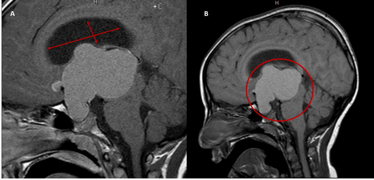
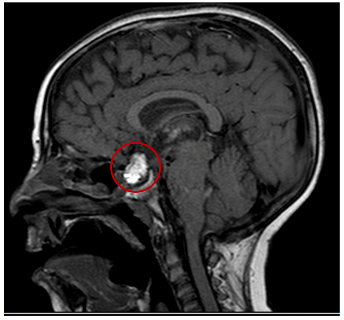
His past medical history includes craniopharyngioma, which was diagnosed at the age of 7 Figure 1 and treated with surgical resection 3 times for recurrence, followed by proton-beam radiotherapy after the third time. He had a placement of a ventriculoperitoneal shunt at the age of 8 years. He developed panhypopituitarism following surgery, secondary adrenal insufficiency, central hypothyroidism, central diabetes insipidus, delayed puberty, growth hormone deficiency with very low Insulin-like growth factor 1 (IGF1) and insulin like growth factor binding protein 3 (IGFBP3) levels, and microcytic anemia and has been on hormone replacement since then with hydrocortisone, thyroxine, desmopressin, and testosterone intramuscular injections. He also takes anti-calcitonin gene-related peptide (anti-CGRP) rescue therapy for the migraines and betahistine for the vertigo, which were prescribed after he reported that beta-blocker therapy had not helped with the tremors or the episodes of shaking, sweating, and dizziness, as he was on propranolol 10 mg/kg twice daily.
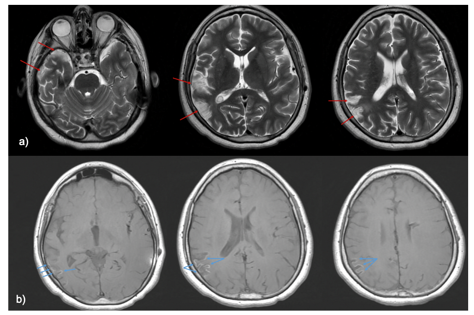
The latest MRI of the brain illustrated T1 hyper-intense signals in the cortex of the right parietal and temporal lobes, suggestive of cortical laminar necrosis Figure 3, as well as subcortical hyper-intense signals in the right parietal and frontal lobes. Optic pathway thinning was seen, which was considered a normal post-operative finding, and mild enlargement of the third ventricle. The rest of the ventricular system showed normal caliber and configuration with hydrocephalus Figure 3. A diagnosis of SMART syndrome was made, and the patient was started on Verapamil with continued clinical monitoring. The decision to initiate our patient on Verapamil is based on its already established benefit in preventing migraines and vasospastic pathophysiology. SMART syndrome is currently thought to involve impaired cerebrovascular autoregulation, radiation-induced vascular dysfunction, and cortical spreading depression, all of which contribute to the presentation with stroke-like and migrainous symptoms [4,5,6,8]. Verapamil has been widely utilized and approved for the prophylactic treatment of migraines by preventing cortical spreading depression and stabilizing vascular tone. Since current literature predicts that these pathophysiological mechanisms also contribute to the development of SMART syndrome, Verapamil was initiated.
Discussion
SMART syndrome is an extremely rare, delayed neurological condition seen following brain radiotherapy. It has a subacute onset with manifestations including migraines with or without aura, stroke-like cortical symptoms such as hemiparesis, neglect, and aphasia, and seizures [1,2,3,4,5,6,7,8,9,10]. The acronym SMART stands for stroke-like migraine attacks following radiation therapy and was first termed by Black et al. [2], although the first ever described cases were by Shuper et al. [9], who described a series of pediatric patients with severe migraines and stroke-like symptoms following radiation.
Pathophysiology is not completely understood, but theories have been postulated. These theories include radiation-induced trigeminovascular endothelial dysfunction [6], impaired cerebrovascular autoregulation [5], cortical spreading depression [4], and disruption of the blood-brain barrier [8]. Radiation-induced mitochondrial dysfunction has also been theorized [1]. Imaging findings seen in SMART syndrome include transient or permanent unilateral gyriform enhancement with or without T2/FLAIR hyperintensity involving the cortex and adjacent white matter in the area that was previously treated with radiation [1,2,3,4,5,6,7,8,9,10]. Diagnostic criteria were initially proposed by Black et al. [2] and included 1) a remote history of external beam cranial irradiation; 2) prolonged, reversible signs and symptoms referable to a unilateral cortical region beginning years after cranial irradiation, including seizure, migraine with or without an aura, and stroke-like symptoms; 3) transient, diffuse, unilateral gyriform enhancement sparing the white matter within a previous radiation field; and 4) not attributed to another disorder [2], but are not the gold standard as further cases of SMART syndrome have been reported that do not strictly adhere to these criteria [12].
Our case represents one of the four reported instances of SMART syndrome following proton beam radiotherapy [5,7,10]. Notably, our patient experienced vertigo, a symptom infrequently observed in SMART syndrome, which was similarly described by Huang et al. [5]. In addition, cerebellar signs, including dysmetria and impaired tandem gait, were documented by Maramattom et al. [7] and were also observed in our patient, although their case involved radiotherapy delivered directly to the brainstem. Huang et al. [5] proposed that proton beam radiotherapy may accelerate the onset of SMART syndrome, as evidenced by their patient’s symptom development within 2 years. In contrast, in the case series by Winter et al. [10], symptoms emerged within 4 years in one patient and 14 months in another. Although this case does not strictly meet the imaging criteria proposed by Black et al. [2] due to the absence of transient gyriform enhancement and the presence instead of cortical laminar necrosis [12], it fits within an increasingly recognized spectrum of atypical SMART syndrome presentations [5,7,10]. Given the patient’s history of proton beam radiotherapy, characteristic clinical symptoms, and exclusion of other etiologies, this case aligns with a broader, evolving definition of SMART syndrome that acknowledges irreversible imaging findings such as laminar necrosis as potential manifestations, especially in severe or delayed onset presentations [12]. This reinforces the need for flexible diagnostic criteria and clinical awareness of SMART syndrome variants in post-radiotherapy patients.
Also, Maramattom et al. [7] reported focal brainstem and leptomeningeal enhancement, a finding previously not seen, and suggested that brainstem SMART syndrome be termed as “Be-SMART syndrome”. It is important to note in our patient that, while he did have cerebellar signs on examination (dysmetria and impaired tandem gait), his MRI findings show no involvement of the brainstem or cerebellum. In our patient, cortical laminar necrosis was observed, which is a recognized complication of proton beam radiotherapy linked to vascular dysfunction [13]. This finding suggests a potential shared pathophysiological pathway between SMART syndrome and radiation necrosis [5,13]. Moreover, Huang et al. [5] have proposed that the presence of radiation necrosis may lower the threshold for developing SMART syndrome, serving as a precipitating factor in its onset. Currently, cortical laminar necrosis in SMART syndrome cases is thought to correlate with permanence of symptoms and lack of meaningful recovery [12], as is the case with our patient, with persistence of symptoms for several years. Previously, SMART syndrome was thought to be reversible but has been described as being permanent in a case series by Black et al. [12].
This case presents a complex and atypical variant of SMART syndrome that doesn’t fit neatly into previously described categories. At the same time, the reported cases involved a 38-year-old man with classic hemispheric SMART symptoms like migraines, transient aphasia, and MRI showing gyriform enhancement after proton beam therapy [5]. The second described a teenage girl with brainstem-only symptoms now referred to as Be-SMART [7], our 18-year-old patient falls somewhere in between. He experiences not only migraines but also crossed limb weakness, tremors, dysmetria, vertigo, and transient speech difficulties during sleep, suggesting more widespread cerebral involvement. His MRI findings are also distinct, showing cortical laminar necrosis and subcortical signal changes, rather than the typical gyriform enhancement. What further sets this case apart is the latency. His symptoms appeared nearly a decade after proton RT, in contrast to the accelerated onset seen in the Winter et al. [10] case series, where the only other SMART cases following proton therapy occurred at 14 months and 4 years post-treatment. Treatment with verapamil was initiated, consistent with current practice, but the addition of CGRP-targeted therapy and betahistine reflects a more tailored approach to his diverse symptoms. Altogether, this case might represent a diffuse or overlapping SMART subtype somewhere between classic and Be-SMART and highlights the need to broaden the clinical and radiologic spectrum by which we understand SMART syndrome.
Conclusion
Our case highlights an atypical presentation of SMART syndrome following proton beam radiotherapy for a craniopharyngioma, marked by episodic vertigo, dysmetria, and impaired tandem gait—symptoms not typically associated with this syndrome. The MRI findings of cortical laminar necrosis and subcortical hyperintensity suggest a potential shared vascular pathophysiology between SMART syndrome and radiation necrosis. Recognizing such atypical features is crucial for clinicians evaluating patients with a history of brain irradiation, as early and accurate diagnosis can lead to targeted treatment and help avoid unnecessary invasive procedures.
中文摘要
放射治疗后脑卒中样偏头痛发作综合征(SMART)伴眩晕、辨距不良与串联步态障碍的病例报告
背景:SMART综合征是脑部放射治疗的罕见、延迟性并发症。SMART是“放射治疗后脑卒中样偏头痛发作”(stroke‑like migraine attacks following radiation therapy)的缩写。本病例是首批描述在颅咽管瘤质子束放射治疗后出现阵发性眩晕、辨距不良和串联步态障碍的案例之一。
目的:报告一例18岁男性在完成颅咽管瘤质子束放射治疗后出现的SMART综合征典型与非典型表现。
方法:患者因偏头痛、上下肢无力、阵发性眩晕、辨距不良及串联步态障碍就诊。其质子束放射治疗在约11岁时完成,用于治疗复发性颅咽管瘤。脑磁共振成像(MRI)显示右顶叶和颞叶皮质呈T1高信号,符合SMART综合征特征;此外,右顶叶、颞叶和额叶皮下亦见高信号。
结果:该病例展示了SMART综合征的非典型表现,提示在放射治疗后出现神经症状的患者中应警惕此综合征,以便及时诊断与治疗。
结论:识别SMART综合征的多样化临床表现对于及时诊断和干预至关重要。
本中文摘要为机器辅助翻译,仅供参考;以英文摘要为准。 Machine-assisted Chinese translation of the English abstract; the English abstract is the version of record.
References
1. Biju RD, Dower A, Moon BG, Gan P. SMART (Stroke-Like Migraine Attacks After Radiation Therapy) Syndrome: A Case Study with Imaging Supporting the Theory of Vascular Dysfunction. Am J Case Rep. 2020: e921795 [PMID: 32221270, https://doi.org/10.12659/AJCR.921795]
2. Black DF, Bartleson JD, Bell ML, Lachance DH. SMART: stroke-like migraine attacks after radiation therapy. Cephalalgia. 2006: 1137 [PMID: 16919065, https://doi.org/10.1111/j.1468-2982.2006.01184.x]
3. Dossin C, Di Perri D, Whenham N, Bocchio AP, Tatar IG. SMART Syndrome: Case Report and Review of the Literature. J Belg Soc Radiol. 2023: 49 [PMID: 37427025, https://doi.org/10.5334/jbsr.3198]
4. Haller V, Dashti D, Peters N. SMART - Not Always Only "Stroke-Like". Headache. 2018: 1465 [PMID: 30311189, https://doi.org/10.1111/head.13415]
5. Huang D, Dixit KS. Stroke-Like Migraine Attacks After Radiation Therapy Syndrome and Radiation Necrosis After Cerebral Proton Beam Radiation: A Case Report of Dual Radiotherapy Complications. Neurohospitalist. 2022: 567 [PMID: 35755215, https://doi.org/10.1177/19418744221102290]
6. Maloney PR, Rabinstein AA, Daniels DJ, Link MJ. Surgically induced SMART syndrome: case report and review of the literature. World Neurosurg. 2014: 240 e7 [PMID: 23314030, https://doi.org/10.1016/j.wneu.2013.01.028]
7. Maramattom BV, Panikar D. Brainstem SMART Syndrome - Be-SMART. Neurol India. 2022: 1731 [PMID: 36076710, https://doi.org/10.4103/0028-3886.355087]
8. Ota Y, Liao E, Shah G, Srinivasan A, Capizzano AA. Comprehensive Update and Review of Clinical and Imaging Features of SMART Syndrome. AJNR Am J Neuroradiol. 2023: 626 [PMID: 37142432, https://doi.org/10.3174/ajnr.A7859]
9. Shuper A, Packer RJ, Vezina LG, Nicholson HS, Lafond D. 'Complicated migraine-like episodes' in children following cranial irradiation and chemotherapy. Neurology. 1995: 1837 [PMID: 7477978, https://doi.org/10.1212/wnl.45.10.1837]
10. Winter SF, Klein JP, Vaios EJ, Karschnia P, Lee EQ, Shih HA, Loebel F, Dietrich J. Clinical Presentation and Management of SMART Syndrome. Neurology. 2021: 118 [PMID: 33947781, https://doi.org/10.1212/WNL.0000000000012150]
11. Rigamonti A, Lauria G, Mantero V, Filizzolo M, Salmaggi A. SMART (stroke-like migraine attack after radiation therapy) syndrome: a case report with review of the literature. Neurol Sci. 2016: 157 [PMID: 26466588, https://doi.org/10.1007/s10072-015-2396-6]
12. Black DF, Morris JM, Lindell EP, Krecke KN, Worrell GA, Bartleson JD, Lachance DH. Stroke-like migraine attacks after radiation therapy (SMART) syndrome is not always completely reversible: a case series. AJNR Am J Neuroradiol. 2013: 2298 [PMID: 23788601, https://doi.org/10.3174/ajnr.A3602]
13. Niemierko A, Schuemann J, Niyazi M, Giantsoudi D, Maquilan G, Shih HA, Paganetti H. Brain Necrosis in Adult Patients After Proton Therapy: Is There Evidence for Dependency on Linear Energy Transfer? Int J Radiat Oncol Biol Phys. 2021: 109 [PMID: 32911019, https://doi.org/10.1016/j.ijrobp.2020.08.058]
Data Availability Statement
Patient data related to this study are not publicly available but can be obtained upon request from the corresponding author.
License
Copyright (c) 2025 Ali Al Yasari, Mahdia Alhaidar, Mohamad Assker

This work is licensed under a Creative Commons Attribution 4.0 International License.
Copyright and Licensing
All articles published in ASIDE Internal Medicine are open access and freely available online immediately upon publication, without embargo.
This journal applies the terms of the Creative Commons Attribution 4.0 International License (CC BY 4.0). Under this license, anyone may copy, redistribute, remix, transform, and build upon the material for any purpose, even commercially, provided that proper credit is given to the original author(s), a link to the license is provided, and any changes made are indicated.
-
License link: https://creativecommons.org/licenses/by/4.0/
-
Machine-readable license metadata is embedded in each article and its DOI record.
Copyright Holder
Copyright in all articles remains with the author(s). ASIDE Internal Medicine is published and owned by the American Society for Inclusion, Diversity, and Equity in Healthcare (ASIDE). By submitting a manuscript, authors agree to grant the publisher a non-exclusive license to publish, distribute, and archive the work under the terms of the CC BY 4.0 license.
Third-party Material
Any material (e.g., images, figures, or tables) not covered by CC BY 4.0 will be clearly indicated in the article. Users must obtain permission directly from the copyright holder for reuse of such third-party material.
Author Rights and Self-Archiving
Authors are permitted to deposit all versions of their work (preprint, author’s accepted manuscript, and version of record) in institutional or subject repositories of their choice immediately upon publication, without embargo, provided that the DOI and license information are included.

Published
Issue
Section
Categories
How to Cite
Article history
- Received
- 19 Apr 2025
- Received in revised form
- 30 Jul 2025
- Accepted
- 31 Jul 2025
- Published
- 29 Aug 2025