
Spontaneous External Jugular Vein Pseudoaneurysm: A Case Report
DOI:
https://doi.org/10.71079/ASIDE.CR.020526394Abstract
Jugular venous pseudoaneurysm represents an exceedingly rare vascular abnormality and may present as an atraumatic neck mass. We report the clinical presentation, imaging findings, operative management under local anesthesia, anticoagulation strategy, and follow-up of a patient with a thrombosed jugular venous pseudoaneurysm after exclusion of common secondary causes. Duplex ultrasonography and CT angiography confirmed the diagnosis and guided surgical planning; complete excision with preservation of venous continuity was performed without perioperative complications, and follow-up duplex at 3 months demonstrated no recurrence.
A jugular venous pseudoaneurysm should be considered in the differential diagnosis of atraumatic neck swelling; management should be individualized based on symptoms, thrombus burden, and imaging findings, with surgery favored for symptomatic or thrombosed lesions.
Keywords:
External jugular vein, Venous pseudoaneurysm, Neck swelling, Surgical management, Case reportFull Text
Introduction
Venous pseudoaneurysms are uncommon, and their occurrence in the jugular vein is exceptionally rare. Unlike arterial pseudoaneurysms, venous pseudoaneurysms are infrequently reported in the literature [1,2,3,4]. We present a case of a 39-year-old female with a jugular venous pseudoaneurysm, detailing the clinical presentation, diagnostic workup, and successful management. Most reported cases are associated with trauma, central venous catheterization, or prior neck surgery, whereas spontaneous presentations are distinctly uncommon.
Case Report
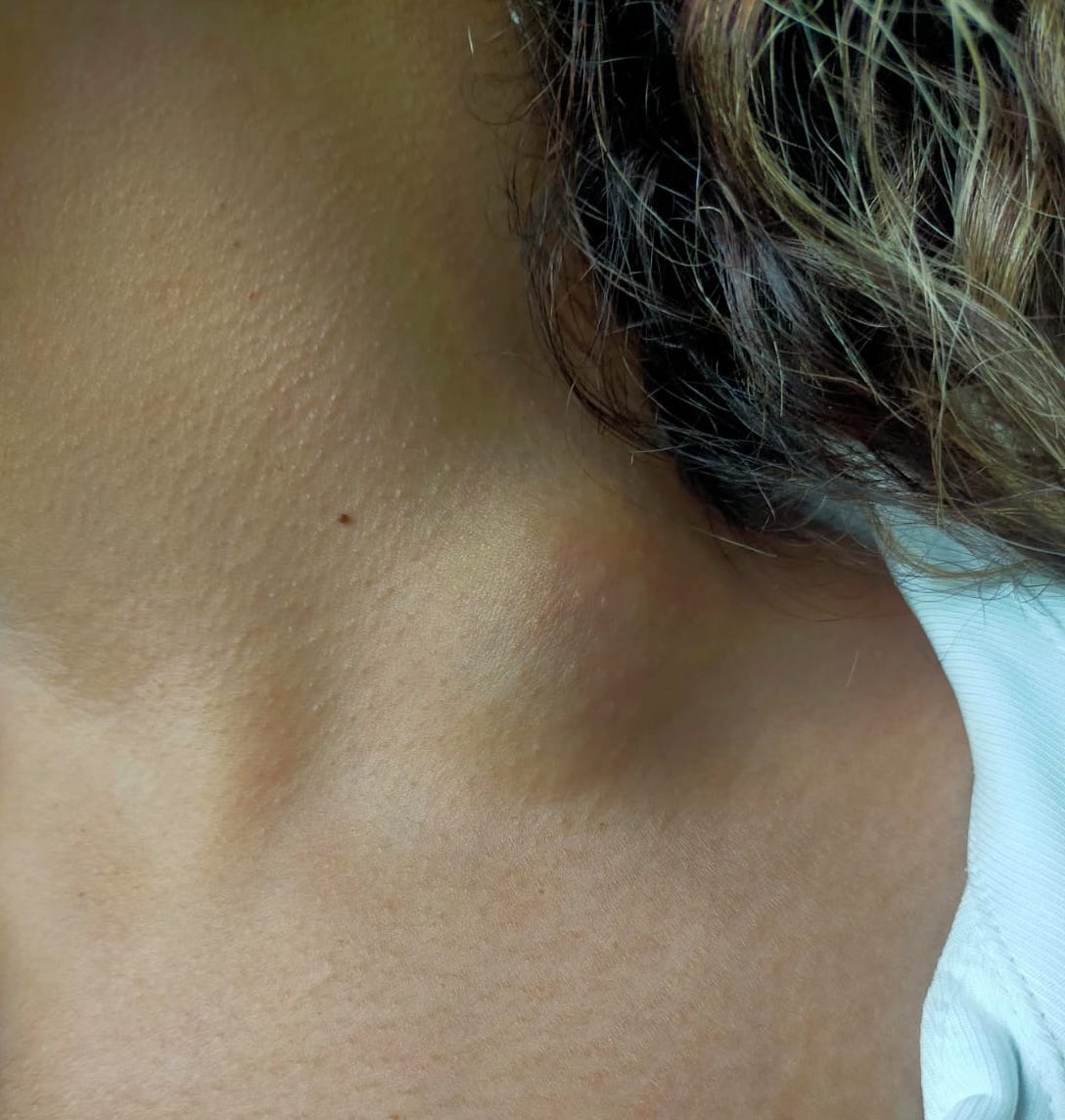
A 39-year-old female presented to our institution with a chief complaint of swelling (Figure 1) and pain on the left side of her neck. The patient denied any history of trauma or recent medical procedures. The mass was initially soft and relatively large; over several weeks, it gradually became firmer and smaller. Physical examination revealed a non-pulsatile and tender mass in the left jugular region (Figure 1). A thorough medical history and imaging studies were performed to characterize the swelling. Baseline laboratory evaluation, including complete blood count, coagulation profile (PT, aPTT, INR), inflammatory markers, and thrombophilia screening, was unremarkable. There was no history or clinical evidence of connective tissue disease, infection, or prior central venous access.
Duplex ultrasonography and contrast-enhanced computed tomography (CT) angiography were conducted to assess the anatomy and hemodynamics of the neck vessels. The imaging studies revealed a jugular venous pseudoaneurysm (Figure 2), characterized by a thrombosed saccular outpouching of the jugular vein wall. The total duration of symptom evolution was 11 weeks. The lesion measured approximately 3 cm in diameter, was non-compressible, and did not increase in size with the Valsalva maneuver. Cranial nerve examination (IX–XII) was normal.
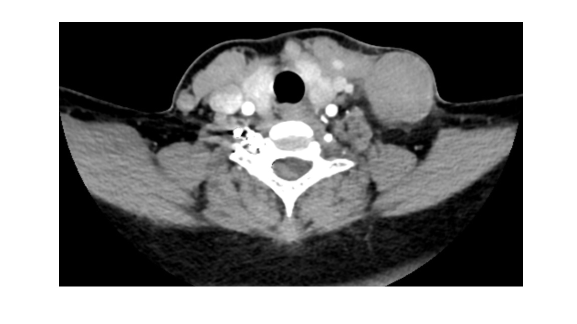
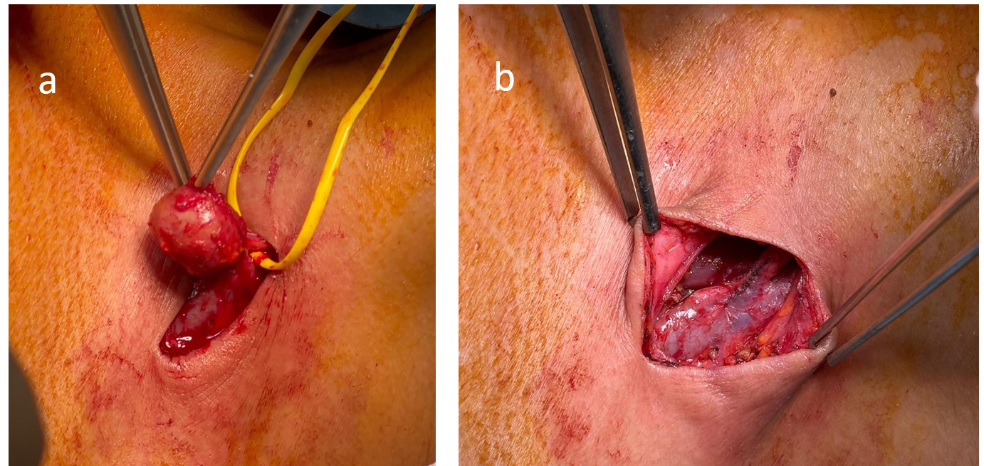
Given the risk of embolization and cosmetic and pain complaints of the patient, surgical intervention was deemed necessary. Although the pseudoaneurysm was predominantly thrombosed, the presence of residual venous flow and friable thrombotic material raised concern for potential embolization. The patient underwent pseudoaneurysm resection and primary vascular repair with local anesthesia. Local anesthesia was achieved with layered infiltration. A total of 20 mL of solution, consisting of 10 mL of 2% prilocaine (200 mg) and 10 mL of 0.5% bupivacaine (50 mg), was administered via layered local infiltration without sedation or epinephrine. Intraoperatively, the pseudoaneurysm was carefully excised (Figure 3a), and the adjacent jugular vein was repaired primarily (Figure 3b). The patient tolerated the procedure well, and postoperative recovery was uneventful. She was discharged home the next day with an oral anticoagulation regimen (apixaban 5mg, twice daily) for at least 3 months. Histopathological examination confirmed the diagnosis of venous pseudoaneurysm, demonstrating disruption of the venous wall architecture with organized thrombus.
The patient was closely monitored postoperatively for any signs of complications or recurrence. Anticoagulation was initiated due to partial thrombosis and theoretical embolic risk. A minimum duration of three months was selected based on venous thrombotic disease practices and planned reassessment with follow-up duplex imaging. Follow-up duplex ultrasonography confirmed successful repair of the jugular vein, with no evidence of residual pseudoaneurysm or vascular flow compromise. The patient reported resolution of symptoms, and no complications were observed during follow-up. At 3 months of follow-up, the patient reported complete resolution of symptoms and maximum satisfaction with the cosmetic outcome.
Timeline: Symptom onset → imaging at week 6 and 9 → surgery at week 11 → discharge on postoperative day 1 → follow-up at 3 months.
Discussion
Jugular venous pseudoaneurysms are exceedingly rare vascular abnormalities. Most reported cervical venous pseudoaneurysms are traumatic or iatrogenic, whereas spontaneous presentations without a clear precipitating factor are distinctly uncommon [1,2,3,4].
The diagnostic workup for venous pseudoaneurysms typically involves imaging studies, with duplex ultrasonography and contrast-enhanced computed tomography (CT) angiography playing pivotal roles in delineating the anatomy and hemodynamics of the lesion [1,2,3]. Detailed identification of the pseudoaneurysm with modern radiologic diagnostic modalities, as seen in our patient, aids in confirming the diagnosis.
Venous pseudoaneurysm should be considered in the differential diagnosis of atraumatic neck swelling, and management should be individualized based on clinical and imaging findings [5]. Differential diagnoses, including lymphadenopathy, thyroid nodules, cystic lesions, and soft tissue tumors, were considered and excluded based on clinical presentation and imaging characteristics [6]. In our case, the absence of solid components, lack of arterial enhancement, and venous continuity on duplex and CT angiography allowed the exclusion of lymphadenopathy, thyroid nodules, cystic lesions, and soft-tissue tumors.
Surgical intervention is generally indicated for jugular venous pseudoaneurysms due to the associated risks of thrombosis, embolization, and rupture [7]. The choice of intervention, whether surgical or endovascular, depends on the specific characteristics of the pseudoaneurysm and the patient’s overall health [1,2,3,4]. In our case, pseudoaneurysm resection and primary vascular repair were performed to relieve pain and cosmetic concerns, providing a direct and effective approach to the pathology. Conservative management with surveillance can be appropriate for selected asymptomatic, nonthrombotic jugular venous lesions; however, surgical excision is typically preferred in symptomatic, enlarging, or thrombosed cases [8].
The successful outcome in our patient emphasizes the importance of timely and definitive management. The absence of complications during the postoperative period and the resolution of symptoms highlight the efficacy of the chosen interventional approach [1,2,3,4]. However, long-term follow-up remains crucial to monitor for any potential recurrence or late-onset complications.
Despite the successful management of this case, the underlying etiology of jugular venous pseudoaneurysms, particularly in the absence of trauma or iatrogenic factors, remains unclear. Further research is warranted to elucidate the pathophysiological mechanisms contributing to the spontaneous formation of venous pseudoaneurysms [1,3,4]. Additionally, the development of standardized diagnostic and therapeutic guidelines for this rare entity can enhance the overall understanding and management of jugular venous pseudoaneurysms.
This represents the only case of jugular venous pseudoaneurysm treated at our institution over the past 10 years. As a single-case report, the findings may not be generalizable, particularly regarding the feasibility of local anesthesia and outpatient management.
Conclusions
This case report contributes to the limited body of literature on jugular venous pseudoaneurysms, emphasizing the importance of a comprehensive diagnostic approach and timely surgical intervention. Continued research is essential to enhance our understanding of the etiology, optimal diagnostic strategies, and management approaches for this rare vascular anomaly.
Conflicts of Interest
The authors declare no competing interests that could have influenced the objectivity or outcome of this research.
Funding Source
The authors declare that no specific grant or funding was received for this research from any public, commercial, or not-for-profit funding agency.
Acknowledgments
None.
Informed Consent
Written informed consent for publication of clinical details and images was obtained from the patient.
Large Language Model
Generative artificial intelligence (ChatGPT, OpenAI) was used solely for language refinement. The authors reviewed and approved the final manuscript and take full responsibility for its content.
Authors Contribution
DMO, YEA, YO, and MU all contributed substantially to the conception and design of the study, data acquisition, and interpretation of clinical findings. DMO and MU drafted the manuscript. DMO, YEA, YO, and MU critically revised the manuscript for important intellectual content. All authors participated in patient management, approved the final version of the manuscript,
Data Availability
The data supporting the findings of this case report are contained within the article. No new datasets were generated or analyzed. Additional clinical details cannot be shared publicly to protect patient privacy; limited clarifications may be provided by the corresponding author upon reasonable request, subject to institutional and ethical approval.
References
- Boutakioute B., Benzalim M., Chehboun A., Ouali M., Ganouni Ncie. Spontaneous external jugular vein pseudoaneurysm: A rare cause of neck swelling. Radiol Case Rep. 2022;17(12):4790-4794. doi:10.1016/j.radcr.2022.09.017 PMID: 36238217 PMCID: PMC9550843
- Dogan S. N., Kazci Z. N., Kavuncu S. B., Bayrak A. H.. Spontaneous external jugular vein pseudoaneurysm treated by thrombin injection: Case report and literature review. J Clin Ultrasound. 2020;48(5):294-297. doi:10.1002/jcu.22816 PMID: 31981367
- Pollard J., Abu-Yousef M.. Internal jugular vein pseudoaneurysm. Ultrasound Q. 2014;30(3):225-7. doi:10.1097/RUQ.0000000000000095 PMID: 25148499
- Schummer W., Schummer C., Voigt R., Heyne J., Steenbeck J.. [Pseudoaneurysm--a rare complication of internal jugular vein cannulation: two case reports in liver transplant patients]. Anasthesiol Intensivmed Notfallmed Schmerzther. 2003;38(8):542-6. doi:10.1055/s-2003-41191 PMID: 12905112
- Bartholomew J. R., Smolock C. J., Kirksey L., Lyden S. P., Badrinathan B., Whitelaw S., Bhandari R.. Jugular Venous Aneurysm. Ann Vasc Surg. 2020;68:567 e5-567 e9. doi:10.1016/j.avsg.2020.04.075 PMID: 32439525
- El Youbi S., Naouli H., Jiber H., Bouarhroum A.. External jugular vein spontaneous aneurysm: a case report. J Surg Case Rep. 2022;2022(5):rjac134. doi:10.1093/jscr/rjac134 PMID: 35586482 PMCID: PMC9110015
- Papes D., Cavar S.. External jugular vein pseudoaneurysm in a 2-year-old child. J Vasc Surg Venous Lymphat Disord. 2023;11(6):1288. doi:10.1016/j.jvsv.2023.05.012 PMID: 37863552
- Vermeer N. C., Elshof J. W., Vriens P. W.. Clinical presentation, diagnosis, and treatment of venous aneurysms. J Vasc Surg Venous Lymphat Disord. 2014;2(3):349-353 e3. doi:10.1016/j.jvsv.2014.02.007 PMID: 26993399
References
1. Boutakioute B, Benzalim M, Chehboun A, Ouali M, Ganouni N. Spontaneous external jugular vein pseudoaneurysm: A rare cause of neck swelling. Radiol Case Rep. 2022;17(12):4790-4. [PMID: 36238217, PMCID: PMC9550843, https://doi.org/10.1016/j.radcr.2022.09.017].
2. Dogan SN, Kazci ZN, Kavuncu SB, Bayrak AH. Spontaneous external jugular vein pseudoaneurysm treated by thrombin injection: Case report and literature review. J Clin Ultrasound. 2020;48(5):294-7. [PMID: 31981367, https://doi.org/10.1002/jcu.22816].
3. Pollard J, Abu-Yousef M. Internal jugular vein pseudoaneurysm. Ultrasound Q. 2014;30(3):225-7. [PMID: 25148499, https://doi.org/10.1097/RUQ.0000000000000095].
4. Schummer W, Schummer C, Voigt R, Heyne J, Steenbeck J. [Pseudoaneurysm–a rare complication of internal jugular vein cannulation: two case reports in liver transplant patients]. Anasthesiol Intensivmed Notfallmed Schmerzther. 2003;38(8):542-6. [PMID: 12905112, https://doi.org/10.1055/s-2003-41191].
5. Bartholomew JR, Smolock CJ, Kirksey L, Lyden SP, Badrinathan B, Whitelaw S, Bhandari R. Jugular Venous Aneurysm. Ann Vasc Surg. 2020;68:567 e5-567 e9. [PMID: 32439525, https://doi.org/10.1016/j.avsg.2020.04.075].
6. El Youbi S, Naouli H, Jiber H, Bouarhroum A. External jugular vein spontaneous aneurysm: a case report. J Surg Case Rep. 2022;2022(5):rjac134. [PMID: 35586482, PMCID: PMC9110015, https://doi.org/10.1093/jscr/rjac134].
7. Papes D, Cavar S. External jugular vein pseudoaneurysm in a 2-year-old child. J Vasc Surg Venous Lymphat Disord. 2023;11(6):1288. [PMID: 37863552, https://doi.org/10.1016/j.jvsv.2023.05.012].
8. Vermeer NC, Elshof JW, Vriens PW. Clinical presentation, diagnosis, and treatment of venous aneurysms. J Vasc Surg Venous Lymphat Disord. 2014;2(3):349-53 e3. [PMID: 26993399, https://doi.org/10.1016/j.jvsv.2014.02.007].
Data Availability Statement
The data supporting the findings of this case report are contained within the article. No new datasets were generated or analyzed. Additional clinical details cannot be shared publicly to protect patient privacy; limited clarifications may be provided by the corresponding author upon reasonable request, subject to institutional and ethical approval.
License
Copyright (c) 2026 Didem Melis Oztas, Yunus Emre Akpinar, Yilmaz Onal, Murat Ugurlucan

This work is licensed under a Creative Commons Attribution 4.0 International License.
Copyright and Licensing
All articles published in ASIDE Case Reports are open access and freely available online immediately upon publication, without embargo.
This journal applies the terms of the Creative Commons Attribution 4.0 International License (CC BY 4.0). Under this license, anyone may copy, redistribute, remix, transform, and build upon the material for any purpose, even commercially, provided that proper credit is given to the original author(s), a link to the license is provided, and any changes made are indicated.
-
License link: https://creativecommons.org/licenses/by/4.0/
-
Machine-readable license metadata is embedded in each article and its DOI record.
Copyright Holder
Copyright in all articles remains with the author(s). ASIDE Case Reports is published and owned by PubPorta Publishing LLC. By submitting a manuscript, authors agree to grant the publisher a non-exclusive license to publish, distribute, archive, and identify itself as the original publisher of the work under the terms of the CC BY 4.0 license.
Academic oversight and scholarly guidance are provided by the American Society for Inclusion, Diversity, and Equity in Healthcare (ASIDE). Copyright and licensing arrangements are administered by the publisher.
Third-party Material
Any material (e.g., images, figures, or tables) not covered by CC BY 4.0 will be clearly indicated in the article. Users must obtain permission directly from the copyright holder for reuse of such third-party material.
Author Rights and Self-Archiving
Authors are permitted to deposit all versions of their work (preprint, author’s accepted manuscript, and version of record) in institutional or subject repositories of their choice immediately upon publication, without embargo, provided that the DOI and license information are included.

Published
Issue
Section
Categories
How to Cite
Article history
- Received
- 22 Nov 2025
- Received in revised form
- 14 Dec 2025
- Accepted
- 24 Dec 2025
- Published
- 5 Feb 2026