
Motion-Artifact ‘Pseudo–Type A Dissection’ on Ungated CTA in a Morbidly Obese Patient with Resolution on ECG-Gated CTA: A Case Report
DOI:
https://doi.org/10.71079/ASIDE.CR.012126279Abstract
We present a case of acute chest pain in a 48-year-old morbidly obese female with multiple comorbidities that raised suspicion for Acute Aortic Syndrome (AAS). The initial ungated PE-protocol CT angiogram (CTA) revealed a linear low-attenuation band in the ascending aorta consistent with a motion artifact-induced pseudo-flap. This finding led to two Transesophageal Echocardiograms (TEEs): the first, performed under conscious sedation, was positive; the second, performed under general anesthesia (GA) to minimize artifact, was negative. To definitively assess the findings, a confirmatory ECG-gated CTA was performed immediately after heart rate control, which was negative for both pulmonary embolism (PE) and aortic dissection. The final diagnosis was Acute Decompensated Heart Failure (ADHF), and the patient was discharged on Guideline-Directed Medical Therapy. This case illustrates that the ungated PE-protocol CTA is critically suboptimal for the ascending aorta and underscores the need to use a dedicated ECG-gated CTA when AAS is suspected, particularly in obese patients prone to motion artifacts, to prevent diagnostic error and unnecessary surgical intervention.
Keywords:
Aortic dissection, ECG-gated CTA, Obesity, Case reportFull Text
Introduction
Motion in the ascending aorta can create imaging artifacts that may be mistaken for a false channel or an intimal flap [1]. These artifacts are often seen in emergency department (ED) scans because the imaging is typically ungated. The primary purpose of these scans is to detect pulmonary embolism, not to screen for acute aortic syndromes, which can lead to misinterpretation. Recognizing that these artifacts may be masquerading as an acute aortic emergency is crucial, especially in the ED. This awareness can prevent unnecessary further testing and even surgical interventions. Additionally, morbid obesity is increasingly recognized as a challenge that can negatively impact the diagnostic accuracy of various imaging methods, including CT scans. These challenges can include Increased Noise (Quantum Mottle), X-ray beam weakening, Truncation/Cropping Artifacts, and Motion Artifacts [2].
| Parameter | Initial CTA (Ungated PE Protocol) | Confirmatory CTA (ECG-Gated Aortic) |
|---|---|---|
| Scanner Model | Siemens Somatom Sensation 64 | Siemens Somatom Sensation 64 |
| Gating Status | Non-Gated | Retrospectively ECG-Gated |
| Tube Voltage (kVp) | 120 | 100 |
| Effective Current (mAs) | 200 | 350 |
| Pitch | 1.2 | 0.3 |
| Rotation Time (s) | 0.5 | 0.33 |
| Contrast Volume / Rate | 75 mL at 4.5 mL/s | 100 mL at 5.0 mL/s |
| HR at Acquisition (bpm) | 95 | 68 (Beta-Blockade utilized) |
CTA, Computed Tomography Angiography.
Imaging for suspected AAS is critical and performed rapidly, with CTA of the chest/abdomen/pelvis as the modality of choice for hemodynamically stable patients (Class I recommendation). The ACR Appropriateness Criteria also rates CTA as “Usually Appropriate” for definitive diagnosis. A key technical caution, however, is that ungated PE-protocol CTA can produce ascending aortic motion artifacts, potentially obscuring or simulating the intimal flap of a critical Type A dissection due to the heart’s cyclical movement. Therefore, dedicated aortic or ECG-gated CTA protocols are generally preferred, especially for evaluating the ascending aorta [3,4].
Case Presentation
A 48-year-old female presented to a rural hospital ED complaining of the sudden onset of chest pain and respiratory distress. Her past medical history included morbid obesity, pulmonary hypertension, obstructive sleep apnea, chronic diastolic heart failure, cardiomegaly, COPD, hypertension, and a history of pulmonary embolism on anticoagulation with apixaban.
Her initial vital signs were as follows: Temp 97.4F, HR 97, RR 24, BP was mildly elevated on arrival but without hypertensive emergency (145/84 mmHg), and SpO2 97% on room air. Her BMI was 73.76 kg/m2. Laboratory parameters did not highlight any acute abnormalities. Troponin level was normal. Physical examination revealed a morbidly obese woman in acute respiratory distress. Auscultation revealed distant heart sounds with no audible murmurs; bibasilar crackles on lung exam; and abdominal examination was difficult due to body habits. Pulses were 2+ in all 4 extremities with slightly lower extremity edema. No focal neurological deficits were noted.
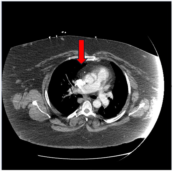
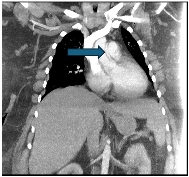
A 12-lead ECG was requested, which showed sinus tachycardia without any acute ST/T changes. The patient underwent a STAT CTA of the chest to evaluate for PE, per the protocol in Table 1. Her Serum Creatinine was 1.7 mg/dL pre-CTA. Although there were no findings suggestive of PE, the CTA was remarkable for a linear low-attenuation band in the ascending aorta, consistent with a ‘pseudo-flap’ that did not extend to the aortic root, especially since the ascending aorta on ungated CTA is artifact-prone. This finding was strongly suggestive of a Type-A aortic dissection; Figure 1 and Figure 2.
Subsequently, the cardiology team performed an initial TEE (TEE#1) under conscious sedation. The procedure utilized a Philips X7-2t probe operating at 5.0–7.0 MHz. Visualization utilized 2D, M-mode, and Color Flow Doppler from the Upper Esophageal to Deep Transgastric windows. During TEE#1, the HR was 95 bpm, and the BP was 142/90 mmHg. Image quality was noted to be sub-optimal in the mid-esophageal (ME) windows due to the patient’s morbid obesity and increased esophageal peristalsis related to conscious sedation. The report confirmed an intimal flap suggestive of ascending aortic dissection not involving the aortic root. She was transferred to a tertiary care center for a cardiothoracic surgery evaluation and possible operative repair.
Upon arrival, the surgical team decided to perform a repeat TEE (TEE#2) using a similar probe to confirm the initial reported findings. This TEE was performed under GA with mechanical ventilation to minimize movement artifact and allow for a more comprehensive esophageal and transgastric survey. Improved image quality was achieved, particularly in the ME windows, due to the absence of esophageal peristalsis under GA. The CT surgical team independently reviewed these images and, after a core review of both TEE#1 and TEE#2, found no convincing evidence of an intimal flap or aortic dissection in any views.
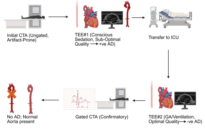
The discordance in findings was adjudicated by the attending cardiothoracic surgeon, who concluded that the findings in TEE#1 were most likely due to image artifact (reverberation) exacerbated by sub-optimal imaging planes and patient movement under conscious sedation, which were subsequently negated by the superior imaging quality and comprehensive assessment of TEE#2 performed under GA. To definitively rule out Type-A dissection, a confirmatory ECG-gated CTA of the chest was immediately recommended, and IV esmolol was started as a 500 g/kg bolus followed by a 50 g/kg/min continuous infusion. A STAT serum creatinine test was performed, revealing a level of 2.2 mg/dL. The ECG-gated CTA was performed to limit pulsation artifact; the protocol is in Table 1. The report was negative for PE or aortic dissection, confirming the final TEE findings and allowing for extubation and transfer out of the ICU within hours. A timeline representing the diagnostic evaluation is provided Figure 3.
| Time after Presentation (Days) | BNP Level (pg/mL) |
|---|---|
| Admission | 1260 |
| 24 hours (Day 1) | 1112 |
| 48 hours (Day 2) | 967 |
| 72 hours (Day 3) | 854 |
| 96 hours (Day 4) | 761 |
| Discharge (Day 5) | 682 |
BNP, B-type Natriuretic Peptide
The patient was extubated without complications, and a trial of 1mg IV bumetanide, along with 50mg/250mL nitroglycerin, was given. One hour later, the patient reported decreasing chest pain and feeling better. BNP level was measured at 1260 pg/mL. The diagnosis of ADHF was presumed, and the patient continued to receive bumetanide and nitroglycerin with BNP level measurement daily for 5 consecutive days: Table 2.
The patient was discharged on day 5 post-presentation. Discharge medications included Furosemide 40mg BID, Sacubitril/Valsartan 49/51mg BID, Carvedilol 3.125mg BID, Dapagliflozin 10mg QD, and Spironolactone 12.5mg QD. They were instructed to follow up within a week at the Cardiology attending clinic.
Discussion
CT scans are a routine method for evaluating AAS in patients presenting with chest pain or related symptoms. However, when used for the chest, this imaging technique is unfortunately more prone to errors and artifacts than when it’s used to examine other parts of the body [5].
When AAS, particularly Type A Aortic Dissection involving the ascending aorta, is suspected, the definitive imaging modality is ECG-gated CTA of the thoracic aorta according to the guidelines. Ungated PE-protocol CTA is suboptimal for the ascending aorta because the movement of the beating heart causes significant motion artifact in the aortic root and proximal ascending aorta. This artifact can obscure a true intimal tear or, conversely, create a ‘pseudo-flap’ (a false-positive dissection diagnosis) due to blurring. ECG-gated imaging, by synchronizing image acquisition with the quietest phases of the cardiac cycle, minimizes this artifact and provides superior visualization of the inner and outer aortic walls, leading to improved edge depiction and greater confidence in measurements; Table 3 [3].
| Feature | Ungated CTA (e.g., PE Protocol) | ECG-Gated CTA (Dedicated Aortic Protocol) |
|---|---|---|
| AAS Utility | Suboptimal, often reserved for ruling out PE | Standard of Care (Preferred for suspected AAS) |
| Image Quality (Ascending Aorta) | Poor, highly susceptible to motion artifact | Superior, minimizes cardiac motion artifact |
| Risk of ‘Pseudo-Flaps’ | High (artifact can mimic dissection flap) | Low |
| Procedure Time | Faster (Single acquisition, non-gated) | Slower (Requires synchronization, planning, and multiple reconstruction phases) |
| Radiation Dose | Typically lower (Non-gated acquisition) | Higher (Uses prospective or retrospective gating, increasing effective mAs/dose) |
AAS, Acute Aortic Syndrome; PE, Pulmonary Embolism; CTA, Computed Tomography Angiography.
Additionally, imaging the obese patient population presents significant technical challenges for CT, primarily due to limitations in scanner hardware and image quality degradation. The increased soft tissue diameter often exceeds the physical constraints of the gantry bore and the Scan Field of View. In contrast, the greater soft-tissue volume leads to severe photon starvation, as the X-ray beam is highly attenuated. This results in unacceptable image noise and quantum mottle. To mitigate these issues and ensure diagnostic quality, acquisition protocols must be tailored. Key strategies include using scanners with high-capacity tables and wide bores, increasing the tube current (mAs) and voltage (kVp) to boost beam penetration, and employing advanced post-processing methods, such as Iterative Reconstruction algorithms, which effectively reduce image noise and enable dose reduction without sacrificing image clarity. For anatomical regions susceptible to motion, like the thoracic aorta, dedicated ECG-gating remains essential to prevent motion artifacts that can obscure or mimic pathology [6,7,2].
Conclusion
Although a routine CT scan is highly effective for identifying aortic dissections, ungated PE-protocol CTA mimics type A dissection, particularly for those located in the ascending aorta, especially in patients with high BMI. To mitigate this risk and prevent unnecessary surgery, urgent ECG-gated CTA is the recommended confirmatory test when AAS suspicion persists, as it produces high-quality, motion-free images by synchronizing with the patient’s cardiac cycle.
Conflicts of Interest
The authors declare no competing interests that could have influenced the objectivity or outcome of this research.
Funding Source
The authors declare that no specific grant or funding was received for this research from any public, commercial, or not-for-profit funding agency.
Acknowledgments
None.
Informed Consent
The authors confirm that patient written consent forms have been obtained for this article, including all accompanying clinical data and images.
Large Language Model
None.
Authors Contribution
All authors contributed equally. All authors reviewed and approved the final manuscript.
Data Availability
All data generated or analyzed during this case report are included within the published article. No additional datasets were generated or analyzed. Due to the nature of a single-patient case report and to protect patient privacy, no raw or identifiable data are publicly available. Any further information is available from the corresponding author upon reasonable request and subject to ethical and privacy considerations.
References
- Qanadli S. D., El Hajjam M., Mesurolle B., Lavisse L., Jourdan O., Randoux B., Chagnon S., Lacombe P.. Motion artifacts of the aorta simulating aortic dissection on spiral CT. J Comput Assist Tomogr. 1999;23(1):1-6. doi:10.1097/00004728-199901000-00001 PMID: 10050797
- Uppot R. N.. Technical challenges of imaging & image-guided interventions in obese patients. Br J Radiol. 2018;91(1089):20170931. doi:10.1259/bjr.20170931 PMID: 29869898 PMCID: PMC6223172
- Isselbacher E. M., Preventza O., Hamilton Black J., 3rd, Augoustides J. G., Beck A. W., Bolen M. A., Braverman A. C., Bray B. E., Brown-Zimmerman M. M., Chen E. P., Collins T. J., DeAnda A., Jr., Fanola C. L., Girardi L. N., Hicks C. W., Hui D. S., Schuyler Jones W., Kalahasti V., Kim K. M., Milewicz D. M., Oderich G. S., Ogbechie L., Promes S. B., Gyang Ross E., Schermerhorn M. L., Singleton Times S., Tseng E. E., Wang G. J., Woo Y. J., Peer Review Committee Members. 2022 ACC/AHA Guideline for the Diagnosis and Management of Aortic Disease: A Report of the American Heart Association/American College of Cardiology Joint Committee on Clinical Practice Guidelines. Circulation. 2022;146(24):e334-e482. doi:10.1161/CIR.0000000000001106 PMID: 36322642 PMCID: PMC9876736
- Expert Panel on Cardiac Imaging, Kicska G. A., Hurwitz Koweek L. M., Ghoshhajra B. B., Beache G. M., Brown R. K. J., Davis A. M., Hsu J. Y., Khosa F., Kligerman S. J., Litmanovich D., Lo B. M., Maroules C. D., Meyersohn N. M., Rajpal S., Villines T. C., Wann S., Abbara S.. ACR Appropriateness Criteria(R) Suspected Acute Aortic Syndrome. J Am Coll Radiol. 2021;18(11S):S474-S481. doi:10.1016/j.jacr.2021.09.004 PMID: 34794601
- Bankier A. A., O'Donnell C. R., Boiselle P. M.. Quality initiatives. Respiratory instructions for CT examinations of the lungs: a hands-on guide. Radiographics. 2008;28(4):919-31. doi:10.1148/rg.284085035 PMID: 18635620
- Fursevich D. M., LiMarzi G. M., O'Dell M. C., Hernandez M. A., Sensakovic W. F.. Bariatric CT Imaging: Challenges and Solutions. Radiographics. 2016;36(4):1076-86. doi:10.1148/rg.2016150198 PMID: 27232505
- Modica M. J., Kanal K. M., Gunn M. L.. The obese emergency patient: imaging challenges and solutions. Radiographics. 2011;31(3):811-23. doi:10.1148/rg.313105138 PMID: 21571658
References
1. Qanadli SD, El Hajjam M, Mesurolle B, Lavisse L, Jourdan O, Randoux B, Chagnon S, Lacombe P. Motion artifacts of the aorta simulating aortic dissection on spiral CT. J Comput Assist Tomogr. 1999;23(1):1-6. [PMID: 10050797, https://doi.org/10.1097/00004728-199901000-00001].
2. Uppot RN. Technical challenges of imaging & image-guided interventions in obese patients. Br J Radiol. 2018;91(1089):20170931. [PMID: 29869898, PMCID: PMC6223172, https://doi.org/10.1259/bjr.20170931].
3. Isselbacher EM, Preventza O, Hamilton Black r J, Augoustides JG, Beck AW, Bolen MA, Braverman AC, Bray BE, Brown-Zimmerman MM, Chen EP, Collins TJ, DeAnda J A, Fanola CL, Girardi LN, Hicks CW, Hui DS, Schuyler Jones W, Kalahasti V, Kim KM, Milewicz DM, Oderich GS, Ogbechie L, Promes SB, Gyang Ross E, Schermerhorn ML, Singleton Times S, Tseng EE, Wang GJ, Woo YJ, Peer Review Committee M. 2022 ACC/AHA Guideline for the Diagnosis and Management of Aortic Disease: A Report of the American Heart Association/American College of Cardiology Joint Committee on Clinical Practice Guidelines. Circulation. 2022;146(24):e334-482. [PMID: 36322642, PMCID: PMC9876736, https://doi.org/10.1161/CIR.0000000000001106].
4. Expert Panel on Cardiac I, Kicska GA, Hurwitz Koweek LM, Ghoshhajra BB, Beache GM, Brown RKJ, Davis AM, Hsu JY, Khosa F, Kligerman SJ, Litmanovich D, Lo BM, Maroules CD, Meyersohn NM, Rajpal S, Villines TC, Wann S, Abbara S. ACR Appropriateness Criteria(R) Suspected Acute Aortic Syndrome. J Am Coll Radiol. 2021;18(11S):S474-81. [PMID: 34794601, https://doi.org/10.1016/j.jacr.2021.09.004].
5. Bankier AA, O'Donnell CR, Boiselle PM. Quality initiatives. Respiratory instructions for CT examinations of the lungs: a hands-on guide. Radiographics. 2008;28(4):919-31. [PMID: 18635620, https://doi.org/10.1148/rg.284085035].
6. Fursevich DM, LiMarzi GM, O'Dell MC, Hernandez MA, Sensakovic WF. Bariatric CT Imaging: Challenges and Solutions. Radiographics. 2016;36(4):1076-86. [PMID: 27232505, https://doi.org/10.1148/rg.2016150198].
7. Modica MJ, Kanal KM, Gunn ML. The obese emergency patient: imaging challenges and solutions. Radiographics. 2011;31(3):811-23. [PMID: 21571658, https://doi.org/10.1148/rg.313105138].
Data Availability Statement
All data generated or analyzed during this case report are included within the published article. No additional datasets were generated or analyzed. Due to the nature of a single-patient case report and to protect patient privacy, no raw or identifiable data are publicly available. Any further information is available from the corresponding author upon reasonable request and subject to ethical and privacy considerations.
License
Copyright (c) 2026 Alok Arora, Ahmed Elsayed, Wadah Jason Ayoub, Yousif Elsayed, Yuvraj Arora

This work is licensed under a Creative Commons Attribution 4.0 International License.
Copyright and Licensing
All articles published in ASIDE Case Reports are open access and freely available online immediately upon publication, without embargo.
This journal applies the terms of the Creative Commons Attribution 4.0 International License (CC BY 4.0). Under this license, anyone may copy, redistribute, remix, transform, and build upon the material for any purpose, even commercially, provided that proper credit is given to the original author(s), a link to the license is provided, and any changes made are indicated.
-
License link: https://creativecommons.org/licenses/by/4.0/
-
Machine-readable license metadata is embedded in each article and its DOI record.
Copyright Holder
Copyright in all articles remains with the author(s). ASIDE Case Reports is published and owned by PubPorta Publishing LLC. By submitting a manuscript, authors agree to grant the publisher a non-exclusive license to publish, distribute, archive, and identify itself as the original publisher of the work under the terms of the CC BY 4.0 license.
Academic oversight and scholarly guidance are provided by the American Society for Inclusion, Diversity, and Equity in Healthcare (ASIDE). Copyright and licensing arrangements are administered by the publisher.
Third-party Material
Any material (e.g., images, figures, or tables) not covered by CC BY 4.0 will be clearly indicated in the article. Users must obtain permission directly from the copyright holder for reuse of such third-party material.
Author Rights and Self-Archiving
Authors are permitted to deposit all versions of their work (preprint, author’s accepted manuscript, and version of record) in institutional or subject repositories of their choice immediately upon publication, without embargo, provided that the DOI and license information are included.
Published
Issue
Section
Categories
How to Cite
Article history
- Received
- 22 Sep 2025
- Received in revised form
- 5 Jan 2026
- Accepted
- 18 Jan 2026
- Published
- 21 Jan 2026