
Fatal Septic Shock with Presumed Urinary Source in a Patient with Clinically Suspected, Previously Undiagnosed Von Hippel–Lindau Disease: A Case Report and Literature Review
DOI:
https://doi.org/10.71079/ASIDE.IM.062426791Abstract
Background: Von Hippel-Lindau disease is a rare form of autosomal dominant cancer syndrome that affects multiple organs, such as the retina and brain hemangioblastoma, renal disease, and other visceral malignancies. Delayed diagnosis is a significant reason for avoidable mortality and morbidity.
Case Presentation: We present a case of a 47-year-old male presenting with fever, vomiting, hypotension, and pus discharge from his urethral catheter, developing into septic shock with multiple organ dysfunction syndrome. He had several decades of progressive symptoms clinically compatible with possible VHL syndrome, consisting of bilateral blindness secondary to globe enucleations performed since childhood, multifocal spinal cord lesions resulting in quadriparesis and neurogenic bladder, and a family history of retinoblastoma, brain tumors, and renal lesions. MRI of the spine showed extensive multifocal intramedullary enhancing lesions, from the cervicomedullary junction to T11, associated with dilated cord due to cystic changes, compression of the posterior medulla leading to obstructive hydrocephalus, and an occipital lesion. These findings favored multiple spinal hemangioblastomas. A corticomedullary cyst of the right kidney was seen. Despite intensive supportive therapy and appropriate broad-spectrum antibiotics, he succumbed to septic shock complicated by a presumed urinary source on hospital day 4.
Conclusion: This particular case illustrates the potential of unidentified genetic tumors resulting in significant neurological problems and infections leading to death. Early detection of multifocal hemangioblastomas and retinal problems can lead to early identification of patients at risk of developing VHL disease.
Keywords:
Von Hippel–Lindau disease, Hemangioblastoma, Urosepsis, Septic shock, Spinal cord tumor, Retinal hemangioblastoma, Phthisis bulbi, Delayed diagnosis, Hereditary tumor syndromeFull Text
Introduction
Von Hippel-Lindau syndrome is a rare autosomal dominant multiorgan neoplasm characterized by involvement of all races with a penetrance rate of nearly 100% by 65 years of age [1,2]. The estimated prevalence rate is 1 out of 36,000 births. Its occurrence results from a mutation in the VHL gene, which encodes the VHL protein, discovered by Latif et al. in 1993 [3] and mapped to locus 3p25 – 26 on the short arm of chromosome 3. This protein plays an indispensable role in mediating the hypoxic pathway, in which, under normoxic conditions, it ubiquitinates the HIF- subunit, leading to its proteasome-dependent degradation. Lack of the protein results in persistent HIF- stabilization and increased levels of gene products associated with angiogenesis and mitosis, a mechanism that was awarded the Nobel Prize in Physiology or Medicine in 2019 [4].
VHL disease clinical presentation includes hemangioblastomas of the CNS and retina, clear cell RCC, renal cysts, pheochromocytomas/paragangliomas, endolymphatic sac tumors, pancreatic cysts/neuroendocrine tumors, and epididymal cystadenomas [5]. High phenotypic variability exists, even among individuals with the same germline mutations, with the average age at symptom onset ranging from 20 to 40 years [2,6]. The diagnostic criteria for VHL disease were initially proposed by Melmon and Rosen (1964) and later modified. In patients without a family history, diagnosis requires two VHL lesions, with at least one being a hemangioblastoma, whereas a single lesion suffices in those with a positive family history [7,6].
Misdiagnosis of the VHL syndrome is still considered an important cause of preventable morbidity and mortality due to the lack of recognition of early symptoms as markers of a hereditary condition affecting multiple organs of the body. In the current case presentation, a 47-year-old man was reported to have had a series of manifestations caused by VHL syndrome throughout several decades before dying from septic shock. Posthumous diagnosis of VHL syndrome in the current patient was suspected clinically in accordance with well-known criteria for this condition.
Literature Review
Study Design and Data Extraction
The subsequent section highlights a review of the literature collected through a search of the PubMed and Google Scholar databases from inception until February 2026 utilizing the key terms “von Hippel-Lindau Disease,” “VHL Hemangioblastoma,” “Retinal Hemangioblastoma,” “Spinal Hemangioblastoma,” “VHL Renal Manifestations,” and “VHL Diagnostic Criteria,” with no date constraints applied. Additional sources were gathered from references cited within selected articles. This study is not a systematic or scoping review. Both research studies and systematic, narrative, or case reviews relevant to VHL syndrome were included.
Epidemiology and Genetics
VHL is an autosomal dominant genetic disorder characterized by high penetrance with a birth prevalence of 1 in 36,000 to 91,000 cases [1,2]. Twenty percent of VHL patients harbor de novo mutations; therefore, the absence of family history cannot be used to rule out VHL [8]. Clinical penetrance reaches 90% by age 65 years, and symptoms become apparent between ages 10 and 30 years [2]. The gene alterations include missense, nonsense, frameshift, and large-deletion mutations. There is a phenotype-genotype correlation whereby adrenal mass development (VHL type 1) is unlikely with truncating mutations, and some missense mutations are predictive of pheochromocytoma (VHL types 2A, 2B, and 2C) [5,9].
CNS Hemangioblastomas
The prevalence rate of CNS hemangioblastoma in VHL syndrome is between 60% and 80%. Hemangioblastomas in the central nervous system are grade I vascularized tumors that arise from stromal cells [10,11]. The common sites in VHL-associated tumors are cerebellum (44-72%), spinal cord (14-50%), and brain stem [10,12]. The onset of hemangioblastomas associated with VHL syndrome occurs earlier compared to sporadic tumors and also has a high incidence of multiple tumors [12]. The signs and symptoms of VHL-associated hemangioblastoma include progressive myelopathy with sensory and motor deficits, pain, and myeloradiculopathy; large hemangioblastomas may lead to significant syringomyelia [13,12]. The presence of brainstem lesions in VHL-associated hemangioblastoma in the cervicomedullary region or posterior medulla causes obstruction of the fourth ventricle, resulting in hydrocephalus [14]. Surgical treatment is the standard treatment for VHL-associated CNS hemangioblastomas; timely treatment should be performed to avoid permanent disabilities [12]. Belzutifan, an inhibitor of HIF-2, was FDA-approved in 2021 as the first pharmacological therapy for non-surgical VHL-associated CNS hemangioblastomas [15,16].
Ocular Manifestations
Retinal hemangioblastomas occur in about 50% of patients with VHL, with presentation usually in the second decade of life, although cases below age 10 have been reported [17]. Retinal hemangioblastomas originate as highly vascular tumors with dilated supplying and draining vasculature in the retina: exudative changes, retinal traction, secondary glaucoma, and neovascularization cause retinal detachment and blindness. Bilateral involvement leading to enucleation is infrequent; however, a recent prospective cohort study of 406 patients with VHL found unilateral enucleation in 8.2% of cases [18]. Bilateral enucleation, as seen in the current patient, is rare and reflects the outcome of bilaterally severe retinal hemangioblastoma, complicated by end-stage glaucoma and phthisis bulbi secondary to an uncontrollable disease process [18,17]. Lens dislocation is a non-classical VHL manifestation; however, in advanced stages of ocular disease, it is a mechanical effect of increased intraocular pressure.
Renal and Visceral Manifestations
The clear cell variant of RCC is seen in more than 70% of VHL patients and represents the most common reason for VHL-related deaths. In some studies, renal cysts, which may be either simple or complicated, have been reported in up to 60% of patients with or before the occurrence of an RCC. The average age of diagnosis for RCC in VHL patients is between 33 and 37 years [5]. One must bear in mind that simple renal cysts are often a coincidental finding in middle-aged individuals without VHL disease; thus, their interpretation should be evaluated within the clinical context before assigning any diagnostic relevance to VHL syndrome. Other visceral symptoms are adrenal pheochromocytomas, pancreatic cysts, and endolymphatic sac tumors.
Diagnostic Criteria and Screening
The criteria for VHL disease include the discovery of two or more hemangioblastomas, or of one hemangioblastoma with an organ manifestation in those lacking a family history [7,6]. A solitary finding suffices in diagnosing the condition in individuals with positive family history. Multisystem surveillance, according to existing guidelines, should be instituted from early childhood among at-risk individuals. MRI of the brain and spine, ophthalmologic examination, abdominal imaging, and plasma/urine analysis for catecholamines form the recommended imaging studies [7]. Definitive genetic testing identifies the condition and helps conduct cascade testing among relatives [5]. In the current scenario, the presence of bilateral eye disease leading to bilateral enucleation, extensive segmental multi-foci involvement of the spinal cord along with positive family history fits into the diagnostic criteria for VHL disease as defined by Melmon & Rosen (1964) and later modified by Binderup et al. (2022) [7,6]; the sole limitation being the lack of histopathological or genetic evidence in support of these lesions.
Case Presentation
Patient History and Background
A 47-year-old man came to the ED with complaints of fever for five days and nausea with vomiting for one day. Multiple diseases complicated the patient’s previous medical history throughout decades. He developed blindness in the left eye from childhood at the age of approximately ten years, which progressed to complete loss of vision and enucleation of the left eye. Whether the cause of blindness was retinal hemangioblastoma, which is the probable diagnosis on ophthalmic consultation, could not be confirmed because the pathological findings from the removed eye were unavailable. The right lens dislocated at approximately 20 years of age and recurred after 5 years, leading to removal of the lens and, subsequently, the entire globe of the right eye. Also, HCV infection was detected in his thirties Table 1.
There has been increasing quadriplegia for 3 years before admission, which eventually led to long-term urinary catheterization, indicative of developing neurogenic bladder dysfunction. There are no reports of neurological or spinal imaging studies during this period. About 20 days before the present presentation, there was a case of T3-T4 laminectomy with removal of a spinal space-occupying lesion due to a fall; however, the indication for the procedure, neurological examination, post-operative report, and pathology reports of the specimen removed are lacking. It cannot be confirmed whether a urinary catheter was placed before the procedure or later, as a pre-existing neurogenic bladder was known; further, the type of catheter used and how long it was in place before admission are not known.
The family history, which was collected by interviewing the patient without corroborating information from medical records, revealed that the patient’s younger sister had a right occipital brain tumor, the patient’s older sister had vision impairment due to a brain tumor, and the patient’s mother had a renal tumor. It was unknown whether any of the patient’s relatives had ever been evaluated for cancer, genetic testing, or cascade testing. There was no documentation that the patient had ever attended genetic counseling or hereditary tumor screening programs.
| Age (yr) | Approx. Year | Event | Investigation / Intervention | Outcome |
|---|---|---|---|---|
| ∼10 | ∼1986 | Sudden-onset left visual loss progressing to complete blindness | Ophthalmological evaluation; retinal diagnosis not documented as hemangioblastoma in available records | Left globe enucleation |
| ∼20 | ∼1996 | Right lens dislocation; Hepatitis C virus (HCV) diagnosis | Surgical lens correction; HCV confirmed (viral load, fibrosis staging, and treatment records unavailable) | Partial right visual preservation; HCV managed expectantly |
| ∼25 | ∼2001 | Recurrent right lens dislocation; progressive right visual decline | Surgical lens removal | Right globe enucleation; bilateral visual loss established |
| ∼44 | ∼2020 | Onset of progressive quadriparesis | No neurological investigation or spinal imaging documented from this period | Gradual decline to quadriplegia; urinary catheter dependence established |
| 47 | ∼Day −20 | Fall; spinal lesion identified on prior imaging | T3–T4 laminectomy; excision of spinal space-occupying lesion; histopathology unavailable; preoperative vs postoperative neurological status undocumented | Postoperative course undocumented; catheter dependence continued |
| 47 | Day 0 | 5-day fever; 1-day nausea/vomiting; septic shock on arrival | ED presentation; orotracheal intubation; urethral catheterization (purulent output); ABG, CBC, coagulation screen; MRI brain and spine; abdominal USS. Urine/blood cultures and lactate unavailable. | Empiric piperacillin-tazobactam; norepinephrine initiated; ICU admission |
| 47 | Days 1–3 | Refractory hemodynamic instability; escalating vasopressor requirements | Sequential addition of dobutamine and dopamine; standard dosing in mcg/kg/min unavailable (recorded as drops/min); MAP targets, fluid balance, ventilation parameters, and SOFA score not documented | Persistent septic shock; progressive multi-organ dysfunction |
| 47 | Day 4 | Critical hemodynamic deterioration; terminal ABG: pH elevated, 70%, bicarbonate elevated, base excess +5.5 | Family declined CPR and further escalation; no post-mortem examination documented; germline VHL mutation testing not performed | Death from refractory septic shock and multi-organ failure; posthumous clinical diagnosis of clinically suspected VHL disease |
ABG, arterial blood gas; CBC, complete blood count; CPR, cardiopulmonary resuscitation; ED, emergency department; HCV, hepatitis C virus; MAP, mean arterial pressure; MRI, magnetic resonance imaging; SOFA, Sequential Organ Failure Assessment; USS, ultrasound; VHL, von Hippel–Lindau.
Examination and Initial Investigation
On presentation, the patient was unstable with a heart rate of 130 beats per minute and blood pressure of 90/30 mmHg. The need to improve his clinical status led to orotracheal intubation and starting him on mechanical ventilation. The purulent discharge from the urethral catheter indicated a urinary infection. Results of the urinalysis, urine and blood cultures, and identification of the organism could not be obtained because these tests were not performed. Serum lactate and procalcitonin levels were not documented.
The laboratory tests revealed leukocytosis with neutrophilia, relative lymphopenia, and increased red cell distribution width. Increased activated partial thromboplastin time suggested the presence of coagulopathy, although the platelet count, PT/INR, fibrinogen level, and D-dimer test were not conducted, making it impossible to score SIC and DIC. The renal function tests revealed normal serum creatinine and electrolyte levels, but because serial creatinine levels were unavailable, progression to AKI was ruled out.
The arterial blood gas (ABG) obtained during the initial examination showed acidaemia, increased pCO2 and pO2, a base deficit of -4, and a normal bicarbonate level, suggesting respiratory and metabolic acidosis. The second ABG result, obtained close to the time of death, showed alkalotic pH, dangerously low pO2 (with an oxygen saturation of only 70%), high bicarbonate, and a base excess of +5.5. This means that there was a mixed metabolic alkalosis with severe hypoxemia due to terminal multi-organ dysfunction with advanced pulmonary decompensation.
Differential Diagnosis of Sepsis Source
Differential diagnosis in relation to the cause of septic shock at presentation consisted of four main factors: (1) a catheter-associated UTI, due to the presence of purulent urination along with urinary catheterization; (2) an infection arising at the site of surgery, which included infection related to the laminectomy done at T3-T4 20 days back or involving the area deeper than the skin; (3) a hospital-acquired or aspiration pneumonia because of orotracheal intubation in the context of significant neurological injury; and (4) a central venous catheter-associated bloodstream infection. In the absence of cultures of urine and blood, and any cross-sectional images or chest X-ray, a definitive determination of the source was not possible; a urological source seemed most likely, given purulent urination.
Imaging Findings
The previous CT angiography of the peripheral vascular system revealed several dilated and tortuous vessels of the spinal cord at the T2 level, supplied by the anterior and posterior spinal arteries, and an intramedullary enhancing nodule at T5, which were indicative of spinal hemangioblastoma with its associated vascular structure. The actual radiology report from the above examination was not accessible for inclusion in this case study. Another possibility, ependymoma, remained a consideration in the differential diagnosis.
The patient had a right renal corticomedullary cyst on ultrasound examination. No dimensions, complexity grading, or Bosniak category was stated; hence, this abnormality may be considered one of the visceral manifestations of VHL disease, although it cannot be taken to have any diagnostic value without more information.
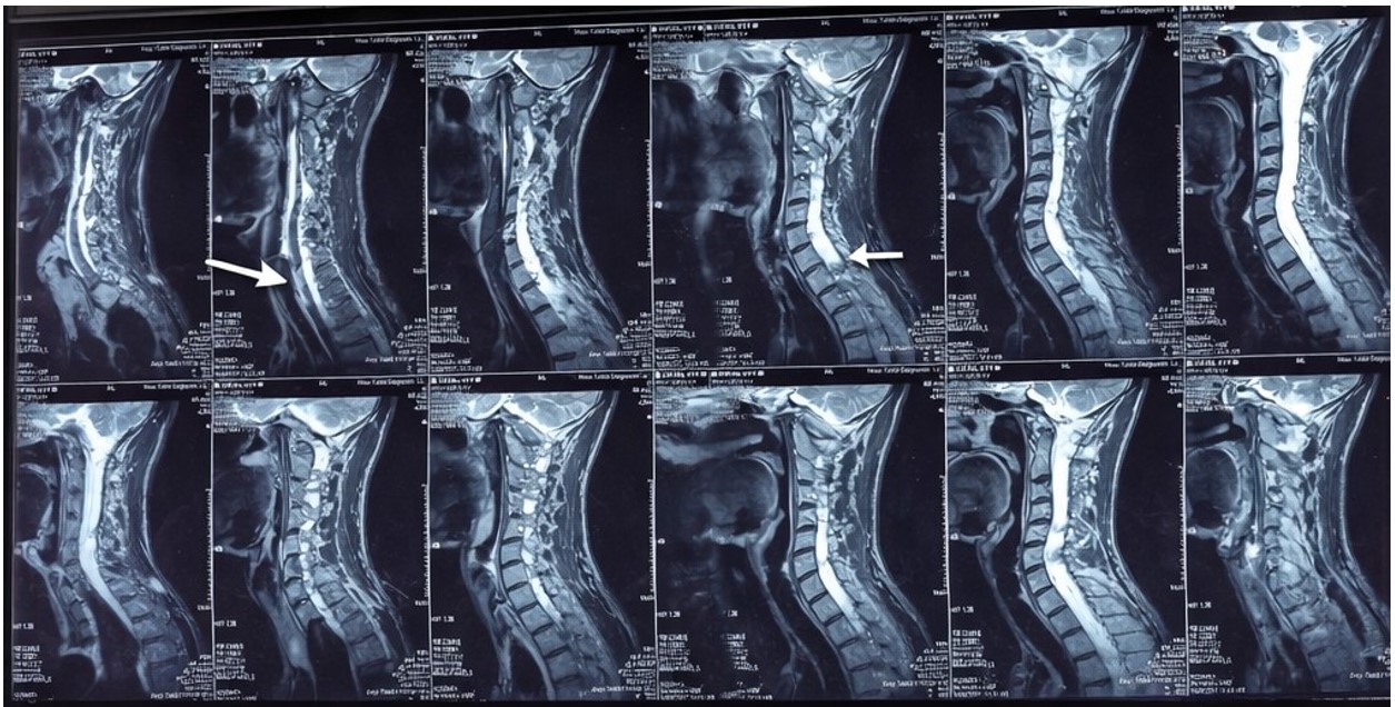
MRI examination of brain and spine revealed bilateral phthisis bulbi with abnormal signal intensity; an enhancing mass lesion of the right eye 1.78 × 1.6 cm was noted, though pathology could not be determined. The seventh and eighth cranial nerves were within normal range. An intramedullary lesion extending from the posterior medulla of the cervicomedullary junction down to the T11 level was found. Nodular enhancement lesions were seen on the cystic cord expansion from C1 – C2 level (1.5 × 1.0 cm), C2 – C3 level (6 × 4 mm), and C4 level (1.8 × 1.6 cm). Mass effect of posterior medullary lesion of 1.5 × 1.6 × 2.4 cm size was exerted on the fourth ventricle, leading to ventricular dilatation and mild transependymal CSF exudation consistent with obstructive hydrocephalus.
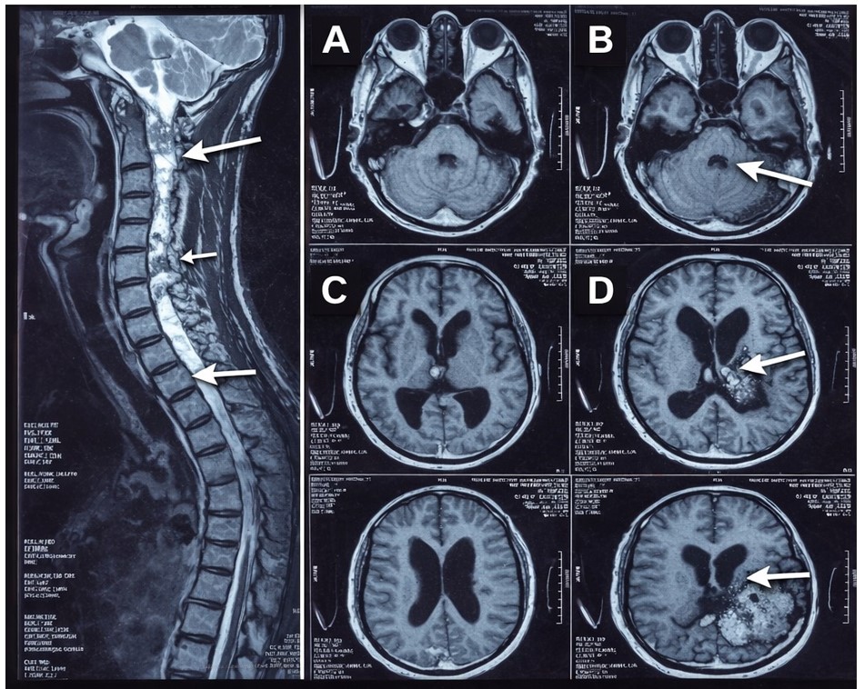
An additional enhancing lesion within the left cerebellar hemisphere was noted as well. Based on the entire imaging appearance, namely the presence of multiple enhancing nodules with cystic degeneration in the spinal cord, the findings were compatible with VHL-related multifocal hemangioblastomas. Still, another possible differential diagnosis was also considered: ependymoma Figure 1 and Figure 2 .
VHL visceral studies, consisting of adrenal and pancreatic scanning and catecholamine study for pheochromocytoma, were not done due to the urgency of the case. Assessment for endolymphatic sac tumor was likewise not done.
Clinical Course and Outcome
The empirical therapy of broad-spectrum antibiotics, including IV piperacillin-tazobactam, was started based on the diagnosis of catheter-associated UTI leading to septic shock. Previous history of antibiotic exposure, laboratory data from microbial cultures, and possible acquisition of health care-associated pathogens were unknown.
Resuscitation of the patient’s hemodynamics began with the administration of 1 L of normal saline intravenously, followed by norepinephrine infusion. During the following 72 hours, vasopressor therapy was increased with the use of the maximum dosage of dobutamine and dopamine. The dosing was done in drops per minute; thus, it was impossible to compare dosages using target values for weight-adjusted dosages. Targets for mean arterial pressure and fluid balance information were unavailable. Renal and liver dysfunction was not tracked on the trend chart. On the fourth day of hospitalization, progressive hemodynamic deterioration ensued.
As a result of a refusal by the patient’s family for CPR or escalation of therapy after four days, death occurred from septic shock with multi-organ failure. Based on the presentation of the patient’s neurological deficits that were worsening, the initial infection is suspected to be urological in nature.
Given the comprehensive analysis of all relevant medical records and the history provided, a retrospective clinical diagnosis of VHL syndrome was suggested according to the clinical criteria set forth by Melmon and Rosen (1964) and updated by Binderup et al. (2022) [7,6]. Testing for mutations in the VHL gene was not performed.
Discussion
In the current case, the patient’s clinical profile was indicative of clinically diagnosed VHL syndrome. This diagnosis was made posthumously based on clinical criteria, although it took many years for him to develop symptoms. Three main aspects should be considered: first, the immediate cause of death, which was sepsis due to multi-organ dysfunction associated with a suspected urological origin; second, the contributory effect of neurological disability, caused by the growth of hemangioblastomas within the brainstem and spinal cord and leading to dependence on a catheter and thus to urological infection; and third, the problem of delayed syndromic diagnosis, which could have affected the natural history of the neurological disease.
The presumptive diagnosis for the source of urological sepsis was made in light of the presence of purulent urine in a patient with chronic catheterization. However, various potential sources of infection coexisted at the same time, such as the T3-T4 laminectomy that was conducted 20 days earlier, which posed the risk of postoperative wound infection, deep spinal infection, or infection related to an implanted device; hospital-acquired pneumonia or aspiration pneumonia secondary to recent orotracheal intubation along with neurological dysfunction; and bloodstream infection secondary to the presence of a central venous catheter. Given the absence of culture tests, imaging studies, and radiologic evidence, it may be prudent not to attribute the origin of sepsis to the urinary tract and label it urosepsis.
The timeline of retinopathy becomes paramount in the discussion of the diagnostic process. Retinal hemangioblastomas bilaterally at an early age are the hallmarks of VHL syndrome [18,17]. Left globe enucleation at around age 10 years, right eye ocular involvement, and bilateral phthisis bulbi can be linked with the predicted pattern of a long-standing, severe case of bilateral retinal hemangioblastoma with associated glaucoma; even though there were no retinal hemangioblastoma pathology reports during the initial presentation, this case history plays a vital role in the diagnostic approach retrospectively. Lensectomy at 20 years of age is not part of the standard signs of VHL syndrome but is a possible complication due to long-standing intraocular pressure.
The hemangioblastomas of the spinal cord were large and included enhancing intramedullary nodules ranging from the cervicomedullary junction to T11. The presence of multifocal nodules, a cystic cord lesion surrounding them, and posterior medullary involvement causing obstructive hydrocephalus are features of VHL-associated multifocal hemangioblastoma. However, ependymoma cannot be ruled out without histological or molecular analysis. Quadriplegia developing over a period of three years, which eventually led to the development of neurogenic bladder and the need for catheterization, was the crucial consequence of the spinal and brainstem hemangioblastomas. Therefore, the way the spinal cord hemangioblastomas caused death was indirectly through neurological deficits and the need for catheterization.
The detection of the right renal cortico-medullary cyst in abdominal ultrasonography can be considered an additional possible visceral manifestation of VHL syndrome. However, it is worth mentioning that the presence of simple renal cysts is frequent among subjects who are 40 years old or above and do not suffer from VHL syndrome, so caution should be observed while interpreting this result due to the lack of information concerning its size and the Bosniak Classification, as well as the absence of any renal mass or RCC. A thorough evaluation of visceral involvement in VHL syndrome, which would have included testing for adrenal, pancreatic, and catecholamine abnormalities, was not possible due to the acute nature of the case and its rapid progression.
Hepatitis C infection, identified during the patient’s third decade of life, constitutes an important comorbidity within the framework of septic shock. No details were provided on viral load, stage of hepatic fibrosis, liver function test results, or immunosuppression. The degree to which HCV played a role in the severity or progression of septic shock remains unknown based on current information.
This delay in diagnosing what appears to be VHL syndrome, identified after the fact, could be explained by multiple factors operating at the systemic level. First, there were the issues of poor access to genetic counseling and hereditary cancer screening services, lack of multidisciplinary monitoring, and limited access to germline testing at the institution where the patient received care for many years. Furthermore, there was never any record suggesting that cascade testing, genetic counseling, and referral of the patient as a syndrome suspect had been conducted before, despite the reported family history of tumors in the retina, brain, and kidneys. This family history, including a sibling with an occipital brain tumor, another sibling with vision loss associated with tumors, and a parent with a kidney tumor, was established according to the patient’s self-report.
The drug Belzutifan, which was granted FDA approval in 2021 for the treatment of non-surgical VHL-associated central nervous system hemangioblastomas [15,16], is briefly mentioned here as a contemporary treatment option for VHL syndrome. It should be noted that this therapy is dependent on molecular diagnosis, clinical indication, and a level of clinical stability not present in the acute context of this case.
Limitations
Some of the major limitations of this case presentation include the lack of histopathological investigation of the lesion excised and the lack of germline testing of the VHL gene; as a result, the diagnosis of VHL remains purely clinical, and the possibility of ependymoma cannot be ruled out. Documented records did not support the patient’s initial retinal diagnosis. Data such as microbiology results, urine cultures, blood cultures, and sensitivities could not be obtained to help determine the source of the infection and the causative organism. Information regarding the ICU management, coagulation profile, HCV viral loads, and hepatic involvement could not be obtained.
Conclusion
In this report, the patient has a medical history that suggests clinically diagnosed VHL syndrome, with no need for genetic analysis or histopathology, as the diagnosis was made postmortem based on clinical parameters. The leading cause of death is sepsis with organ dysfunction due to an infection of undetermined microbial origin involving a urological source. The significant risk factor for mortality in this case is urinary catheterization due to neurological deficits caused by multiple hemangioblastomas in the spinal cord and brain stem that have not been medically addressed. This report highlights the significance of hemangioblastomas, particularly bilateral or early-onset retinal involvement, in the diagnosis of hereditary cancer syndromes. When resources are available, family genetic screening in accordance with clinical guidelines is recommended.
Key finding
This particular case illustrates the potential of unidentified genetic tumors resulting in significant neurological problems and infections leading to death. Early detection of multifocal hemangioblastomas and retinal problems can lead to early identification of patients at risk of developing VHL disease.
From the authors' abstract.
中文摘要
伴尿源疑似感染的致命脓毒性休克:一例临床疑似、先前未诊断的Von Hippel–Lindau疾病患者的病例报告与文献综述
背景:Von Hippel–Lindau疾病是一种罕见的常染色体显性癌症综合征,影响多器官,如视网膜和脑血管瘤、肾脏疾病及其他内脏恶性肿瘤。诊断延迟是可避免死亡和发病率的重要原因。
病例介绍:我们报告一例47岁男性,出现发热、呕吐、低血压及尿道导管排出脓液,随后发展为脓毒性休克伴多器官功能障碍综合征。他有数十年的进展性症状,临床符合可能的VHL综合征,包括自童年起因球状眼球切除导致的双眼失明、多发性脊髓病变导致四肢瘫痪和神经性膀胱,以及家族史中存在视网膜母细胞瘤、脑肿瘤和肾脏病变。脊柱磁共振显示从颈髓交界到T11的广泛多发性脊髓内增强病灶,伴有因囊性改变导致的脊髓扩张,压迫后脑干引起阻塞性脑积水,以及枕部病灶。这些发现提示多发性脊髓血管瘤。右肾皮质-髓质囊肿亦被发现。尽管给予了密集的支持治疗和适当的广谱抗生素,他于住院第4天因疑似尿源性脓毒性休克而不幸去世。
结论:此病例说明未被识别的遗传肿瘤可导致显著的神经系统问题和致死感染。早期发现多发性血管瘤和视网膜问题,可促使早期识别有发展VHL疾病风险的患者。
本中文摘要为机器辅助翻译,仅供参考;以英文摘要为准。 Machine-assisted Chinese translation of the English abstract; the English abstract is the version of record.
References
1. Lonser RR, Glenn GM, Walther M, Chew EY, Libutti SK, Linehan WM, Oldfield EH. von Hippel-Lindau disease. Lancet. 2003: 2059 [PMID: 12814730, https://doi.org/10.1016/S0140-6736(03)13643-4]
2. Maher ER, Neumann HP, Richard S. von Hippel-Lindau disease: a clinical and scientific review. Eur J Hum Genet. 2011: 617 [PMID: 21386872, https://doi.org/10.1038/ejhg.2010.175]
3. Latif F, Tory K, Gnarra J, Yao M, Duh FM, Orcutt ML, Stackhouse T, Kuzmin I, Modi W, Geil L, et al. Identification of the von Hippel-Lindau disease tumor suppressor gene. Science. 1993: 1317 [PMID: 8493574, https://doi.org/10.1126/science.8493574]
4. Kaelin WG, Jr. The VHL Tumor Suppressor Gene: Insights into Oxygen Sensing and Cancer. Trans Am Clin Climatol Assoc. 2017: 298 [PMID: 28790514].
5. Van Leeuwaarde RS, Ahmad S, van Nesselrooij B, Giles RH. Von Hippel-Lindau Syndrome. GeneReviews [Internet]. Seattle (WA): University of Washington; 2024.
6. Melmon KL, Rosen SW. Lindau's Disease. Review of the Literature and Study of a Large Kindred. Am J Med. 1964: 595 [PMID: 14142412, https://doi.org/10.1016/0002-9343(64)90107-x]
7. Louise MBM, Smerdel M, Borgwadt L, Beck Nielsen SS, Madsen MG, Moller HU, Kiilgaard JF, Friis-Hansen L, Harbud V, Cortnum S, Owen H, Gimsing S, Friis Juhl HA, Munthe S, Geilswijk M, Rasmussen AK, Moldrup U, Graumann O, Donskov F, Gronbaek H, Stausbol-Gron B, Schaffalitzky de Muckadell O, Knigge U, Dam G, Wadt KA, Bogeskov L, Bagi P, Lund L, Stochholm K, Ousager LB, Sunde L. von Hippel-Lindau disease: Updated guideline for diagnosis and surveillance. Eur J Med Genet. 2022: 104538 [PMID: 35709961, https://doi.org/10.1016/j.ejmg.2022.104538]
8. Crossey PA, Eng C, Ginalska-Malinowska M, Lennard TW, Wheeler DC, Ponder BA, Maher ER. Molecular genetic diagnosis of von Hippel-Lindau disease in familial phaeochromocytoma. J Med Genet. 1995: 885 [PMID: 8592333, https://doi.org/10.1136/jmg.32.11.885]
9. Varshney N, Kebede AA, Owusu-Dapaah H, Lather J, Kaushik M, Bhullar JS. A Review of Von Hippel-Lindau Syndrome. J Kidney Cancer VHL. 2017: 20 [PMID: 28785532, https://doi.org/10.15586/jkcvhl.2017.88]
10. Acharya A, Panigrahi S, Mahapatra AK, Deo RC, Senapati SB. Von-Hippel Lindau (VHL) syndrome with bilateral cerebellar hemangioblastomas and retinal angiomas: A rare presentation. Int J Surg Case Rep. 2023: 108188 [PMID: 37080144, https://doi.org/10.1016/j.ijscr.2023.108188]
11. Setyawan NH, Hartanto RA, Malueka RG, Dwianingsih EK, Dharma DP. Dual manifestations: spinal and cerebellar hemangioblastomas indicative of von Hippel-Lindau syndrome. Radiol Case Rep. 2024: 5000 [PMID: 39247472, https://doi.org/10.1016/j.radcr.2024.07.158]
12. Kreatsoulas DC, Lonser RR. Spinal cord hemangioblastomas in von Hippel-Lindau disease. Neurooncol Adv. 2024: iii66 [PMID: 39430395, https://doi.org/10.1093/noajnl/vdad153]
13. Kim OH. Multifocal Spinal Hemangioblastoma in von Hippel-Lindau Syndrome: A Case Report and Literature Review. Journal of the Korean Society of Radiology. 2015: [https://doi.org/10.3348/jksr.2015.72.3.171]
14. Nguyen TH, Pham T, Strickland T, Brewer D, Belirgen M, Al-Rahawan MM. Von Hippel-Lindau with early onset of hemangioblastoma and multiple drop-metastases like spinal lesions: A case report. Medicine (Baltimore). 2018: e12477 [PMID: 30278534, https://doi.org/10.1097/MD.0000000000012477]
15. Fallah J, Brave MH, Weinstock C, Mehta GU, Bradford D, Gittleman H, Bloomquist EW, Charlab R, Hamed SS, Miller CP, Dorff SE, Chambers WA, Mixter BD, Dinin J, Pierce WF, Ricks TK, Tang S, Donoghue M, Pazdur R, Amiri-Kordestani L, Ibrahim A, Beaver JA. FDA Approval Summary: Belzutifan for von Hippel-Lindau Disease-Associated Tumors. Clin Cancer Res. 2022: 4843 [PMID: 35727604, https://doi.org/10.1158/1078-0432.CCR-22-1054]
16. Jonasch E, Donskov F, Iliopoulos O, Rathmell WK, Narayan VK, Maughan BL, Oudard S, Else T, Maranchie JK, Welsh SJ, Thamake S, Park EK, Perini RF, Linehan WM, Srinivasan R, Investigators MK. Belzutifan for Renal Cell Carcinoma in von Hippel-Lindau Disease. N Engl J Med. 2021: 2036 [PMID: 34818478, https://doi.org/10.1056/NEJMoa2103425]
17. Karimi S, Arabi A, Shahraki T, Safi S. Von Hippel-Lindau Disease and the Eye. J Ophthalmic Vis Res. 2020: 78 [PMID: 32095212, https://doi.org/10.18502/jovr.v15i1.5950]
18. Chew EY. Ocular manifestations of von Hippel-Lindau disease: clinical and genetic investigations. Trans Am Ophthalmol Soc. 2005: 495 [PMID: 17057815].
License
Copyright (c) 2026 Talha Khan, Hasnain Wajeeh Saqib, Syed Tahir Hussain, Ahmad Mustafa Butt, Muhammad Hassaan Javaid, Awais Hussain Kazim, Aqsa Ibrahim, Saif Ali Khan, Noor Ül-Izza, Abdullah Abbas

This work is licensed under a Creative Commons Attribution 4.0 International License.
Copyright and Licensing
All articles published in ASIDE Internal Medicine are open access and freely available online immediately upon publication, without embargo.
This journal applies the terms of the Creative Commons Attribution 4.0 International License (CC BY 4.0). Under this license, anyone may copy, redistribute, remix, transform, and build upon the material for any purpose, even commercially, provided that proper credit is given to the original author(s), a link to the license is provided, and any changes made are indicated.
-
License link: https://creativecommons.org/licenses/by/4.0/
-
Machine-readable license metadata is embedded in each article and its DOI record.
Copyright Holder
Copyright in all articles remains with the author(s). ASIDE Internal Medicine is published and owned by the American Society for Inclusion, Diversity, and Equity in Healthcare (ASIDE). By submitting a manuscript, authors agree to grant the publisher a non-exclusive license to publish, distribute, and archive the work under the terms of the CC BY 4.0 license.
Third-party Material
Any material (e.g., images, figures, or tables) not covered by CC BY 4.0 will be clearly indicated in the article. Users must obtain permission directly from the copyright holder for reuse of such third-party material.
Author Rights and Self-Archiving
Authors are permitted to deposit all versions of their work (preprint, author’s accepted manuscript, and version of record) in institutional or subject repositories of their choice immediately upon publication, without embargo, provided that the DOI and license information are included.

Published
Issue
Section
Categories
How to Cite
Article history
- Received
- 23 Apr 2026
- Received in revised form
- 16 May 2026
- Accepted
- 6 Jun 2026
- Published
- 24 Jun 2026