
Boerhaave Syndrome in an Elderly Woman: A Case Report
DOI:
https://doi.org/10.71079/ASIDE.CR.030426414Abstract
Boerhaave syndrome is a rare emergency characterized by spontaneous esophageal perforation secondary to severe vomiting. Diagnosis is often difficult due to the nonspecific nature of the symptoms and their similarity to other emergencies. Mortality is high, and it increases if rapid diagnosis and treatment are not provided.
A 76-year-old woman presented to the emergency department after an episode of syncope, hematemesis, and severe retrosternal chest pain. Initial laboratory evaluation showed leukocytosis (12.7 ×10³/µL), elevated C-reactive protein (2.66 mg/dL), and preserved renal function (creatinine 0.68 mg/dL). An initial upper gastrointestinal endoscopy showed detached esophageal mucosa with adherent clots. Contrast-enhanced chest CT scan confirmed esophageal perforation associated with esophageal wall pneumatosis and extensive pneumomediastinum—surgical management with left thoracotomy and esophageal exclusion, mediastinal drainage, and cervical esophagostomy. On postoperative day seven, the patient developed acute kidney injury (creatinine 2.36 mg/dL), metabolic acidosis (HCO₃⁻ 13.4 mmol/L), and hyperlactatemia (4.54 mmol/L), followed by hemodynamic instability and respiratory failure. The patient died 10 days after surgery.
Boerhaave syndrome occurs due to a sudden increase in esophageal intraluminal pressure during vomiting. Its presentation can range from asymptomatic to shock. Diagnosis requires a high index of suspicion, imaging studies, and sometimes upper gastrointestinal endoscopy. Surgery remains the mainstay of treatment, with high mortality if diagnosis and treatment are delayed.
This case highlights the importance of early recognition and multidisciplinary management in patients with severe vomiting, hematemesis, and acute chest pain. Multidisciplinary coordination is key to optimizing the prognosis.
Keywords:
Aged, Boerhaave Syndrome, Case Reports, Esophageal Diseases, Spontaneous PerforationFull Text
Introduction
Boerhaave syndrome is a rare medical-surgical emergency that causes esophageal perforation associated with severe vomiting, first described in 1724 [1,2]. This condition is associated with high mortality, and its nonspecific clinical presentation often delays timely diagnosis and treatment. Although its incidence is low, it is a highly lethal condition, with mortality rates that increase in the absence of early diagnosis and treatment [3,4].
Clinical recognition is challenging because the symptoms can mimic other emergencies, particularly cardiovascular or intra-abdominal pathologies, which require healthcare professionals to maintain a high index of suspicion and to use complementary tests promptly to properly determine its etiology [3,4]. Given its low frequency and high mortality rate, the publication of clinical cases contributes to strengthening knowledge about its diagnostic characteristics, therapeutic options, and clinical outcomes. In this context, we present the case of a 76-year-old patient diagnosed with Boerhaave syndrome, whose outcome highlights the need for a timely and multidisciplinary approach.
Case Presentation
A 76-year-old female patient with a history of hypertension and cataract surgery in her left eye with intraocular lens implantation was present. She was being treated after cataract surgery with corticosteroids (prednisone 20 milligrams daily for an unknown time). She presented at the emergency department after a syncopal episode lasting approximately 10 minutes, with full recovery of consciousness. Initial evaluation in the emergency department included vital signs assessment and laboratory tests. No electrocardiogram abnormalities were reported, and orthostatic hypotension was clinically suspected in the context of hypovolemia secondary to hematemesis. A complete syncope work-up was not performed: no echocardiography, cardiac monitoring, or neuroimaging studies were obtained. This was accompanied by ten episodes of vomiting preceded by nausea: initially with food and traces of blood, later progressing to frank hematemesis. Simultaneously, she experienced oppressive, retrosternal chest pain, rated 10/10 on the visual analog scale (VAS), which resolved spontaneously.
On physical examination upon admission, the patient was awake, with a Glasgow Coma Scale score of 14/15 (Eye Opening: 4, Verbal Response: 4, Motor Response: 6), blood pressure 131/77 mmHg, heart rate 68 bpm, respiratory rate 18 breaths per minute, temperature 36.5 °C, and oxygen saturation 95% on room air. Additionally, pale conjunctivae, oral mucosa with signs of bleeding, a tender neck without subcutaneous emphysema, decreased breath sounds bilaterally at the lung bases, tenderness to palpation in the hypogastrium, and a negative digital rectal examination for gastrointestinal bleeding were noted.
Paraclinical findings showed leukocytosis with a white blood cell count of 12.72 ×10³/µL (reference range [RR]: 4.0 – 10.0 ×10³/µL), neutrophilia of 87.9% (RR: 40 – 75%), hemoglobin 15.3 g/dL (RR: 13.0 – 17.0 g/dL), hematocrit 45.8% (RR: 40 – 50%), and platelet count 224 ×10³/µL (RR: 150 – 450 ×10³/µL). Coagulation parameters were within normal limits, with prothrombin time 10.0 s (RR: 9 – 12 s), activated partial thromboplastin time 20.8 s (RR: 25 – 35 s), and international normalized ratio 1.10. Inflammatory markers revealed an elevated C-reactive protein of 2.66 mg/dL (RR: <0.5 mg/dL). High-sensitivity troponin T was 3.31 ng/L (RR: <14 ng/L). Serum creatinine was 0.68 mg/dL (RR: 0.7 – 1.2 mg/dL), with no evidence of acute kidney injury. Serum lactate and arterial blood gas analysis were not available, limiting full assessment of sepsis severity.
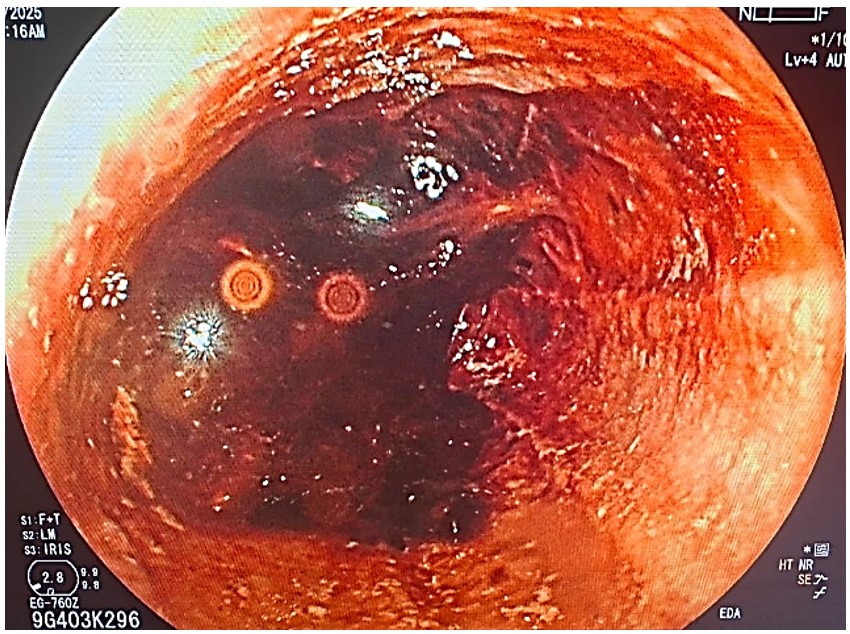
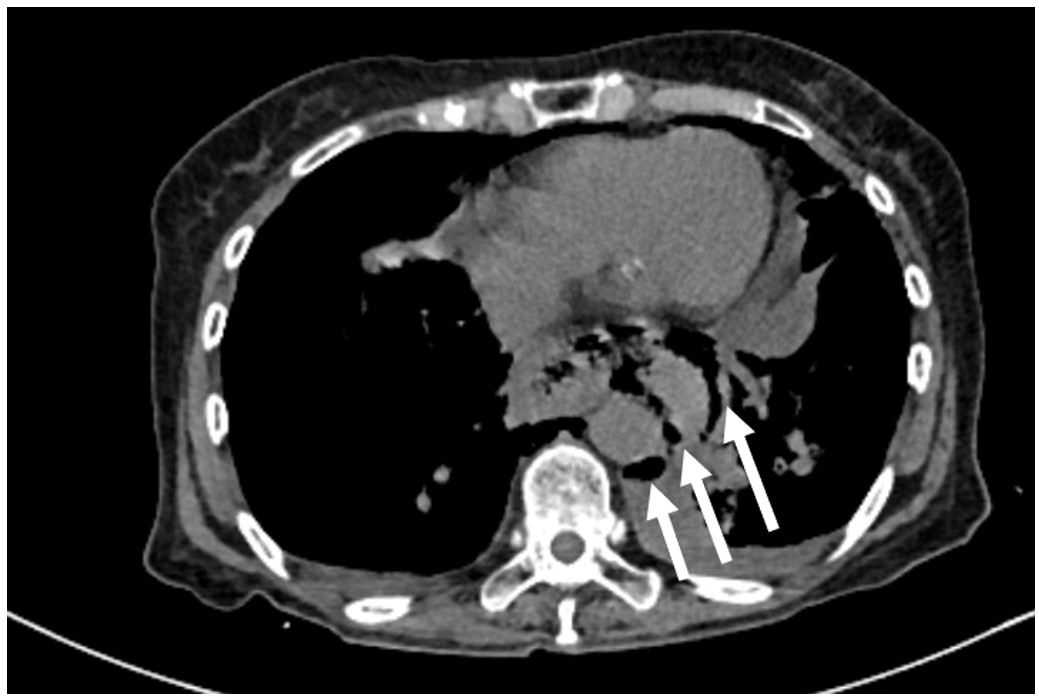
In this context, an initial upper gastrointestinal endoscopy was performed under suspicion of upper gastrointestinal bleeding of unknown cause, revealing food and blood debris along the esophageal lumen, with multiple adherent clots. Upon displacement, the mucosa was found to be frayed, erosive, and friable, with insufficient visualization of its surface Figure 1. A chest CT (unenhanced and contrast-enhanced with iodinated contrast) was subsequently performed, which demonstrated a marked circumferential and asymmetric thickening of the infracarinal esophagus, involving the distal esophagus, gastroesophageal junction, and proximal stomach, with esophageal dilatation up to 44 mm and heterogeneous intraluminal content compatible with blood, gas, and probable food debris. A full-thickness esophageal wall discontinuity was identified at the distal third, with extravasation of esophageal contents into the posterior inferior mediastinum (without a report of the exact height of the esophageal perforation). This was associated with intramural esophageal pneumatosis and extensive pneumomediastinum involving all mediastinal compartments, extending into the cervical and right dorsal soft tissues. Bilateral pleural effusions were present, predominating on the left side, with associated basal laminar atelectasis. No mediastinal abscess, pleural empyema, pulmonary consolidation, or pericardial effusion was identified Figure 2. Given the risk of mediastinitis, antibiotic therapy with ceftriaxone 1 gram every 12 hours and clindamycin 300 milligrams every 6 hours was initiated on hospital day 2 and continued until death, for a total duration of 8 days. No cultures were performed.
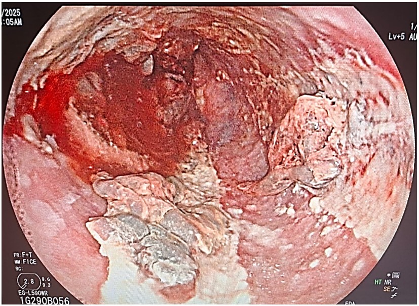
Based on the results, the patient was jointly evaluated by gastroenterologists and cardiothoracic surgeons, who agreed to perform a second upper gastrointestinal endoscopy the following day, revealing an esophageal perforation 26 cm from the upper dental arch Figure 3. Given its thoracic location and the impossibility of doing endoscopic treatment, open surgery was performed. Firm adhesions were found between the aorta and the lower third of the esophagus in the mediastinal pleura, as well as adhesions from the lower pulmonary lobe to the mediastinum and firm pleuropulmonary adhesions at the level of the lower lobe. An esophageal perforation of approximately 3 cm in diameter was evident, with paraesophageal and periaortic mucus, a frayed anterior esophageal wall with signs of necrosis, and perilesional phlegmon formation. Based on these findings, a left thoracotomy with esophageal exclusion was performed, along with release of mediastinal adhesions, a left lateral cervicotomy, creation of a cervical esophagostomy, placement of two 28 FR chest drains, and creation of a Stamm feeding jejunostomy. The patient initially showed a favorable evolution, with adequate control of postoperative pain and good tolerance to enteral nutrition through the jejunostomy; however, on the seventh postoperative day the patient experienced a deterioration of the general condition. The laboratory findings shown a renal dysfunction met AKIN stage 2 criteria, with a rise in serum creatinine to 2.36 mg/dL (reference range [RR]: 0.7 – 1.3 mg/dL), and associated azotemia (urea 116 mg/dL, RR: 15 – 45 mg/dL). Metabolic derangement was confirmed by arterial blood gas analysis showing reduced bicarbonate (HCO 13.4 mmol/L, RR: 22 – 26 mmol/L) with compensatory hypocapnia (PCO 21.9 mmHg, RR: 35 – 45 mmHg) and preserved pH (7.405), consistent with metabolic acidosis. Tissue hypoperfusion was objectively supported by hyperlactatemia (lactate 4.54 mmol/L, RR: <2.0 mmol/L). Additionally, leukocytosis with neutrophilia (12.13 /μL, neutrophils 11.45 /μL) and markedly elevated D-dimer (3.57 mg/L FEU, RR: <0.5 mg/L FEU) were present, supporting a systemic inflammatory and septic process. Alongside these findings, the patient developed hemodynamic instability and respiratory distress, with an oxygen saturation of 90% despite an inspired oxygen fraction (FiO) of 100%. Despite resuscitative measures, the patient died 10 days after surgery. A summary of the clinical timeline is provided in Table 1.
| Event | Time from arrival | Details |
|---|---|---|
| Vomiting and retrosternal pain | 0 hours | Onset of hematemesis. |
| First upper gastrointestinal endoscopy | 8 hours | Visualization of frayed, erosive, and friable esophageal mucosa, with insufficient visualization. |
| CT scan | 10 hours | Demonstration of discontinuity in the distal third of the esophagus, pneumatosis, and pneumomediastinum. |
| Second upper gastrointestinal endoscopy | 30 hours | Visualization of an esophageal perforation 26 cm from the upper dental arch (distal thoracic esophagus). |
| Surgery | 72 hours | Left thoracotomy with esophageal exclusion was performed, and a Stamm feeding jejunostomy was created. |
| Clinical deterioration | 7 days | Hemodynamic instability, respiratory failure, metabolic acidosis, and acute renal failure. |
| Death | 10 days | Despite supportive and resuscitation measures. |
CT, Computerized Tomography.
Discussion
Esophageal perforations can be classified as iatrogenic, traumatic, due to foreign bodies, secondary to other pathologies, or spontaneous. Within this last group is Boerhaave syndrome, considered a surgical emergency first described by the Dutch physician Herman Boerhaave in 1724 [1].
Boerhaave syndrome is an injury caused by increased esophageal intraluminal pressure against a closed cricopharyngeal cartilage within the context of a normal underlying esophagus, usually associated with frequent and severe vomiting. This increased pressure exerts force on the esophagus at its weakest point, which can lead to esophageal perforation, which is most frequently occurring on the left posterolateral aspect of the distal esophagus [2].
Boerhaave syndrome is the second most frequent cause of esophageal perforations, representing 15% of cases, with an incidence of 3.1 cases per million people per year, surpassed only by iatrogenic perforations [1,2]. Furthermore, Boerhaave syndrome is more prevalent in males, with a ratio ranging from 2:1 to 5:1, and a predominant incidence between the sixth and seventh decades of life; it has an approximate mortality rate of 40% [3,2].
Clinical manifestations typically range from completely asymptomatic patients to presentations with irreversible septic shock [3]. The classic clinical presentation of the syndrome is Mackler’s triad, which includes vomiting, subcutaneous emphysema, and chest pain; however, only about 14% of patients usually present all symptoms simultaneously [3,4]. Due to the nonspecific nature of its symptoms, complementary examinations are frequently performed, including laboratory tests, to differentiate Boerhaave syndrome from other conditions such as acute pancreatitis, acute myocardial infarction, and others. Imaging studies, such as CT scans or esophagograms, are also commonly used to determine the site of perforation [3]. On the other hand, the role of upper gastrointestinal endoscopy is controversial due to the risk of increased perforation, so it should be reserved for specific cases and performed by experts [2]. Endoscopy was initially performed under the working diagnosis of upper gastrointestinal bleeding of unknown origin. A second endoscopy was subsequently undertaken to better delineate the perforation after radiological confirmation, given the need to define the lesion prior to surgical planning. This diagnostic strategy, while clinically justified, may have contributed to delayed definitive management and is acknowledged as a limitation of the case, underscoring the importance of prioritizing cross-sectional imaging when Boerhaave syndrome is suspected.
Mortality from Boerhaave syndrome can decrease to 25% if diagnosis and treatment are performed within the first 24 hours of the onset of symptoms; however, it can rise dramatically if both are delayed beyond 48 hours of the onset of symptoms [4]. Surgery with primary esophageal repair and debridement remains the first treatment option in Boerhaave syndrome, regardless of the time of presentation, while esophagectomy is considered an option in cases where the diagnosis has been delayed, there is extensive inflammation, or contamination in the rupture area [5,4]. Other treatment options for esophageal rupture include endoscopic clipping, pleural drainage, antibiotic therapy, and nutritional support; however, management usually requires a combination of multiple treatment approaches [2,4].
Several aspects of this case merit critical discussion. Boerhaave syndrome classically involves the distal esophagus. In our patient, a discrepancy was observed between endoscopic and radiological localization of the perforation. While upper gastrointestinal endoscopy identified the lesion at 26 cm from the upper dental arch, contrast-enhanced CT demonstrated involvement of the infracarinal and distal thoracic esophagus. This discordance is likely explained by luminal distortion secondary to mediastinal inflammation, esophageal wall necrosis, and paraesophageal contamination, which may affect endoscopic distance estimation. Cross-sectional imaging therefore provided a more reliable assessment of the anatomical level of perforation. These findings also explain the severity of the clinical course and the need for esophageal exclusion rather than primary repair, given the extensive mediastinal inflammation and tissue necrosis observed intraoperatively. Additionally, while primary repair is generally recommended when feasible, the decision to perform esophageal exclusion rather than primary repair was based on intraoperative findings of extensive tissue necrosis, paraesophageal contamination, and dense mediastinal adhesions, which precluded safe suturing and favored damage-control surgery.
Conclusion
This case underscores three main lessons. First, the combination of severe vomiting, hematemesis, and acute chest pain (particularly in elderly patients) should guide the diagnosis of esophageal perforation, even in the absence of classic findings such as subcutaneous emphysema. Second, contrast-enhanced chest computed tomography should be prioritized as the diagnostic modality of choice, as early endoscopy may be misleading and carries procedural risks when Boerhaave syndrome is not initially suspected. Third, surgical strategy must be individualized: in the presence of extensive esophageal necrosis and mediastinal contamination, esophageal exclusion may represent the safest option, although it is associated with a high risk of systemic complications as seen in the case. Early recognition and expedited multidisciplinary decision-making remain critical determinants of outcome.
Conflicts of Interest
The authors declare no competing interests that could have influenced the objectivity or outcome of this research
Funding Source
The authors declare that no specific grant or funding was received for this research from any public, commercial, or not-for-profit funding agency.
Acknowledgments
None.
Informed Consent
Informed consent was obtained from the patient for the publication of this case report before the surgery, and therefore before her death. It was well explained and guaranteed the patient’s confidentiality and anonymity.
Large Language Model
The authors declare that generative artificial intelligence (AI) tools (ChatGPT, OpenAI) were used to facilitate translation and grammar review during the preparation of this manuscript. The authors reviewed and verified all content and assumed full responsibility for the integrity and accuracy of the manuscript.
Authors Contribution
JAR was responsible for conceptualization, investigation, methodology, and supervision. NSC, JRT, and EAM contributed to data curation, investigation, methodology, and writing the original draft. BMS contributed to data curation, visualization, and writing of the review and editing.
Data Availability
All relevant clinical and imaging data are contained within the manuscript. No additional datasets were generated or analyzed for this case report.
References
- Hauge T., Abu Hejleh A., Nilsson M., Schroder W.. Boerhaave syndrome. Br J Surg. 2024;111(9). doi:10.1093/bjs/znae216 PMID: 39225330
- Turner A. R., Collier S. A., Turner S. D.. Boerhaave Syndrome [Internet]. 2025.
- Triantafyllou T., Lamb P., Skipworth R., Couper G., Deans C.. Surgical treatment of Boerhaave syndrome in the past, present and future: updated results of a specialised surgical unit. Ann R Coll Surg Engl. 2024;106(6):509-514. doi:10.1308/rcsann.2024.0020 PMID: 38563067 PMCID: PMC11214861
- Tzeng C. H., Chen W. K., Lu H. C., Chen H. H., Lee K. I., Wu Y. S., Lee F. Y.. Challenges in the diagnosis of Boerhaave syndrome: A case report. Medicine (Baltimore). 2020;99(2):e18765. doi:10.1097/MD.0000000000018765 PMID: 31914100 PMCID: PMC6959951
- Kooij C. D., Boptsi E., Weusten Blam, de Vries D. R., Ruurda J. P., van Hillegersberg R.. Treatment of Boerhaave syndrome: experience from a tertiary center. Surg Endosc. 2025;39(4):2228-2238. doi:10.1007/s00464-025-11540-8 PMID: 39930122 PMCID: PMC11933238
References
1. Hauge T, Abu Hejleh A, Nilsson M, Schroder W. Boerhaave syndrome. Br J Surg. 2024;111(9). [PMID: 39225330, https://doi.org/10.1093/bjs/znae216]. DOI: https://doi.org/10.1093/bjs/znae216
2. Turner AR, Collier SA, Turner SD. Boerhaave Syndrome [Internet]; 2025. Updated 2023 Dec 4. In: StatPearls. Treasure Island (FL): StatPearls Publishing; Available from: https://www.ncbi.nlm.nih.gov/books/NBK430808/.
3. Triantafyllou T, Lamb P, Skipworth R, Couper G, Deans C. Surgical treatment of Boerhaave syndrome in the past, present and future: updated results of a specialised surgical unit. Ann R Coll Surg Engl. 2024;106(6):509-14. [PMID: 38563067, PMCID: PMC11214861, https://doi.org/10.1308/rcsann.2024.0020]. DOI: https://doi.org/10.1308/rcsann.2024.0020
4. Tzeng CH, Chen WK, Lu HC, Chen HH, Lee KI, Wu YS, Lee FY. Challenges in the diagnosis of Boerhaave syndrome: A case report. Medicine (Baltimore). 2020;99(2):e18765. [PMID: 31914100, PMCID: PMC6959951, https://doi.org/10.1097/MD.0000000000018765]. DOI: https://doi.org/10.1097/MD.0000000000018765
5. Kooij CD, Boptsi E, Weusten B, de Vries DR, Ruurda JP, van Hillegersberg R. Treatment of Boerhaave syndrome: experience from a tertiary center. Surg Endosc. 2025;39(4):2228-38. [PMID: 39930122, PMCID: PMC11933238, https://doi.org/10.1007/s00464-025-11540-8]. DOI: https://doi.org/10.1007/s00464-025-11540-8
Data Availability Statement
All relevant clinical and imaging data are contained within the manuscript. No additional datasets were generated or analyzed for this case report.
License
Copyright (c) 2026 Jaysoom Willeem Abarca Ruiz, Mauricio Nicolás Suárez Caicedo, Edgar Josué Redrobán Tufiño, Estefanía Adrianela Arteaga Morocho, Byron Rubén Martínez Salazar

This work is licensed under a Creative Commons Attribution 4.0 International License.
Copyright and Licensing
All articles published in ASIDE Case Reports are open access and freely available online immediately upon publication, without embargo.
This journal applies the terms of the Creative Commons Attribution 4.0 International License (CC BY 4.0). Under this license, anyone may copy, redistribute, remix, transform, and build upon the material for any purpose, even commercially, provided that proper credit is given to the original author(s), a link to the license is provided, and any changes made are indicated.
-
License link: https://creativecommons.org/licenses/by/4.0/
-
Machine-readable license metadata is embedded in each article and its DOI record.
Copyright Holder
Copyright in all articles remains with the author(s). ASIDE Case Reports is published and owned by PubPorta Publishing LLC. By submitting a manuscript, authors agree to grant the publisher a non-exclusive license to publish, distribute, archive, and identify itself as the original publisher of the work under the terms of the CC BY 4.0 license.
Academic oversight and scholarly guidance are provided by the American Society for Inclusion, Diversity, and Equity in Healthcare (ASIDE). Copyright and licensing arrangements are administered by the publisher.
Third-party Material
Any material (e.g., images, figures, or tables) not covered by CC BY 4.0 will be clearly indicated in the article. Users must obtain permission directly from the copyright holder for reuse of such third-party material.
Author Rights and Self-Archiving
Authors are permitted to deposit all versions of their work (preprint, author’s accepted manuscript, and version of record) in institutional or subject repositories of their choice immediately upon publication, without embargo, provided that the DOI and license information are included.
Published
Issue
Section
Categories
How to Cite
Article history
- Received
- 6 Dec 2025
- Received in revised form
- 13 Feb 2026
- Accepted
- 15 Feb 2026
- Published
- 4 Mar 2026