Dysphagia Megalatriensis Secondary to Rheumatic Severe Mitral Regurgitation with Giant Left Atrium: A Case Report
DOI:
https://doi.org/10.71079/ASIDE.CV.091126846Abstract
Dysphagia megalatriensis is a rare condition caused by extensive left atrial dilatation, usually due to chronic rheumatic mitral valvular heart disease. It causes esophageal compression, which can be mistaken for primary esophageal conditions.
A 45-year-old female patient with a 10-year history of rheumatic heart disease presented with gradually progressive dysphagia for solids and liquids, worsening breathlessness, orthopnea, poor appetite, and weight loss. Physical examination revealed signs of severe mitral regurgitation and chronic heart failure. Echocardiography revealed severe rheumatic mitral regurgitation with substantial leaflet thickening, commissural fusion, subvalvular involvement, giant left atrium (87 mm; left atrial volume index 148 mL/m²), atrial fibrillation, and pulmonary hypertension. No significant mitral stenosis was found. Quantitative assessment of mitral valve area and mean transmitral gradient was unavailable because comprehensive Doppler assessment was not completed before referral. Contrast-enhanced computed tomography revealed signs of a markedly enlarged left atrium with external compression of the thoracic esophagus. In contrast, barium swallow confirmed smooth extrinsic compression of the esophagus without any obstructive luminal mass. The patient underwent medical optimization with diuretics, rate control, anticoagulation, nutritional support, and initiation of secondary prophylaxis for rheumatic fever before being referred to a cardiothoracic center for planned mitral valve replacement with left atrial reduction plasty.
Dysphagia megalatriensis should be considered in patients with dysphagia and chronic rheumatic mitral valve disease after exclusion of primary esophageal pathology. Multimodality imaging is essential for diagnosis. Medical stabilization followed by referral for definitive surgical management is appropriate; however, the planned operation had not yet been performed, and postoperative outcomes were unavailable at the time of manuscript preparation.
Keywords:
Dysphagia Megalatriensis, Giant Left Atrium, Rheumatic Heart Disease, Mitral Regurgitation, Esophageal Compression, Left Atrial EnlargementFull Text
Introduction
Acute rheumatic fever (ARF) is an immune-mediated inflammatory disease that develops following pharyngeal infection with Group A Streptococcus (Streptococcus pyogenes), typically two to three weeks after the initial infection.[1] The disease may affect the joints, skin, heart, and central nervous system, with diagnosis based on the Jones criteria.[1] Cardiac involvement most commonly manifests as valvulitis affecting the mitral and aortic valves, leading to valvular insufficiency and, over time, stenosis.[2] Recurrent episodes of ARF may result in progressive fibrosis, commissural fusion, and distortion of the mitral valve, ultimately leading to chronic rheumatic heart disease (RHD).
Giant left atrium (GLA) is a recognized but rare consequence of RHD of the mitral valve, resulting from sustained volume and pressure loads that cause progressive left atrial dilatation. Giant left atrium is classically defined as a parasternal long axis diameter of the left atrium >65 mm or a left atrial volume index of greater than 100 mL/m², with left atrial dimensions greater than 80 mm seen in more advanced and prolonged cases.[3,4] The massive increase in size of the left atrium causes mass effects on nearby structures. Compression of the respiratory airways leads to carina splaying and atelectasis; compression of the recurrent left laryngeal nerve causes hoarseness (Ortner’s syndrome); posterior displacement of the thoracic esophagus causes dysphagia or dysphagia megalatriensis.[3,5] Transesophageal echocardiography is associated with high procedure-related risks in cases of esophageal compression; hence, it should be performed if its benefits clinically justify the risk and other imaging techniques are not applicable.[5]
Though the occurrence of RHD has declined worldwide due to the availability of more antibiotics, the disease continues to be a serious problem in developing nations, where late diagnosis and inadequate prevention systems result in the development of complications like GLA.[6] Dysphagia megalatriensis is observed very infrequently today, with fewer than a couple of dozen cases being mentioned in the literature.[3,4] In most cases, the patients are adults who have rheumatic mitral disease that has been left untreated for long periods of time.
There are several educational aspects related to this particular case: it describes the problem of differential diagnosis between cardiac esophageal compression and intrinsic esophageal disease; it draws attention to the risks associated with the delayed diagnosis of RHD as well as the lack of secondary prophylaxis in a country where RHD is endemic; and it demonstrates that a complex diagnostic procedure including echocardiography, HRCT, and barium swallow should be used to make an accurate diagnosis. This report has been written in accordance with the CARE criteria.[7]
Case Presentation
Patient History and Timeline
A 45-year-old woman came to our cardiology ward with complaints of progressive breathlessness on exercise as well as at rest, poor appetite, orthopnea, and non-productive cough. Of importance was the progressive dysphagia, starting from solids to fluids, which had occurred within two to three weeks, along with weight loss.
She had been diagnosed with rheumatic heart disease about 10 years ago when she first presented with symptoms of exertional dyspnea. She recalled an episode of fever associated with migratory polyarthritis during childhood, although no medical documentation was available. In the course of the next 10 years, her symptoms had gradually increased even though she had been intermittently receiving medications like diuretics at the peripheral health center. She had never been put on secondary prophylaxis using benzathine penicillin G throughout this period. She had no complaints of throat infection, joint pain, skin rashes, or neurological symptoms during the present admission. A summary chronological timeline is presented in Table 1.
Physical Examination
The patient appeared chronically ill and cachectic, with bilateral temporal wasting. Physical examination results were as follows: blood pressure 110/70 mm Hg; pulse rate 88 beats per minute, with the pulse being irregularly irregular; respiratory rate 18 breaths per minute; oxygen saturation 96% on room air; body temperature 37.1°C. The BMI was not measured; however, mid-upper arm circumference was reduced.
The findings from the cardiovascular assessment included the apex impulse located laterally to the sixth intercostal space in the anterior axillary line, along with an apical palpable thrill. Auscultation revealed a grade 4/6 systolic murmur at the apex that radiated to the left axilla during all respiratory activities, indicative of severe mitral valve regurgitation. Bilateral crackles up to the mid-zones and mild expiratory wheezes bilaterally were heard on auscultation. There was no elevated jugular venous pressure, but there was mild bilateral pedal edema.
Imaging Investigations
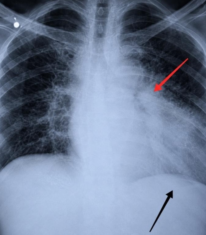
Chest X-ray (Figure 1) showed gross cardiomegaly with splitting of the tracheal carina, which is typical of giant left atrial dilatation. Cardiothoracic ratio exceeded 0.65. Bilateral perihilar haziness was noted, indicative of increased pulmonary venous pressure.
A 12-lead ECG confirmed atrial fibrillation with a ventricular rate of approximately 90 beats per minute. There were no features of pre-excitation or acute ischemia. P waves suggestive of left atrial enlargement had been noted on an earlier recording in 2014, but were no longer visible because of atrial fibrillation. Left ventricular hypertrophy by voltage criteria was not confirmed. The exact duration of atrial fibrillation before admission could not be determined, although the patient reported palpitations for at least two years.
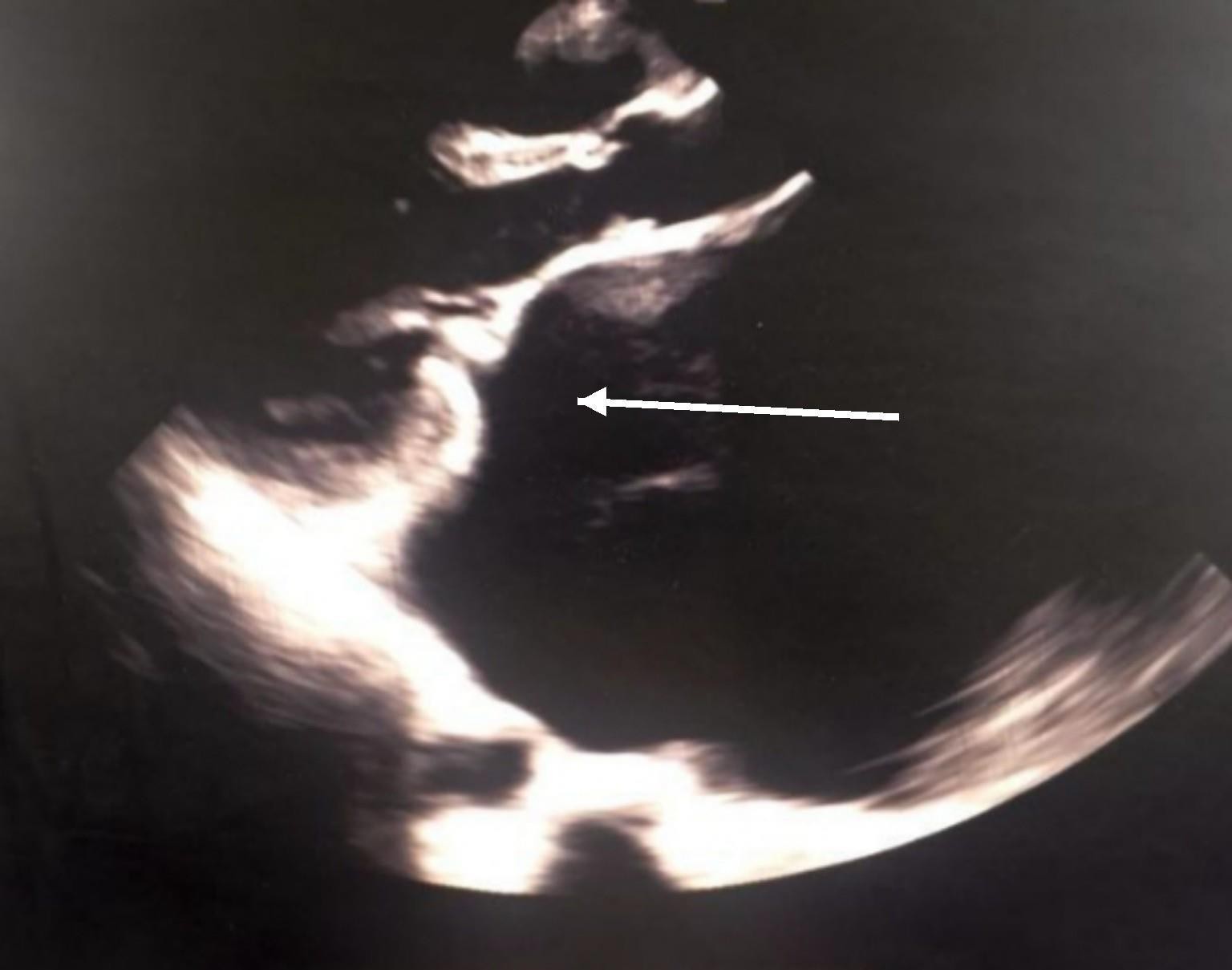
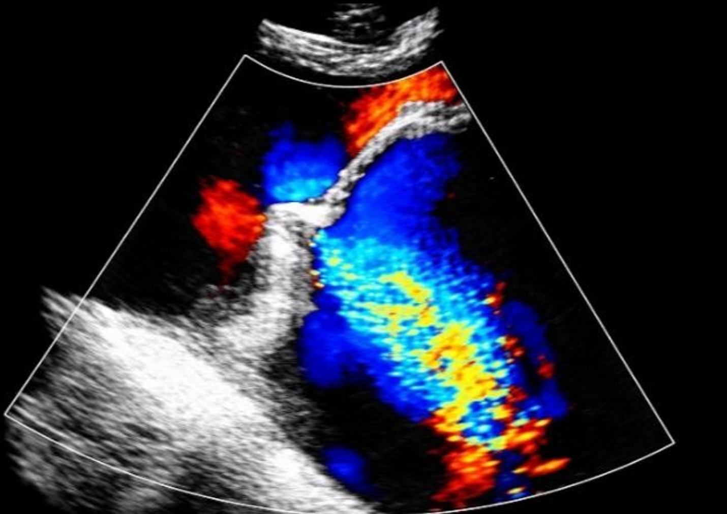
Transthoracic echocardiography (Figure 2 and Figure 3) demonstrated a giant left atrium measuring 87 mm in the parasternal long-axis view with a left atrial volume index of 148 mL/m². The mitral valve showed diffuse leaflet thickening, commissural fusion, and subvalvular thickening consistent with chronic rheumatic involvement. Severe mitral regurgitation was present with an effective regurgitant orifice area of 0.52 cm², regurgitant volume of 75 mL/beat, vena contracta width of 8 mm, and a wide eccentric posterolateral regurgitant jet on color Doppler. Left ventricular end-diastolic diameter measured 62 mm with preserved systolic function (LVEF 53%). Moderate pulmonary hypertension was present with an estimated right ventricular systolic pressure of 62 mm Hg. No clinically significant mitral stenosis was demonstrated. Quantitative assessment of mitral valve area and mean transmitral gradient was unavailable because comprehensive Doppler assessment was not completed before referral.
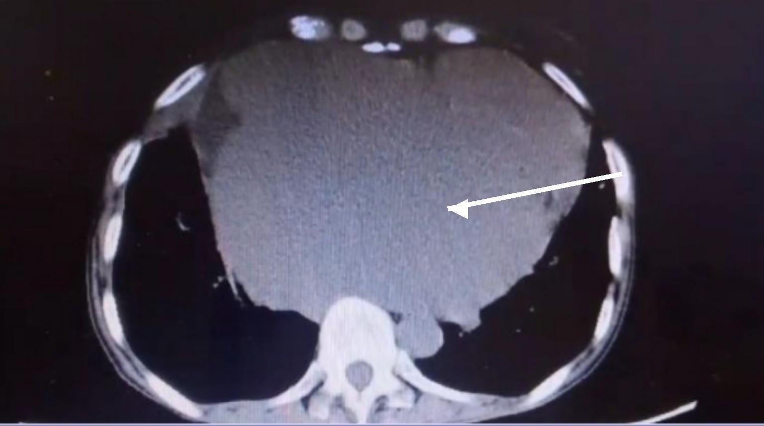
The chest CT scan with contrast (Figure 4) revealed an extremely enlarged left atrium, resulting in mass effect in the posterior mediastinum, causing anterior displacement and external compression of the mid-to-lower esophagus. The condition correlated exactly with the clinical presentation of dysphagia.
The barium swallow demonstrated smooth extrinsic compression of the mid-esophagus without an obstructing intraluminal lesion. These findings supported cardiac compression as the cause of dysphagia; however, intrinsic mucosal disease could not be completely excluded because upper gastrointestinal endoscopy was deferred owing to the patient’s clinical condition and the anticipated procedural risk.
Laboratory Investigations
Selected laboratory values are summarized in Table 2.
Transesophageal echocardiography (TEE) was considered for further evaluation of mitral valve anatomy and left atrial appendage thrombus. However, it was deferred because of the increased procedural risk associated with significant esophageal compression by the enlarged left atrium. Following multidisciplinary discussion, alternative imaging modalities, including repeat transthoracic echocardiography and cardiac magnetic resonance imaging, were planned at the receiving tertiary center if clinically indicated before surgery.
Management
After admission, management of decompensated heart failure was undertaken using the below-mentioned strategy. Intravenous administration of furosemide 40 mg once daily, followed by oral therapy, to obtain a net negative fluid balance, in addition to careful monitoring of kidney function tests and electrolyte levels. Administration of spironolactone 25 mg once daily, which is an aldosterone antagonist that works on neurohormonal blocking in heart failure. Once-daily administration of losartan 25 mg was used to reduce afterload by inhibiting angiotensin II receptor activity. However, losartan was preferred because of its better tolerability compared with ACE inhibitors, given the risk of cough. Nebivolol 2.5 mg was administered once daily to control the rate of atrial fibrillation. However, since there was a borderline state of decompensation, it was administered cautiously while monitoring hemodynamics and development of bronchospasm. Warfarin was started at a target INR of 2.0–3.0 to prevent blood clot formation due to AF in a patient with rheumatic mitral valve disease. INR monitoring was performed every 48 hours during the inpatient period.
Digoxin 0.125 mg once daily was added due to the poor effect of nebivolol alone on ventricular rate control. The reduced dose was chosen in view of the patient’s low body weight, normal-to-mildly reduced renal function, and hypoalbuminemia. Because the admission serum potassium was 3.8 mmol/L, electrolyte replacement was prescribed with a target serum potassium level above 4.0 mmol/L during treatment to reduce the risk of digoxin toxicity.
The patient had never received secondary prophylaxis against recurrent Streptococcus infections before now, even though the patient had been suffering from this condition for 10 years. During this admission, benzathine penicillin G 1.2 million units intramuscularly was administered every 4 weeks, and plans for further secondary prophylaxis were coordinated with the tertiary surgical facility and the patient’s local clinic. Penicillin allergy was excluded by history.
With the rapid progression of both solids and liquids dysphagia, presence of cachexia with serum albumin level of 28 g/L, and high aspiration risk due to almost total esophageal blockage, it was decided to consult the dietitian and speech pathology as soon as possible. All oral intake was stopped until safe swallowing could be assessed. Feeding via nasogastric tube was initiated on Day 3 of admission with a high-protein, high-calorie formula to deliver 1,800–2,000 kcal/day and 1.5 g protein/kg/day to overcome severe malnutrition before surgery. Aspiration precautions, including head-of-bed elevation (30–45°), confirmation of nasogastric tube placement, and avoidance of oral intake, were implemented. Nutritional reassessment was planned at the receiving surgical center as part of preoperative optimization.
After hemodynamic stabilization and multidisciplinary review involving cardiology, cardiothoracic surgery, anesthesia, and nutrition teams, the patient was referred to a tertiary cardiothoracic center for planned mitral valve replacement with left atrial reduction plasty. The procedure had not yet been performed at the time of manuscript preparation; therefore, postoperative outcomes were unavailable.
Discussion
Giant left atrium is a well-recognized but rare condition seen later in cases of chronic mitral valve disease associated with rheumatic heart disease. The continuous volume overload from mitral regurgitation, combined with rheumatic inflammatory remodeling of the atrial wall, leads to dilation. Excessive enlargement of the left atrium (diameter ≥65–80 mm) leads to mechanical pressure effects on adjacent mediastinal structures, resulting in left main bronchus compression with resultant carina deviation and atelectasis, recurrent left laryngeal nerve compression with subsequent hoarseness of voice (Ortner’s syndrome), and posterior displacement of the thoracic esophagus resulting in mechanical dysphagia known as dysphagia megalatriensis.[4,8,9] At the same time, left atrial fibrosis-related atrial fibrillation, pulmonary hypertension, and the risks of thromboembolism add to the disease severity.[10,11]
The difficulty with diagnosing dysphagia megalatriensis lies in the symptomatology being very similar to primary esophageal disorders. In the past, it was mistaken for achalasia, esophageal cancer, and even mediastinal tumors. Furthermore, the enlargement of the left atrium can give the impression of an aortic aneurysm or pleural effusion on a plain chest X-ray examination.[4,12] It is therefore important to adopt a systematic multimodality approach to diagnosing this condition. Two-dimensional echocardiography will confirm the presence of the valve disorder and left atrial enlargement.[4,9] Barium swallow is useful for demonstrating smooth extrinsic esophageal compression. However, it does not exclude intrinsic mucosal disease, and upper gastrointestinal endoscopy should be considered when clinically appropriate. In our patient, endoscopy was deferred because of clinical instability and concern regarding procedural risk associated with severe esophageal compression.[3] As has been pointed out above, transesophageal echocardiography carries a high procedural risk when there is significant esophageal compression, and its use should be limited to patients who stand to benefit from the information provided by the test, such as mitral valve shape and the ability to repair.[3,5]
The rheumatic cause of valvular pathology in this patient is evidenced by the concordance of echocardiographic valve anatomy (thickening of leaflets, commissural fusion, subvalvular involvement), a 10-year illness duration, a childhood history of fever with arthritis, and an elevated ASO titer. An elevated ASO titer indicates previous exposure to Group A Streptococcus but is not diagnostic of chronic rheumatic heart disease. In this patient, the diagnosis was supported primarily by the characteristic echocardiographic findings together with the clinical history of previous acute rheumatic fever and chronic valvular disease.
Management of GLA requires two phases: first, medical stabilization of hemodynamic abnormalities, followed by surgical correction of the structural defect. Medical management, which includes loop diuretics, afterload reduction, rate control in AF, and anticoagulant therapy, helps mitigate the effects of heart failure and reduce the risk of thromboembolic complications. However, it does not help correct the mechanical compression of the esophagus caused by the dilated atrium.[13] There has not been any substantial evidence of successful non-surgical treatment for GLA, nor of significant improvement in symptoms of dysphagia megalatriensis without correcting the structural defects. Surgical management, typically consisting of mitral valve repair or replacement with concomitant left atrial reduction plasty, has been reported to relieve esophageal compression and improve symptoms in appropriately selected patients.[8,12] Series performed in dedicated centers show a perioperative mortality of 2–3%, a significant reduction from the previous value of around 20%, due to improvements in surgical techniques, cardioplegic strategies, and perioperative management.[8] Despite this considerable reduction, the risk associated with surgery in a cachectic and malnourished patient with pulmonary hypertension and chronic AF is still not insignificant and should be decided on an individual basis after optimizing the patient preoperatively.
There are several points in this case worth elaborating upon. Firstly, the lack of secondary prevention for a decade likely played a significant role in the recurrence of subclinical rheumatism and the subsequent deterioration of valvular structures. Benzathine penicillin G was administered during this hospitalization in accordance with standard treatment protocols for RHD and will be continued after surgery. Secondly, the condition of malnutrition, hypoproteinemia, and severe dysphagia is a factor not only relevant but potentially overlooked in terms of preoperative risks in GLA. Nutritional optimization with enteral feeding before surgery is appropriate and was achieved in this case. Thirdly, the digoxin dose was deliberately reduced from 0.25 mg to 0.125 mg based on the patient’s low body weight and normal-to-mildly reduced renal function; this demonstrates the appropriateness of individualization when prescribing drugs to high-risk individuals and underscores the importance of clearly stating the dose used in case reports.
This current case contributes to the small but increasing body of literature regarding dysphagia megalatriensis. It serves as an additional case of the late presentation of advanced rheumatic heart disease in South Asia. This illustrates the importance of considering cardiac compression as a cause of dysphagia for practitioners in endemic regions of RHD, especially when routine investigation of the esophagus proves inconclusive.
Conclusion
Dysphagia megalatriensis is a rare but important manifestation of giant left atrial enlargement and should be considered in patients presenting with dysphagia after primary esophageal disease has been excluded. In patients with chronic rheumatic mitral valve disease, multimodality imaging with transthoracic echocardiography, contrast-enhanced computed tomography, and barium swallow can establish the diagnosis and define the extrinsic nature of esophageal compression. Medical stabilization, nutritional optimization, anticoagulation, and secondary prophylaxis for rheumatic fever are essential before referral for definitive surgery. In this case, the patient was stabilized and referred for planned mitral valve replacement with left atrial reduction plasty. Still, the operation had not yet been performed, and postoperative outcomes were unavailable.
| Timepoint | Event |
|---|---|
| Childhood (recalled) | Episode of fever and migratory polyarthritis; no formal diagnosis or documentation available. |
| 2014 | Formal diagnosis of rheumatic heart disease at a peripheral facility; initiated on diuretics. No echocardiography documented at that time. Secondary prophylaxis not commenced. |
| 2014–2024 | Progressive worsening of exertional dyspnea; intermittent diuretic use without specialist follow-up. No secondary prophylaxis throughout this period. |
| ~3 weeks before admission | Onset of rapidly progressive dysphagia, initially to solids then to liquids; associated with anorexia and weight loss. |
| ~4 weeks prior to admission | Worsening dyspnea at rest; onset of orthopnea, non-productive cough. |
| Day 1 (Admission) | Admitted to cardiology unit; physical examination, ECG, chest X-ray, and laboratory investigations performed. |
| Day 2–3 | Transthoracic echocardiography, HRCT chest, and barium swallow performed. Diagnosis of dysphagia megalatriensis established. |
| Day 3–5 | Medical optimization initiated: diuresis, rate control, afterload reduction, anticoagulation. Dietitian and speech-language pathology consulted. Nasogastric feeding commenced. |
| Day 5–7 | Cardiothoracic surgical consultation. Patient and family counseled regarding mitral valve replacement and left atrial reduction plasty. |
| Day 7 (Discharge planning) | Patient clinically stabilized; referred to tertiary cardiothoracic surgery center for elective surgical intervention. |
ECG, electrocardiogram; HRCT, high-resolution computed tomography.
| Parameter | Result | Reference Range |
|---|---|---|
| Hemoglobin | 12.9 g/dL | 12.0–16.0 g/dL |
| Mean corpuscular volume | 88 fL (normocytic) | 80–100 fL |
| Serum creatinine | 0.9 mg/dL | 0.5–1.1 mg/dL |
| eGFR (CKD-EPI) | 78 mL/min/1.73 m² | ≥60 |
| Serum potassium | 3.8 mmol/L | 3.5–5.0 mmol/L |
| Serum sodium | 136 mmol/L | 136–145 mmol/L |
| HbA1c | 6.1% (prediabetes range) | <5.7% (normal) |
| Anti-streptolysin O (ASO) titre | 400 IU/mL (elevated) | <200 IU/mL |
| INR (on warfarin, Day 5) | 2.4 | Target 2.0–3.0 |
| Serum albumin | 28 g/L (low) | 35–50 g/L |
| HBsAg | Non-reactive | — |
| HIV antibody | Non-reactive | — |
| Blood group | AB positive | — |
eGFR, estimated glomerular filtration rate; CKD-EPI, Chronic Kidney Disease Epidemiology Collaboration; HbA1c, glycated hemoglobin; ASO, anti-streptolysin O; INR, international normalized ratio; HBsAg, hepatitis B surface antigen; HIV, human immunodeficiency virus.
Conflicts of Interest
The authors declare no conflicts of interest.
Funding Source
This research received no specific grant from any funding agency in the public, commercial, or not-for-profit sectors.
Institutional Review Board (IRB)
The case was managed in accordance with the Declaration of Helsinki. Formal ethical approval was not required for a single-patient, de-identified case report at the participating institutions.
Informed Consent
Written informed consent was obtained from the patient for publication of this case report and any associated clinical images. All images presented in this report have been reviewed to ensure that patient-identifying information has been removed.
Large Language Model
None
Authors Contribution
JF, AB, MHJ, and AK were responsible for case selection, while JF and AB were responsible for data collection. MHJ and AK wrote the first draft of the manuscript, and OT compiled the references. HUW was responsible for patient follow-up and obtaining informed consent, while OT and HUW contributed to reviewing, editing, and final formatting. All authors reviewed and approved the final manuscript.
Data Availability
Data sharing does not apply to this article as no datasets were generated or analyzed during the current study.
References
- Gewitz MH, Baltimore RS, Tani LY, et al. Revision of the Jones criteria for the diagnosis of acute rheumatic fever in the era of Doppler echocardiography: a scientific statement from the American Heart Association. Circulation. 2015;131(20):1806-1818. doi:10.1161/CIR.0000000000000205 PMID: 25908771
- Cannon J, Roberts K, Milne C, Carapetis JR. Rheumatic heart disease severity, progression and outcomes: a multi-state model. J Am Heart Assoc. 2017;6(3):e003498. doi:10.1161/JAHA.116.003498 PMID: 28255075
- Deschepper C, Devos D, DePauw M. Cardiac dysphagia — a ghost of the past? A case report. Eur Heart J Case Rep. 2021;5(9):ytab338. doi:10.1093/ehjcr/ytab338 PMID: 34527858
- Pandit BN, Aggarwal P, Subramaniyan S, Gujral JS, Nath RK. Largest giant left atrium in rheumatic heart disease. J Cardiol Cases. 2021;24(1):10-13. doi:10.1016/j.jccase.2020.11.022 PMID: 34257753
- Bugra AK, Kadirogullari E, Onan B. Reduction plasty for giant left atrium causing dysphagia: a case report. Gen Thorac Cardiovasc Surg. 2021;69(3):546-549. doi:10.1007/s11748-020-01490-4 PMID: 32965607
- Steer AC, Carapetis JR. Acute rheumatic fever and rheumatic heart disease in indigenous populations. Pediatr Clin North Am. 2009;56(6):1401-1419. doi:10.1016/j.pcl.2009.09.011 PMID: 19962028
- Gagnier JJ, Kienle G, Altman DG, et al. The CARE guidelines: consensus-based clinical case reporting guideline development. J Med Case Rep. 2013;7:223. doi:10.1186/1752-1947-7-223 PMID: 24228906
- Apostolakis E, Shuhaiber JH. The surgical management of giant left atrium. Eur J Cardiothorac Surg. 2008;33(2):182-190. doi:10.1016/j.ejcts.2007.11.003 PMID: 18096399
- Gajanana D, Morris DL, Janzer SF, George JC, Figueredo VM. Giant left atrium causing dysphagia. Tex Heart Inst J. 2016;43(5):469-471. doi:10.14503/THIJ-15-5682 PMID: 27777540
- Patel DA, Lavie CJ, Milani RV, Shah S, Gilliland Y. Clinical implications of left atrial enlargement: a review. Ochsner J. 2009;9(4):191-196. PMID: 21603443
- Zilberszac R, Gleiss A, Massetti M, et al. Left atrial size predicts outcome in severe but asymptomatic mitral regurgitation. Sci Rep. 2023;13(1):3892. doi:10.1038/s41598-023-31163-0 PMID: 36890195
- Santra G, Paul R, Pradhan S, Choudhury PS, Das S. A case of giant left atrium mimicking pleural effusion. Oman Med J. 2014;29(4):e077. doi:10.5001/omj.2014.82 PMID: 30996820
- Greenberg B. Medical management of patients with heart failure and reduced ejection fraction. Korean Circ J. 2022;52(3):173-197. doi:10.4070/kcj.2021.0401 PMID: 35257531
References
1. Gewitz MH, Baltimore RS, Tani LY, et al. Revision of the Jones criteria for the diagnosis of acute rheumatic fever in the era of Doppler echocardiography: a scientific statement from the American Heart Association. Circulation. 2015;131(20):1806-1818. [PMID: 25908771, doi:10.1161/CIR.0000000000000205] DOI: https://doi.org/10.1161/CIR.0000000000000205
2. Cannon J, Roberts K, Milne C, Carapetis JR. Rheumatic heart disease severity, progression and outcomes: a multi-state model. J Am Heart Assoc. 2017;6(3):e003498. [PMID: 28255075, doi:10.1161/JAHA.116.003498] DOI: https://doi.org/10.1161/JAHA.116.003498
3. Deschepper C, Devos D, DePauw M. Cardiac dysphagia - a ghost of the past? A case report. Eur Heart J Case Rep. 2021;5(9):ytab338. [PMID: 34527858, doi:10.1093/ehjcr/ytab338] DOI: https://doi.org/10.1093/ehjcr/ytab338
4. Pandit BN, Aggarwal P, Subramaniyan S, Gujral JS, Nath RK. Largest giant left atrium in rheumatic heart disease. J Cardiol Cases. 2021;24(1):10-13. [PMID: 34257753, doi:10.1016/j.jccase.2020.11.022] DOI: https://doi.org/10.1016/j.jccase.2020.11.022
5. Bugra AK, Kadirogullari E, Onan B. Reduction plasty for giant left atrium causing dysphagia: a case report. Gen Thorac Cardiovasc Surg. 2021;69(3):546-549. [PMID: 32965607, doi:10.1007/s11748-020-01490-4] DOI: https://doi.org/10.1007/s11748-020-01490-4
6. Steer AC, Carapetis JR. Acute rheumatic fever and rheumatic heart disease in indigenous populations. Pediatr Clin North Am. 2009;56(6):1401-1419. [PMID: 19962028, doi:10.1016/j.pcl.2009.09.011] DOI: https://doi.org/10.1016/j.pcl.2009.09.011
7. Gagnier JJ, Kienle G, Altman DG, et al. The CARE guidelines: consensus-based clinical case reporting guideline development. J Med Case Rep. 2013;7:223. [PMID: 24228906, doi:10.1186/1752-1947-7-223] DOI: https://doi.org/10.1186/1752-1947-7-223
8. Apostolakis E, Shuhaiber JH. The surgical management of giant left atrium. Eur J Cardiothorac Surg. 2008;33(2):182-190. [PMID: 18096399, doi:10.1016/j.ejcts.2007.11.003] DOI: https://doi.org/10.1016/j.ejcts.2007.11.003
9. Gajanana D, Morris DL, Janzer SF, George JC, Figueredo VM. Giant left atrium causing dysphagia. Tex Heart Inst J. 2016;43(5):469-471. [PMID: 27777540, doi:10.14503/THIJ-15-5682] DOI: https://doi.org/10.14503/THIJ-15-5682
10. Patel DA, Lavie CJ, Milani RV, Shah S, Gilliland Y. Clinical implications of left atrial enlargement: a review. Ochsner J. 2009;9(4):191-196. [PMID: 21603443]
11. Zilberszac R, Gleiss A, Massetti M, et al. Left atrial size predicts outcome in severe but asymptomatic mitral regurgitation. Sci Rep. 2023;13(1):3892. [PMID: 36890195, doi:10.1038/s41598-023-31163-0] DOI: https://doi.org/10.1038/s41598-023-31163-0
12. Santra G, Paul R, Pradhan S, Choudhury PS, Das S. A case of giant left atrium mimicking pleural effusion. Oman Med J. 2014;29(4):e077. [PMID: 30996820, doi:10.5001/omj.2014.82]
13. Greenberg B. Medical management of patients with heart failure and reduced ejection fraction. Korean Circ J. 2022;52(3):173-197. [PMID: 35257531, doi:10.4070/kcj.2021.0401] DOI: https://doi.org/10.4070/kcj.2021.0401
Data Availability Statement
Data sharing does not apply to this article, as no datasets were generated or analyzed during the current study.
License
Copyright (c) 2026 Dr Jaweria Farman, Dr Alina Batool, Dr Okasha Tahir, Dr Muhammad Hassaan Javaid, Abbas Khan, Hakim Ullah Wazir

This work is licensed under a Creative Commons Attribution 4.0 International License.
Copyright and Licensing
All articles published in ASIDE Cardiovascular are open access and freely available online immediately upon publication, without embargo.
This journal applies the terms of the Creative Commons Attribution 4.0 International License (CC BY 4.0). Under this license, anyone may copy, redistribute, remix, transform, and build upon the material for any purpose, even commercially, provided that proper credit is given to the original author(s), a link to the license is provided, and any changes made are indicated.
-
License link: https://creativecommons.org/licenses/by/4.0/
-
Machine-readable license metadata is embedded in each article and its DOI record.
Copyright Holder
Copyright in all articles remains with the author(s). ASIDE Cardiovascular is published and owned by PubPorta Publishing LLC. By submitting a manuscript, authors agree to grant the publisher a non-exclusive license to publish, distribute, archive, and identify itself as the original publisher of the work under the terms of the CC BY 4.0 license.
Academic oversight and scholarly guidance are provided by the American Society for Inclusion, Diversity, and Equity in Healthcare (ASIDE). Copyright and licensing arrangements are administered by the publisher.
Third-party Material
Any material (e.g., images, figures, or tables) not covered by CC BY 4.0 will be clearly indicated in the article. Users must obtain permission directly from the copyright holder for reuse of such third-party material.
Author Rights and Self-Archiving
Authors are permitted to deposit all versions of their work (preprint, author’s accepted manuscript, and version of record) in institutional or subject repositories of their choice immediately upon publication, without embargo, provided that the DOI and license information are included.
Published
Issue
Section
How to Cite
Article history
- Received
- 12 May 2026
- Received in revised form
- 30 Jul 2026
- Accepted
- 25 Aug 2026
- Published
- 11 Sep 2026