Pancreatic Rest Complicated by Actinomyces Gastric Abscess in a Young Male: A Case Report
DOI:
https://doi.org/10.71079/ASIDE.GI.06012584Abstract
Pancreatic rest, or ectopic pancreatic tissue, is a rare condition. It is characterized by pancreatic tissue outside its usual location, most commonly in the gastric antrum or proximal small intestine. We present a rare case of a 21-year-old male with recurrent epigastric pain, vomiting, and fever. Imaging and endoscopic ultrasound (EUS) identified a subepithelial lesion with features consistent with pancreatic rest. Subsequent fine-needle aspiration (FNA) grew Actinomyces and Streptococcus. Given persistent symptoms and incomplete resolution despite prolonged antibiotics, he underwent partial gastrectomy. This case highlights an unusual infectious complication of pancreatic rest with Actinomyces and underscores the need to consider surgical intervention in cases refractory to medical therapy.
Keywords:
Endoscopic ultrasound (EUS), Gastric abscess, Heterotopic pancreas, Pancreatic restFull Text
Introduction
Pancreatic rest is a condition where pancreatic tissue is located outside its usual anatomical site. It is also called ectopic, heterotopic, or aberrant pancreatic tissue. This tissue lacks an anatomical or vascular connection to the main pancreas yet retains its own separate blood and nerve supply [1,2]. This condition predominantly affects the gastrointestinal tract, particularly the gastric antrum and proximal small intestine, with an incidence ranging from 0.55% to 13% in autopsy series [1]. While pancreatic rests are often asymptomatic and discovered incidentally during endoscopic or imaging studies, common symptoms include nausea, vomiting, epigastric pain, weight loss, gastrointestinal bleeding, and gastric outlet obstruction. Rarely, it can lead to complications such as gastric ulceration, pancreatitis, obstructive jaundice when near the Ampulla of Vater, or even acute perforation due to inflammation in the stomach or duodenum [2,3]. Gastric abscesses secondary to pancreatic rest are particularly rare, with few reported cases describing this phenomenon [4,5]. These abscesses are believed to result from localized inflammation or necrosis of ectopic acinar tissue. Identifying pancreatic rest as the source of a gastric abscess is clinically challenging, particularly when the initial presentation mimics more common gastric subepithelial lesions such as gastrointestinal stromal tumors (GISTs) or duplication cysts [2,6]. Malignant transformation is uncommon, occurring in 0.7% to 1.8% of cases [3]. For example, a case report highlighted a pancreatic rest presenting as a subepithelial nodule, with histological analysis revealing pancreatic intraepithelial neoplasia [7]. We report a rare case of a young male with recurrent gastric abscess formation due to pancreatic rest, complicated by Actinomyces and alpha-hemolytic Streptococcus infection. To our knowledge, this is among the few reported cases of infected pancreatic rest and the first with hematogenous Actinomyces seeding from dental work.
Case Presentation
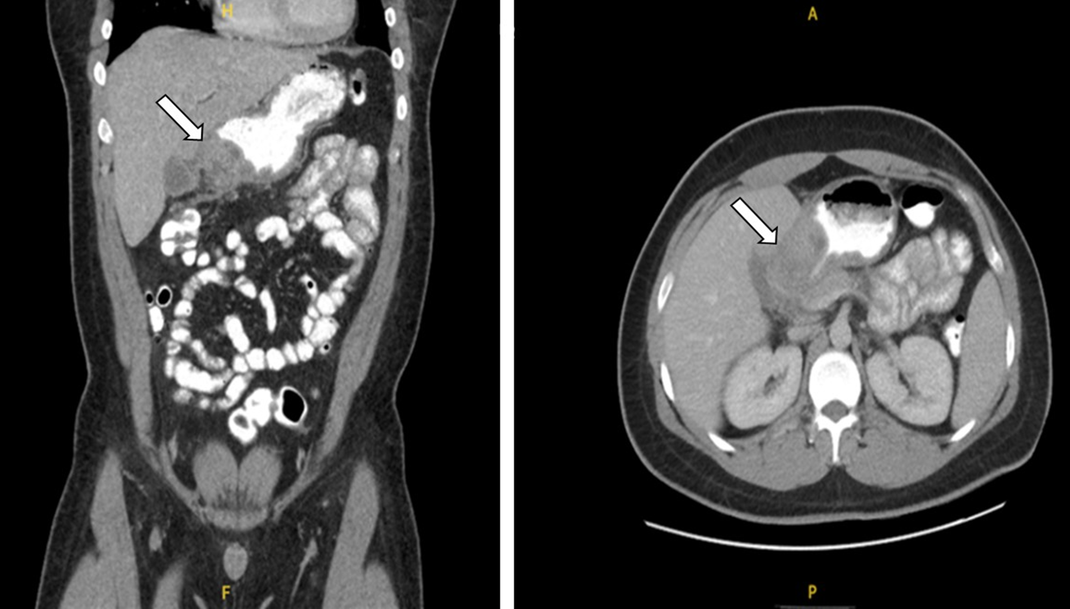
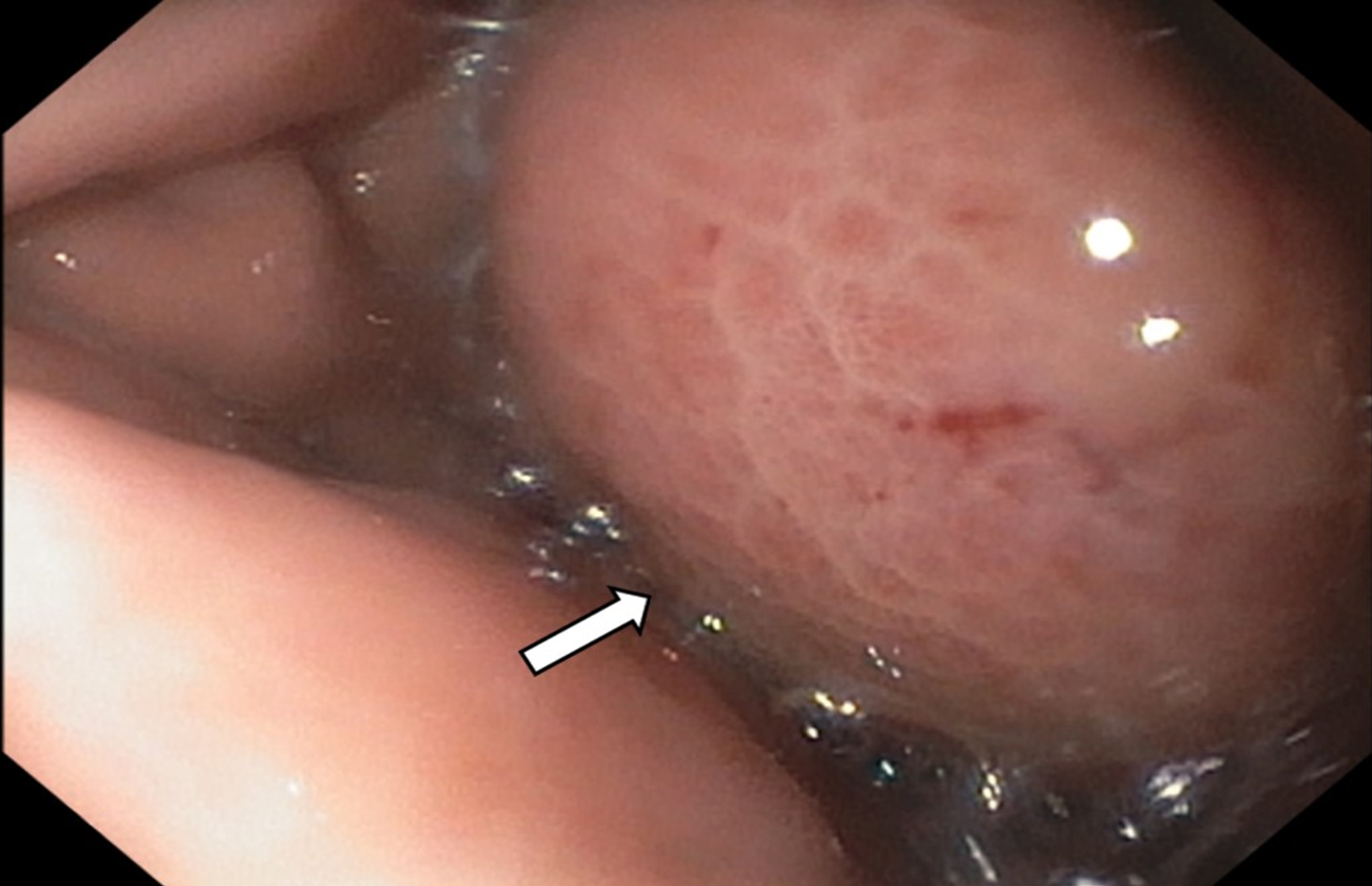
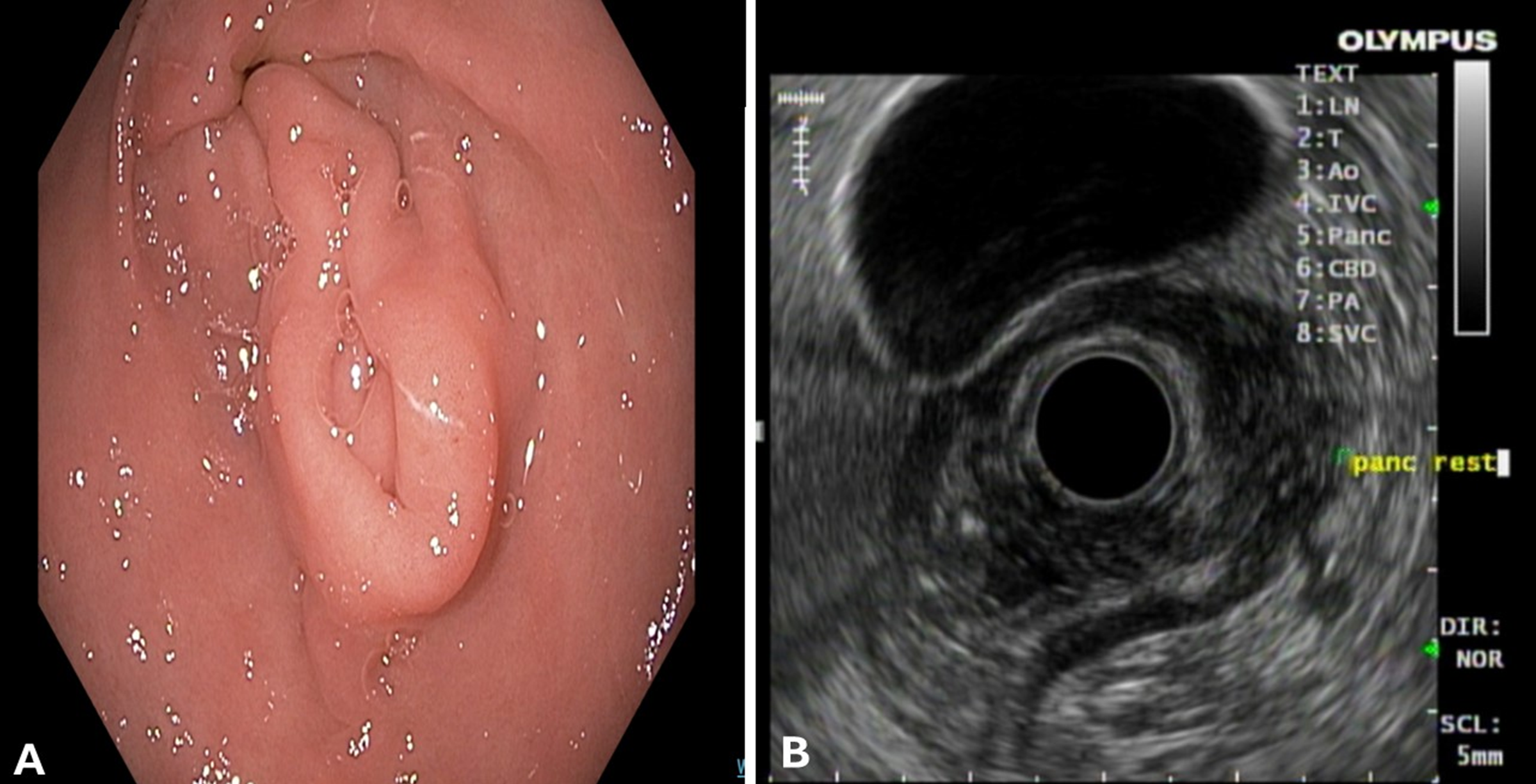
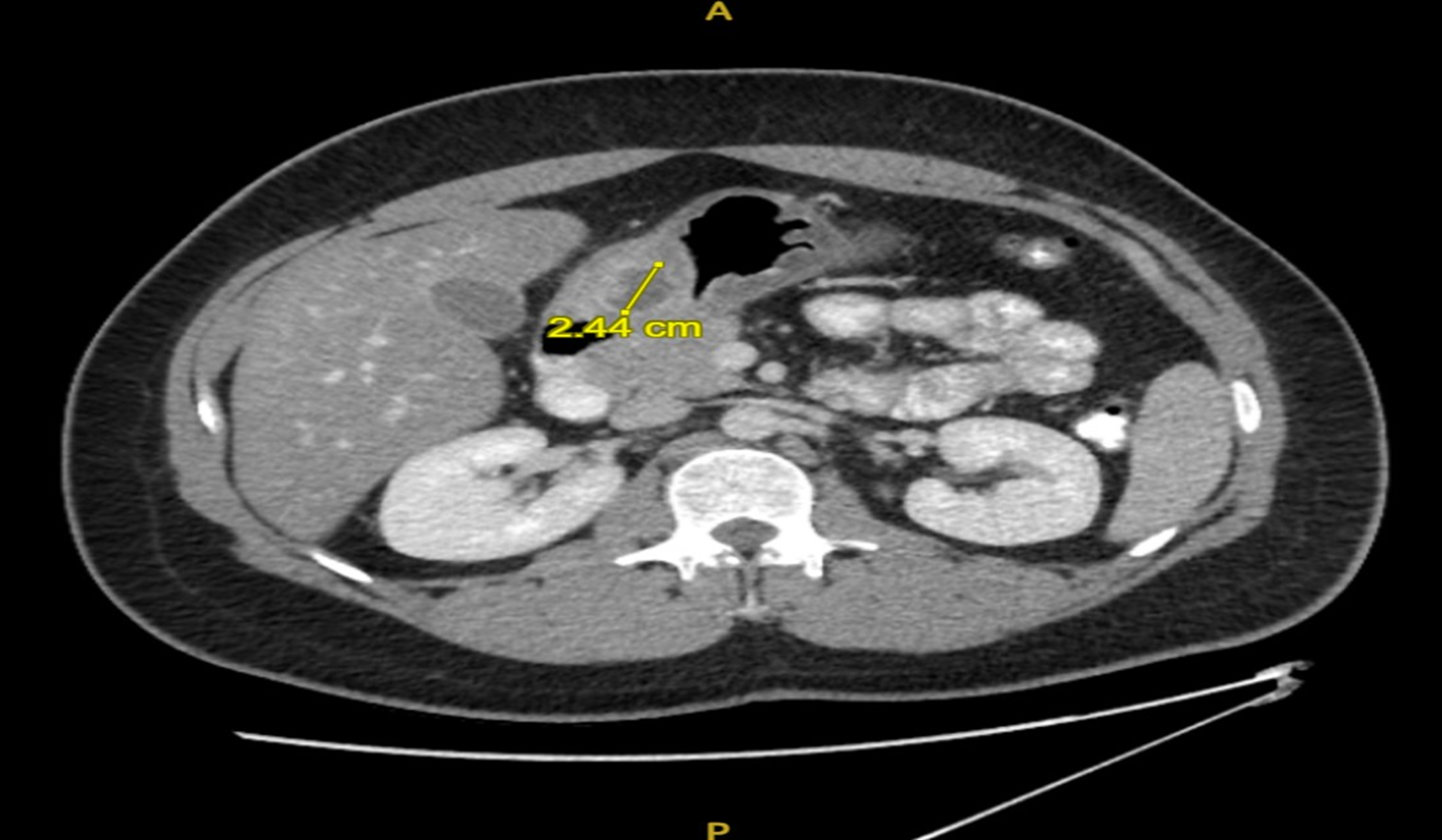
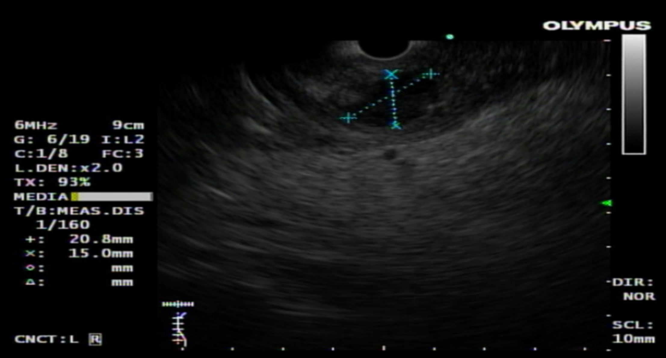
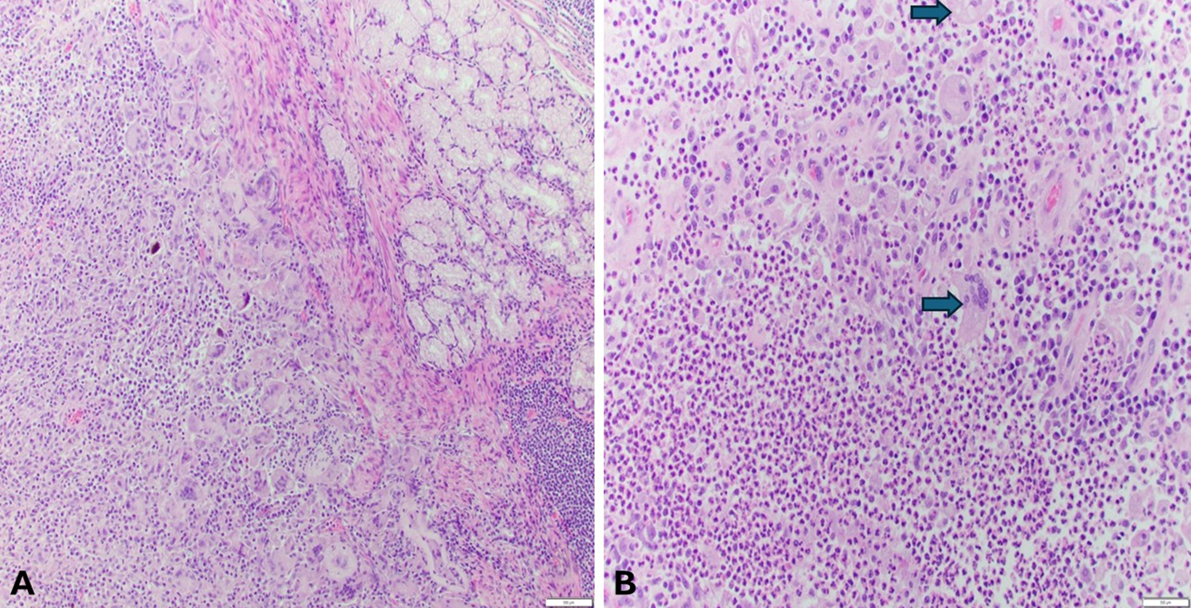
A 21-year-old male with no significant past medical history initially presented to the emergency department (ED) with epigastric abdominal pain radiating to the back, early satiety, and vomiting. Laboratory workup showed white blood cells (WBC) , lipase 26 U/L, and alanine aminotransferase 16 U/L. A computed tomography (CT) scan of the abdomen and pelvis was done, and it showed mild edematous and inflammatory stranding adjacent to the distal stomach. He was discharged with a prescription for omeprazole and metoclopramide and advised to follow up with gastroenterology. One week later, he returned with worsening symptoms and a fever (101°F). Repeat labs showed leukocytosis , Lipase 26 U/L, an alkaline phosphatase of 76 U/L, aspartate aminotransferase of 23 U/L, alanine aminotransferase of 29 U/L, and lipase of 26 U/L. A repeat CT scan with contrast demonstrated mass-like thickening of the gastric antral wall with luminal compression and adjacent stranding, suggesting an inflammatory phlegmon with no drainable collection Figure 1. He was admitted and started on ceftriaxone and Metronidazole. Esophagogastroduodenoscopy (EGD) revealed large subepithelial extrinsic compression in the antrum, with the suspected fluid collection, possibly a walled-off abscess Figure 2. No ulceration was observed, and biopsies were not obtained due to the soft consistency of the lesion. He was discharged on Metronidazole and Cefdinir for 7 days with plans for a repeat CT scan and EGD in 6 weeks. A follow-up CT scan showed interim resolution of the antral mass. The EGD showed a 15 mm submucosal nodule along the greater curvature of the antrum concerning pancreatic rest. EUS revealed an extended area of heterogenous echotexture extending from the muscularis propria to the submucosa, consistent with pancreatic rest Figure 3 . Five months later, the patient presented to the ED with recurrent epigastric pain, nausea, and diarrhea. Labs showed WBC , lipase 26 U/L, and amylase 43 U/L. An abdominal-pelvic CT scan with contrast showed a 2.4 cm fluid-containing structure in the anterior/caudal wall of the gastric antrum near the pylorus, suspicious of an abscess versus a walled-off ulcer Figure 4. Ceftriaxone and metronidazole were reinitiated. A repeat EUS showed an irregular, oval, and anechoic cyst measuring 27 mm x 21 mm, with debris present and well-defined, smooth margins Figure 5. Fine-needle aspiration revealed no malignant cells, with abundant neutrophils and histiocytes. Culture grew alpha-hemolytic Streptococcus and Actinomyces species. He was discharged on a 7-day course of cefpodoxime and metronidazole. A repeat CT scan four weeks later showed reduced size of the gastric wall fluid collection, with no new findings. Due to persistence, he was referred to Infectious Diseases and started on cefdinir and doxycycline. Imaging showed a 2.2 × 1.9 cm gastric antral mass without fluid two months later. Cefdinir was stopped, and doxycycline was continued for six months to treat Actinomyces. He later reported a recent root canal prior to symptom onset, suggesting an odontogenic source. Given the persistence of the lesion despite multiple courses of antibiotics and concerns over prolonged antibiotic use in a young patient, he underwent partial gastrectomy. Preoperative CT showed a persistent submucosal mass measuring 2.3 × 2.0 × 2.0 cm. A surgical biopsy showed a submucosal abscess involving a small bowel and stomach with foreign-body giant cells and clear margins Figure 6. At the one-year follow-up, the patient remained asymptomatic and denied any gastrointestinal symptoms, and no follow-up imaging was performed due to the absence of clinical concerns.
Discussion
Pancreatic rest is typically found incidentally during endoscopic imaging and is considered a gastric subepithelial lesion when found incidentally in the stomach. The differential for gastric sub-epithelial lesions can be broad, but it is important to consider pancreatic rest. Pancreatic rest can be found throughout the gastrointestinal tract; however, it is more commonly found in the stomach, particularly in the distal stomach along the greater curvature of the antrum [7]. Our patient’s endoscopic ultrasound revealed a submucosal nodule with heterogenous echotexture extending from the muscularis propria to the submucosa of the greater curvature of the antrum of the stomach consistent with pancreatic rest. The classic appearance of pancreatic rest on endoscopic ultrasound is described as hypoechoic or mixed echogenicity with heterogeneity within the second, third, or fourth layer (muscularis mucosa, submucosa, and muscularis propria, respectively) [4]. The heterogenous appearance is attributed to the presence of acini within the ectopic pancreatic tissue [7]. EUS is the preferred imaging study of choice to distinguish pancreatic rest from other gastric sub-mucosal lesions, with GISTs and leiomyomas being the most common [5]. The finding within the stomach can commonly be mistaken for malignancy; however, malignant features of an intramural lesion seen on EUS are typically greater than 4 cm in size, echogenic foci greater than 3 mm, cystic areas within the lesion, irregular borders, and the presence of adjacent lymph nodes with malignant pattern [6]. Our patient did not have any of these malignant features. Although pancreatic rest is typically asymptomatic, it can result in complications, including acute or chronic pancreatitis, pancreatic necrosis, pseudocyst, abscess, gastric outlet obstruction, and carcinoma [5,8]. Gastric abscess formation secondary to pancreatic rest has been reported only in a few cases. Alastal et al. reported a patient with an ectopic pancreas complicated by recurrent abscess and pancreatitis, ultimately requiring surgical intervention [9]. Similarly, Berry et al. described a heterotopic pancreas initially mistaken for malignancy, necessitating resection [10]. However, neither case identified Actinomyces or a suspected odontogenic source. Our case is unique in that the abscess was polymicrobial, including Actinomyces, and may have originated from hematogenous spread following a recent root canal procedure, a mechanism not previously reported. Furthermore, while Alastal’s case involved abscesses in the setting of ectopic pancreatitis [9], our patient lacked clinical or imaging features of pancreatitis. Instead, recurrent abscesses formed without enzymatic inflammation, possibly due to localized tissue necrosis and infection within the pancreatic rest itself. Histopathology supported the diagnosis, with foreign-body giant cells reflecting chronic inflammation indicative of a prolonged immune response. Their presence suggests the body’s attempt to wall off or isolate the ectopic pancreatic tissue, further supporting the chronicity and infectious etiology of the lesion. These cells form granulomas that may occur in response to tissue injury in ectopic pancreatic rests[11]. Management of such cases typically begins with antibiotic therapy, as was initially attempted in our patient. However, despite multiple courses of antibiotics and temporary clinical improvement, imaging showed persistence of the lesion, ultimately necessitating partial gastrectomy. This case underscores the importance of recognizing rare infectious complications of pancreatic rest. It demonstrates how multidisciplinary evaluation, including imaging, endoscopy, pathology, infectious disease, and surgical intervention, can lead to successful outcomes. It also contributes to a better understanding of Actinomyces-associated gastric abscesses, which are exceedingly rare and may present in atypical ways. This case report is limited by the lack of histological confirmation of pancreatic acini in the initial diagnosis, although imaging and clinical features were consistent. Additionally, follow-up beyond one year would be beneficial to assess for long-term recurrence.
Conclusions
This case illustrates a rare but clinically significant complication of pancreatic rest and highlights the need for timely recognition and coordinated care. Awareness of atypical infectious presentations can guide appropriate interventions and improve patient outcomes.
中文摘要
胰腺残余并发放线菌胃脓肿的年轻男性病例报告
背景:胰腺残余,亦称异位胰腺组织,是一种罕见疾病。其特征为胰腺组织位于其正常位置之外,最常见于胃幽门部或近端小肠。
目的:报告一例21岁男性复发性上腹痛、呕吐及发热的罕见病例。
方法:影像学及内镜超声(EUS)发现一条与胰腺残余相符的黏膜下病变。随后进行细针穿刺(FNA),培养出放线菌(Actinomyces)和链球菌(Streptococcus)。
结果:尽管长期抗生素治疗,症状持续且未完全缓解,最终患者接受部分胃切除术。
结论:本病例强调了胰腺残余的罕见感染并发症——放线菌胃脓肿,并提示在对药物治疗无效的病例中应考虑外科干预。
本中文摘要为机器辅助翻译,仅供参考;以英文摘要为准。 Machine-assisted Chinese translation of the English abstract; the English abstract is the version of record.
References
Masoodi I, Al-Lehibi A, Almohaimeed K, Hussain S. Pancreatic Rest - An Unusual Cause of Dyspepsia: A Case Report with Literature Review. Saudi J Med Med Sci. 2016;4(3):225-8. [PMCID: PMC6298353, https://doi.org/10.4103/1658-631X.188261].
Kim DU, Lubner MG, Mellnick VM, Joshi G, Pickhardt PJ. Heterotopic pancreatic rests: imaging features, complications, and unifying concepts. Abdom Radiol (NY). 2017;42(1):216-25. [https://doi.org/10.1007/s00261-016-0874-9].
Leung G, Mills J, Bucobo JC, Docimo S. Evaluation and management of a pancreatic rest noted during pre-bariatric surgery screening endoscopy. Surg Endosc. 2021;35(2):536-61. [https://doi.org/10.1007/s00464-020-08040-2].
Humphris JL, Jones DB. Subepithelial mass lesions in the upper gastrointestinal tract. J Gastroenterol Hepatol. 2008;23(4):556-66. [https://doi.org/10.1111/j.1440-1746.2007.05232.x].
Iwahashi S, Nishi M, Yoshimoto T, Kashihara H, Takasu C, Tokunaga T, Miyatani T, Higashijima J, Yoshikawa K, Wada Y, Bando Y, Shimada M. A case of gastric heterotopic pancreas with gastroduodenal invagination. Surg Case Rep. 2019;5(1):110. [PMCID: PMC6620227, https://doi.org/10.1186/s40792-019-0669-7].
Pih GY, Kim DH. Endoscopic Ultrasound-Guided Fine Needle Aspiration and Biopsy in Gastrointestinal Subepithelial Tumors. Clin Endosc. 2019;52(4):314-20. [PMCID: PMC6680013, https://doi.org/10.5946/ce.2019.100].
Bahirwani J, Duarte-Chavez R, Stoll L, Matin A. Endoscopic Mucosal Resection of Pancreatic Rest Presenting as a Sub-epithelial Nodule in the Gastric Antrum. Cureus. 2023;15(12):e50713. [PMCID: PMC10792705, https://doi.org/10.7759/cureus.50713].
Wawrzynski J, Leon L, Shah SA, Adrain A, Goldstein LJ, Feller E. Gastric Heterotopic Pancreas Presenting as Abdominal Pain with Acute and Chronic Pancreatitis in the Resected Specimen. Case Rep Gastrointest Med. 2019;2019(2021712):2021712. [PMCID: PMC6431401, https://doi.org/10.1155/2019/2021712].
Alastal Y, Khalil B, Singh S, Almadani SB. Ectopic Pancreas in the Gastric Antrum Wall Complicated by Ectopic Pancreatitis and Persistent Gastric Abscess. ACG Case Rep J. 2018;5:e34. [PMCID: PMC5948316, https://doi.org/10.14309/crj.2018.34].
Berry R, Rahal HK, Ho W. Pancreatic Heterotopia Found in the Gastric Antrum Mistaken for Malignancy. ACG Case Rep J. 2019;6(10):e00232. [PMCID: PMC6855548, https://doi.org/10.14309/crj.0000000000000232].
Wang WG, Zhang Y, Wang L, Chen Y, Tian BL. Chronic pancreatic inflammatory granuloma caused by foreign body presenting as a pancreatic pseudotumor: A case report and literature review. Pancreatology. 2015;15(5):573-5. [https://doi.org/10.1016/j.pan.2015.05.474].
Data Availability Statement
All data supporting the findings of this case report are included in the article. No additional datasets were generated or analyzed during the current study. Further inquiries can be directed to the corresponding author.
License
Copyright (c) 2025 Ayah Obeid, Frank Lin, Anuraag Bandi, Loveleen Sidhu, Sinan Kutty, Gurshawn Singh, Lisa Stoll

This work is licensed under a Creative Commons Attribution 4.0 International License.
Copyright and Licensing
All articles published in ASIDE Gastroenterology are open access and freely available online immediately upon publication, without embargo.
This journal applies the terms of the Creative Commons Attribution 4.0 International License (CC BY 4.0). Under this license, anyone may copy, redistribute, remix, transform, and build upon the material for any purpose, even commercially, provided that proper credit is given to the original author(s), a link to the license is provided, and any changes made are indicated.
-
License link: https://creativecommons.org/licenses/by/4.0/
-
Machine-readable license metadata is embedded in each article and its DOI record.
Copyright Holder
Copyright in all articles remains with the author(s). ASIDE Gastroenterology is published and owned by PubPorta Publishing LLC. By submitting a manuscript, authors agree to grant the publisher a non-exclusive license to publish, distribute, archive, and identify itself as the original publisher of the work under the terms of the CC BY 4.0 license.
Academic oversight and scholarly guidance are provided by the American Society for Inclusion, Diversity, and Equity in Healthcare (ASIDE). Copyright and licensing arrangements are administered by the publisher.
Third-party Material
Any material (e.g., images, figures, or tables) not covered by CC BY 4.0 will be clearly indicated in the article. Users must obtain permission directly from the copyright holder for reuse of such third-party material.
Author Rights and Self-Archiving
Authors are permitted to deposit all versions of their work (preprint, author’s accepted manuscript, and version of record) in institutional or subject repositories of their choice immediately upon publication, without embargo, provided that the DOI and license information are included.
Published
Issue
Section
How to Cite
Article history
- Received
- 25 Apr 2025
- Received in revised form
- 13 May 2025
- Accepted
- 22 May 2025
- Published
- 1 Jun 2025