Metabolic Dysfunction-Associated Steatotic Liver Disease (MASLD): A Multisystemic Narrative Review of Cardiovascular and Oncological Implications
DOI:
https://doi.org/10.71079/ASIDE.GI.022626448Abstract
Background: Metabolic dysfunction-associated steatotic liver disease (MASLD) has surpassed viral hepatitis as the primary driver of chronic liver disease globally. While traditionally viewed through the lens of hepatic progression, its clinical trajectory is increasingly defined by extrahepatic complications.
Objective: This narrative review evaluates MASLD as a manifestation of systemic metabolic failure, specifically analyzing its role in accelerating cardiovascular dysfunction and extrahepatic carcinogenesis—the two principal causes of mortality in this population.
Methods: A comprehensive literature synthesis was conducted from 2020 to December 2025, using databases including PubMed, Scopus, and the Egyptian Knowledge Bank (EKB) to identify high-impact studies and international guidelines.
Results: The pathophysiology involves a metabolic cascade whereby hepatic lipid accumulation and insulin resistance trigger systemic inflammatory signaling. Disrupted lipid handling and genetic determinants promote pro-atherogenic and pro-oncogenic environments. The review advocates a transition to risk-stratified approaches using noninvasive biomarkers, such as the FIB-4 index.
Conclusion: Addressing the bidirectional relationship between hepatic steatosis and systemic comorbidities requires a multidisciplinary therapeutic strategy. This framework provides a basis for early intervention to reduce the burden of cardiovascular events and malignancy among patients with MASLD.
Keywords:
MASLD, Cardiovascular disease, Hepatocellular carcinoma, FIB-4 index, Metabolic syndrome, Steatotic liver, Systemic inflammation, ResmetiromFull Text
Introduction
The specific diagnostic criteria and alcohol consumption thresholds for MASLD, alongside other subcategories of steatotic liver disease, are summarized in Table 1 [1,2].
In 2023, a global Delphi consensus reclassified non-alcoholic fatty liver disease (NAFLD) as metabolic dysfunction-associated steatotic liver disease (MASLD) to better reflect the underlying pathophysiological drivers of the condition. The transition from NAFLD to MASLD reflects a clinical effort to remove the stigma associated with the former terminology and to more accurately emphasize the central role of metabolic dysfunction in disease progression.
Globally, MASLD affects more than one-third of the adult population [2]. The diagnostic framework requires the identification of hepatic steatosis in combination with at least one of five cardiometabolic risk factors (CMRF): overweight/obesity, hyperglycemia, hypertension, hypertriglyceridemia, or low HDL-cholesterol. This requirement distinguishes MASLD from other forms of steatotic liver disease, emphasizing the systemic nature of the condition [2,3].
Although hepatic steatosis itself is often clinically silent, MASLD is associated with an increased risk of cirrhosis, hepatocellular carcinoma (HCC), and liver-related mortality. Paradoxically, however, cardiovascular disease remains the leading cause of death in affected individuals. This mortality is driven primarily by the systemic metabolic milieu rather than by hepatic steatosis in isolation [2]. This review aims to outline the current understanding of MASLD pathophysiology, evaluate its relationship with cardiometabolic risk, examine the mechanisms linking MASLD to cardiovascular and oncological outcomes, and summarize contemporary diagnostic and therapeutic strategies [1,2].
| Category | Definition | Alcohol Consumption Thresholds | Cardiometabolic Criteria |
|---|---|---|---|
| MASLD | Hepatic steatosis + 1 CMRF [1,3] | 20g/day (F); 30g/day (M) [3] | Required [3] |
| MetALD | Hepatic steatosis + 1 CMRF + Alcohol [1,3] | 20 – 50g/day (F); 30 – 60g/day (M) [3] | Required [3] |
| ALD | Steatosis driven by alcohol [1] | Not Required [3] | Not Required [3] |
| Cryptogenic SLD | Steatosis with no clear etiology [3] | Minimal to none [3] | None [3] |
Acronyms: ALD, alcohol-associated liver disease; CMRF, cardiometabolic risk factors; F, female; M, male; MASLD, metabolic dysfunction–associated steatotic liver disease; MetALD, metabolic dysfunction–associated with increased alcohol intake; SLD, steatotic liver disease.
Notes: Alcohol consumption thresholds are defined as daily intake [1,3]. Cardiometabolic criteria require the presence of at least one of five specific risk factors (e.g., BMI, fasting glucose, blood pressure, or lipid levels) [3].
Source: Adapted from Adinolfi et al., 2026. [1], based on the 2023 multi-society consensus [3].
Nomenclature and Definitions
To ensure clinical clarity, this review adopts the 2023 Multi-Society Consensus nomenclature: MASLD (Metabolic Dysfunction-Associated Steatotic Liver Disease): Defined by the presence of hepatic steatosis plus at least one of five cardiometabolic risk factors (CMRF), with no other discernible cause and minimal alcohol consumption ( 20g/day for females; 30g/day for males) [1,3].
MetALD (Metabolic Dysfunction and Alcohol-Associated Liver Disease): Describes individuals who meet MASLD criteria but consume greater amounts of alcohol (20-50g/day for females; 30-60 g/day for males) [4,3].
MASH (Metabolic Dysfunction-Associated Steatohepatitis): The inflammatory stage of MASLD, characterized by hepatocyte ballooning and inflammation, with or without fibrosis [5,3].
Literature Search Strategy
To ensure a comprehensive synthesis of the current evidence regarding MASLD and its multisystemic implications, a literature search was conducted in PubMed, Scopus, Elsevier (ScienceDirect), and the Egyptian Knowledge Bank (EKB). A primary search was conducted for 2020 – 2025, with the inclusion of landmark historical studies where appropriate for the pathophysiological context. Keywords and MeSH terms included "MASLD," "steatotic liver disease," "cardiovascular risk," and "extrahepatic malignancy." Selection was prioritized for high-impact longitudinal studies, meta-analyses, and international society guidelines to ensure the clinical relevance and quality of the narrative synthesis.
Epidemiology
Global Prevalence and Trends
MASLD is currently the most prevalent chronic liver disease worldwide. Recent epidemiological data indicate that global adult prevalence has reached approximately 38% [6], a significant increase from the 25% reported in the previous decade. This trend parallels the rising global incidence of obesity and type 2 diabetes mellitus (T2DM). Longitudinal projections suggest that if current metabolic trends persist, global prevalence may exceed 50% by 2040 [7,8].
Regional Heterogeneity
The distribution of MASLD exhibits significant geographic variation, influenced by a combination of genetic predisposition, dietary patterns, and socioeconomic factors: North America: Prevalence is estimated to be between 35% and 40%, attributed to high caloric intake and sedentary lifestyles [6,7,8].
Asia
Historically lower, prevalence is increasing rapidly, currently estimated at 30 – 34%. This region is notable for the "Lean MASLD" phenotype, occurring in individuals with a normal Body Mass Index (BMI) [9,7].
Europe
Estimates range from 25% to 30%. While the overall prevalence is lower than in North America, the aging demographic contributes to a higher burden of advanced hepatic fibrosis [6,8].
MENA and Egypt:
The Middle East and North Africa (MENA) region reports the highest global prevalence, ranging from 36% to 42%. In Egypt, prevalence exceeds 40%, correlating with high regional rates of insulin resistance [6].
Prevalence in High-Risk Cohorts
The burden of MASLD is disproportionately concentrated within populations characterized by pre-existing metabolic dysfunction: Type 2 Diabetes: 65 – 70% of patients with T2DM are affected by MASLD, with a heightened risk for progression to advanced fibrosis [7].
Severe Obesity
In individuals with a BMI > 35 kg/m, prevalence exceeds 90% [7].
Type 1 Diabetes
Recent evidence suggests that MASLD affects approximately 22% of adults with Type 1 Diabetes, highlighting the impact of exogenous insulin-related weight gain and metabolic health in this cohort [4].
Pathophysiology
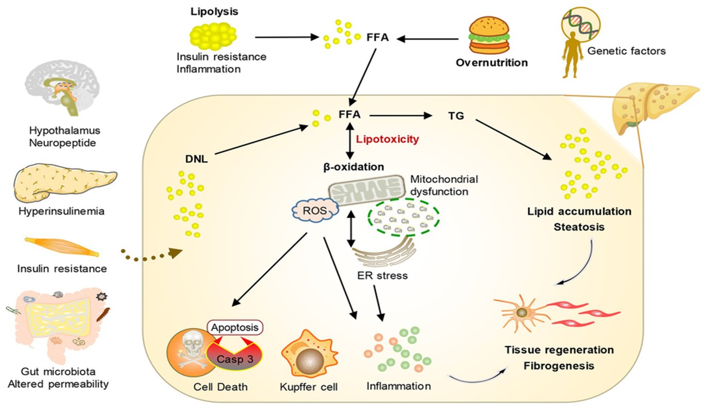
MASLD pathophysiology is characterized by the disruption of hepatic lipid homeostasis, where imbalances in lipid uptake, de novo lipogenesis, -oxidation, and export lead to hepatocellular triglyceride accumulation. Lipotoxic intermediates induce oxidative stress, endoplasmic reticulum stress, and mitochondrial dysfunction, activating proinflammatory and profibrotic signaling pathways. Persistent hepatocyte injury promotes hepatic stellate cell activation and extracellular matrix deposition, driving fibrosis and progressive liver dysfunction. The disease reflects systemic metabolic dysregulation, with contributions from adipose tissue, gut-derived factors, and insulin resistance amplifying hepatic pathology Figure 1 [10].
Hepatic Lipid Metabolism
The disease initiates when at least one cardiometabolic criterion – such as obesity or T2DM – is present, shifting the liver into a state of chronic lipid surplus [3]. Hepatic steatosis develops when the delivery of free fatty acids (FFAs) and internal lipid synthesis surpasses the liver’s capacity for oxidation or export via very-low-density lipoproteins (VLDL) [11,12].
The FFAs supplying this overload originate from adipose-tissue lipolysis (), followed by hepatic DNL (), and dietary intake () [13,14]. In the setting of insulin resistance, the liver paradoxically increases DNL while failing to suppress the flux of fatty acids mobilized from peripheral fat stores [14,15]. This imbalance generates toxic lipid intermediates that trigger oxidative stress and organelle dysfunction, marking the transition from simple storage to active hepatic injury [16,17].
Insulin Resistance
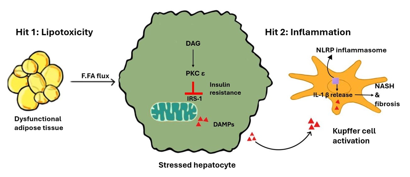
This lipid buildup directly drives insulin resistance through a specific biochemical cycle [18]. As lipids like diacylglycerol (DAG) accumulate in hepatocytes, they activate PKC, which impairs insulin receptor signaling and prevents the liver from regulating glucose production effectively [19,20,21].
Simultaneously, inflammatory signaling pathways – activated by excess saturated fatty acids – further disrupt insulin signaling through the phosphorylation of insulin receptor substrate-1 (IRS-1) [22,23]. This creates a self-sustaining loop: insulin resistance promotes further lipid accumulation, which in turn exacerbates systemic inflammatory signaling and progressive metabolic failure [22,24].
Inflammation and Cellular Injury
Metabolic stress triggers the innate immune system, characterizing the advanced stages of disease progression. Lipotoxicity-induced stress causes mitochondrial dysfunction and the leakage of reactive oxygen species (ROS), resulting in cellular damage and the release of damage-associated molecular patterns (DAMPs) [5,25].
These DAMPs function as a critical bridge in the "multiple hit” cascade Figure 2, linking metabolic lipotoxicity to innate immune activation [5,25,26]. They activate Kupffer cells (resident macrophages) and trigger the NLRP3 inflammasome [5,27]. This immune response produces pro-inflammatory cytokines (IL-1 and IL-18) and promotes distinct modes of cell death Table 2 [28,27]. This chronic cycle of hepatocyte death and inflammatory activation eventually triggers hepatic stellate cells to deposit collagen, driving the progression from simple steatosis to irreversible fibrosis [27].
| Mode of Death | Primary Mechanism | Role in MASLD Progression | Key References |
|---|---|---|---|
| Apoptosis | Caspase-dependent programmed cell death. | Initial response to lipotoxic stress; markers (e.g., CK-18) correlate with disease activity. | [5,28] |
| Pyroptosis | NLRP3 inflammasome-mediated; involves Gasdermin D. | Highly inflammatory; promotes rapid cytokine release (IL-1) and stellate cell activation. | [27] |
| Ferroptosis | Iron-dependent lipid peroxidation. | Driven by ROS and impaired antioxidant capacity; critical in the transition to MASH. | [28] |
| Necroptosis | RIPK1/RIPK3-mediated regulated necrosis. | Promotes massive DAMP release and severe sterile inflammation. | [5] |
Hepatocyte death is the primary driver of progression from simple steatosis to MASH (Metabolic Dysfunction-Associated Steatohepatitis), the inflammatory stage of metabolic liver disease [29,24]. Each pathway contributes differently to liver injury: Apoptosis is a programmed cell death mediated by caspases; Pyroptosis is a highly inflammatory response involving the NLRP3 (NOD-, LRR- and pyrin domain-containing protein 3) inflammasome [25,27]; Ferroptosis is an iron-dependent form of death caused by ROS (Reactive Oxygen Species) [28,26]; and Necroptosis is a regulated necrosis involving RIPK1/RIPK3 that releases DAMPs (Damage-Associated Molecular Patterns) to trigger sterile inflammation [5].
Acronyms: CK-18, cytokeratin-18; MASH, metabolic dysfunction–associated steatohepatitis; RIPK, receptor-interacting protein kinase; ROS, reactive oxygen species.
Notes: This table summarizes the biochemical markers and triggers for various cell death pathways identified in steatotic hepatocytes.
Source: Synthesized from current literature (2020 – 2025).
Risk Factors and Associated Conditions
MASLD arises from a complex synergy of metabolic, genetic, and lifestyle factors. Insulin resistance (IR) serves as the central engine, stimulating hepatic de novo lipogenesis and increasing the flux of free fatty acids from dysfunctional adipose tissue [13,14]. Visceral adiposity exacerbates this state by releasing pro-inflammatory cytokines that impair systemic lipid buffering [12]. Collectively, T2DM, dyslipidemia, and hypertension cluster to amplify the risk of advanced hepatic fibrosis and adverse cardiovascular outcomes [5,6].
Major Risk Factors
Type 2 Diabetes (T2DM)
T2DM independently doubles the risk of hepatocellular carcinoma and liver-related mortality, significantly accelerating the transition from simple steatosis to advanced fibrosis [30,31,32].
Obesity and Dyslipidemia
Excess adiposity and atherogenic dyslipidemia increase the hepatic fatty acid load, triggering chronic oxidative stress [5,33,6].
Genetic Predisposition
Variants such as PNPLA3 (impaired lipid droplet hydrolysis) and TM6SF2 (disrupted VLDL export) significantly influence disease susceptibility and the velocity of fibrosis progression [13,27,34].
Associated Conditions
Dietary Factors
High-calorie diets rich in fructose and saturated fats are potent stimulators of fat synthesis. Fructose is particularly damaging as it bypasses normal metabolic checks to drive triglyceride production [13,16,14].
Gut-Liver Axis (SIBO)
Small-intestinal bacterial overgrowth promotes increased intestinal permeability. This facilitates the translocation of lipopolysaccharides (LPS) into the portal circulation, activating Toll-like receptors and exacerbating hepatocellular injury [25,15].
Obstructive Sleep Apnea
Chronic intermittent hypoxia associated with OSA induces systemic oxidative stress and sympathetic overactivity, which intensify hepatic inflammatory signaling [25,13,19].
Endocrine and Hormonal Factors:
Hypothyroidism: Low thyroid hormones decrease the liver’s ability to oxidize fat and export VLDL [33,6].
Hyperuricemia: High uric acid promotes insulin resistance and stresses liver mitochondria [22,24].
Polycystic Ovary Syndrome (PCOS): Hyperinsulinemia in PCOS promotes direct hepatic fat production [13,19].
The "Lean MASLD" Phenotype
"Lean MASLD" occurs in individuals with a normal Body Mass Index (BMI 18.5 – 24.9 kg/m) who exhibit at least one cardiometabolic risk factor [4,2].
Adipose Tissue Dysfunction: Pathogenesis is frequently driven by the limited expansion capacity of peripheral adipose tissue. When subcutaneous storage thresholds are exceeded, lipids are diverted to ectopic sites, resulting in significant hepatic accumulation [13,12].
Genetic Influence
This phenotype is strongly associated with the PNPLA3 variant, explaining the presence of significant hepatic fat despite lower systemic adiposity [4,13].
Metabolic Drivers of Malignancy
The pro-inflammatory and hyperinsulinemic state of MASLD creates a systemic environment conducive to carcinogenesis.
Established Associations:
Hepatocellular Carcinoma (HCC)
MASLD is a primary driver of primary liver malignancy. Distinctively, HCC in the context of MASLD frequently occurs in the absence of established cirrhosis, suggesting that lipotoxicity-induced DNA damage and chronic oxidative stress can initiate malignant transformation independently of advanced fibrosis [35,36,37,38].
Colorectal Cancer (CRC)
MASLD is associated with a 60% increase in CRC risk [39]. Pathophysiology likely involves the disruption of the gut-liver axis and the systemic elevation of insulin-like growth factor 1 (IGF-1), which promotes colonic epithelial proliferation and inhibits apoptosis [40].
Emerging Clinical Associations
Breast and Gynecological Malignancies:
MASLD is linked to a 20 – 40% higher incidence of breast cancer, particularly in postmenopausal women. The proposed mechanism involves the aromatization of androgens in adipose tissue and the resulting estrogenic stimulation of breast and uterine tissues [41,39].
Thyroid and Pulmonary Malignancies
Recent data indicate an association between MASLD and more aggressive phenotypes of thyroid cancer. Furthermore, systemic inflammatory cytokines – specifically IL-6 and TNF- – may synergize with environmental factors to increase lung cancer risk, though obesity remains a significant confounding variable in these cohorts [41].
Extrahepatic Manifestations
MASLD is a systemic multisystem disorder; its presence serves as a sentinel marker for clinical deterioration in other organ systems. While metabolic factors drive the initial liver injury, the resulting chronic systemic inflammation and shared pro-fibrotic pathways lead to significant complications outside the liver [42,35,34].
Cardiovascular Manifestations
Cardiovascular disease (CVD) is the leading cause of death in MASLD patients, often exceeding liver-related mortality. The liver’s inflammatory state directly contributes to cardiac structural and electrical remodeling:
Heart Failure
MASLD independently raises the risk of incident heart failure, particularly Heart Failure with preserved Ejection Fraction (HFpEF), by approximately 50% [42,43].
Structural Remodeling:
Patients frequently exhibit subclinical changes, including left ventricular hypertrophy and impaired diastolic filling, even in the early stages of liver fat accumulation [42].
Arrhythmias:
There is a robust association with Atrial Fibrillation, likely driven by pro-inflammatory cytokines originating from both the liver and epicardial adipose tissue [42,43,44].
Extrahepatic Malignancies
The pro-oncogenic environment of MASLD – characterized by hyperinsulinemia and chronic low-grade inflammation – extends beyond the liver to several extrahepatic sites [45,35,36]:
Colorectal Cancer (CRC):
MASLD is associated with a 60% higher risk of CRC and a significant increase in colorectal adenomas. This association is strongest in patients with advanced fibrosis, necessitating vigilant screening [40,46,39].
Gastrointestinal and Other Cancers:
Increased risks are noted for esophageal and gastric cancers [41]. Emerging evidence also suggests a higher incidence of breast and thyroid malignancies, often presenting with more aggressive clinical features [35,41].
Renal and Endocrine Manifestations
The crosstalk between the liver and other metabolic organs creates a cycle of systemic decline:
Chronic Kidney Disease (CKD):
MASLD is an independent risk factor for CKD. Pro-inflammatory and pro-fibrotic signals (such as TGF-) released by the liver promote glomerular damage and decline renal function [43,34].
Metabolic Feedback Loops:
While insulin resistance drives MASLD, the presence of hepatic steatosis conversely makes glycemic control more difficult in patients with T2DM, increasing the likelihood of diabetic microvascular complications [47,29].
Diagnosis and Risk Stratification
Diagnosis requires the identification of hepatic steatosis via imaging or histology, the presence of 1 cardiometabolic risk factor (CMRF), and the systematic exclusion of competing etiologies [2,3].
Laboratory Assessment and Differential Diagnosis
Transaminases (ALT/AST) are poor exclusionary tests; a normal ALT does not exclude advanced fibrosis. Clinical suspicion should remain high in patients with T2DM or obesity regardless of enzyme levels [4,33].
Transaminase Thresholds:
While lab ranges vary, an ALT > 30 U/L is increasingly recognized as the threshold for further evaluation in adults [4].
Differential Diagnosis:
To confirm MASLD, the following must be excluded:
Imaging and Non-Invasive Tests
The diagnostic goal is to identify Advanced Fibrosis (F 3), as this is the primary predictor of liver-related mortality.
| Modality | Performance and Thresholds | Limitations |
|---|---|---|
| Ultrasonography | High specificity (98%) for moderate/severe steatosis. | Sensitivity drops if fat content < 20% or BMI > 40 kg/m [6]. |
| FIB-4 Index | < 1.3 (Low Risk): NPV > 90% for F3/F4 [4,33]. 1.3–2.67: Indeterminate [1,3]. > 2.67 (High Risk): Refer to hepatology [1,4]. | Accuracy decreases in patients < 35 or > 65 years old [4]. |
| • VCTE (FibroScan) | < 8 kPa (Low Risk): Rules out advanced fibrosis [33]. 8-12 kPa: Indeterminate [48,3]. > 12–15 kPa (High Risk): Highly suggestive of F3/F4 [4,33]. | Reduced reliability in severe obesity (BMI > 35) without XL probes [33]. |
| • MRI-PDFF | Gold standard for fat quantification (> 5% threshold) [1,48]. | High cost; limited availability for routine monitoring [6]. |
The diagnostic utility of these modalities depends on their ability to accurately stage fibrosis, as increasing stiffness values correlate with higher risks of liver-related complications. FIB-4 (Fibrosis-4 Index) serves as a high-sensitivity triage tool to rule out advanced disease in primary care [4,33], whereas VCTE (Vibration-Controlled Transient Elastography) and MRI-PDFF (Proton Density Fat Fraction) provide quantitative assessments of liver stiffness and hepatic fat content, respectively [1,33,48]. While NITs (Non-Invasive Tests) offer a safe alternative to biopsy, their accuracy is influenced by the NPV (Negative Predictive Value), which remains the primary metric for excluding advanced fibrosis in at-risk populations [48,3].
Acronyms: FIB-4, Fibrosis-4 Index; MRI-PDFF, Magnetic Resonance Imaging Proton Density Fat Fraction; NPV, Negative Predictive Value; VCTE, Vibration-Controlled Transient Elastography; LSM, Liver Stiffness Measurement; CAP, Controlled Attenuation Parameter; ELF, Enhanced Liver Fibrosis.
Notes: Performance ranges are based on evidence-based thresholds; operator dependence and patient obesity are noted as primary limitations [33].
Source: Compiled from AASLD and EASL clinical practice guidelines.
While imaging is central to diagnosis, clinicians must recognize its technical boundaries. Conventional ultrasound has a sensitivity of only 60 – 90% for detecting steatosis, often failing when the fat fraction is below 20% [24,6]. Furthermore, the accuracy of VCTE is highly operator-dependent and significantly limited by the ’skin-to-capsule’ distance in patients with morbid obesity, which can lead to falsely elevated LSM readings [33]. Despite standardized cutoffs, diagnostic uncertainty persists within ’indeterminate’ ranges (e.g., FIB-4 1.3 – 2.67 or VCTE 8 – 12 kPa), where guidelines vary on whether to prioritize immediate secondary testing or longitudinal monitoring [1,48,3] Table 3.
Clinical Referral and Management Pathway
Following the AASLD/EASL 2023 consensus, the clinical pathway follows a tiered approach Figure 3:
Tier 1 (Low Risk)
Calculate FIB-4 for all at-risk patients , including those with T2DM or obesity. If FIB-4 < 1.3, advanced fibrosis is ruled out with high negative predictive value; these patients should repeat non-invasive tests in 2 – 3 years and focus on cardiovascular disease (CVD) risk reduction. [4,33,3].
Tier 2 (Indeterminate Risk)
For FIB-4 1.3 – 2.67, perform a secondary non-invasive test such as VCTE or the Enhanced Liver Fibrosis (ELF) test. If results remain indeterminate, patients should be reassessed annually or referred based on clinical suspicion [4,3].
Tier 3 (Specialist Care)
Patients with FIB-4 > 2.67 or LSM 12 kPa are considered high-risk and require hepatology referral. Confirmed advanced fibrosis (F3/F4) necessitates the initiation of MASH management, biannual HCC screening, and formal variceal risk assessment [4,33,3].
![FIB-4 Risk Stratification Flowchart. A tiered clinical approach for primary care triage. Patients with a FIB-4 score < 1.3 (or < 2.0 for those aged > 65) are considered low risk [33,3]. Those in the Indeterminate Risk category (FIB-4 1.3 – 2.67) require secondary testing [4]. Finally, patients with](https://asidejournals.com/index.php/Gastroenterology/article/download/448/version/447/433/14811/3.jpg)
The Role of Liver Biopsy
Biopsy is no longer first-line but remains indicated when:
Management of Advanced Fibrosis (F3) and Cirrhosis (F4)
Patients identified with advanced fibrosis (F3) or with compensated advanced chronic liver disease (cACLD) (F4) require a structured care bundle to mitigate the risks of decompensation and mortality. This multifaceted approach includes:
HCC Surveillance
Mandatory biannual abdominal ultrasound ( alpha-fetoprotein) for all patients with cirrhosis (F4) and considered for those with F3 based on individual risk [30,35,38].
Portal Hypertension
According to the Baveno VII criteria, screening endoscopy for esophageal varices can be safely avoided in patients with a liver stiffness measurement < 20 kPa AND a platelet count > 150,000/μL. Conversely, those not meeting these criteria require endoscopic screening [4,48,3].
Transplant Referral
Explicit referral for liver transplantation assessment is triggered by a MELD-Na score 15, the first occurrence of hepatic decompensation (ascites, encephalopathy, or jaundice), or HCC within Milan criteria [4,48] Decompensation Monitoring: Regular clinical surveillance for complications such as ascites, hepatic encephalopathy, and variceal hemorrhage is essential for patients cACLD [48,3].
Vaccination
Routine immunization against hepatitis A (HAV), hepatitis B (HBV), and pneumococcus, as well as annual influenza and COVID-19 boosters, is necessary to prevent acute-on-chronic liver failure [1,4,48].
Nutritional Support
Targeted intervention for sarcopenia involves a protein-rich diet (1.2 – 1.5 g/kg/day) and Vitamin D optimization to preserve bone mineral density [48].
Metabolic & Bone Health
Regular screening for osteoporosis/osteopenia and Vitamin D deficiency [1,48].
Management and Treatment
The primary objectives in MASLD management are to achieve histological resolution of MASH and to cease or reverse fibrosis, thereby modifying the risk of cirrhosis, hepatocellular carcinoma, and liver-related mortality [1,4,24,48]. Concurrent management of the cardiometabolic cluster – obesity, T2DM, dyslipidemia, and hypertension – is mandatory to reduce extrahepatic risks, including chronic kidney disease and heart failure [47,2,29].
Lifestyle and Behavioral Interventions
Lifestyle modification remains the non-pharmacological basis of therapy. Weight loss exhibits a dose-response relationship with histological improvement:
Dietary and Physical Activity Protocols
Dietary Patterns
Beyond general weight loss, the Mediterranean Diet is uniquely effective due to its high concentration of monounsaturated fatty acids and antioxidants. These components directly influence hepatic de novo lipogenesis and systemic inflammation, leading to superior metabolic outcomes compared to standard low-fat diets [49,4,48].
Exercise
Guidelines recommend 150 minutes of moderate-intensity aerobic activity or 75 – 150 minutes of vigorous activity weekly. Exercise improves hepatic insulin sensitivity and reduces liver fat even in the absence of significant weight change [1,4,48].
Pharmacotherapy: The 2024 – 2025 Shift
Until recently, pharmacotherapy was limited to off-label use of vitamin E or pioglitazone. The landscape has been transformed by the approval of targeted agents and high-potency incretin therapies Table 4:
Resmetirom
A selective thyroid hormone receptor- (THR-) agonist approved for non-cirrhotic MASH. It achieved MASH resolution in ~30% and fibrosis improvement in 25% of trial participants. [9].
Incretin Mimetics
Semaglutide and Tirzepatide have demonstrated MASH resolution rates exceeding 60%. While primary indications are obesity and T2DM, they significantly improve the metabolic environment and reduce liver stiffness [42,50].
| Agent | Class / Primary Indication | Key Histological Outcome |
|---|---|---|
| Resmetirom | THR- Agonist; MASH with F2 – F3 fibrosis [1,9]. | 25% Fibrosis improvement; 30% MASH resolution [9,48]. |
| Semaglutide | GLP-1 RA; Obesity and T2DM [50,48]. | |
| Tirzepatide | GIP/GLP-1 RA; Obesity and T2DM [9,50]. | 62.8% MASH resolution; signal for fibrosis improvement [50]. |
| Pioglitazone | PPAR- Agonist; T2DM (off-label for MASH) [4,48]. | Improved steatohepatitis; risk of weight gain [4,48]. |
Therapeutic selection is increasingly guided by the patient’s clinical phenotype. Resmetirom is prioritized for patients requiring direct fibrosis regression in non-cirrhotic (F2 – F3) stages, based on the MAESTRO-NASH trial outcomes [9]. Incretin mimetics (GLP-1 and dual GIP/GLP-1 RAs) are favored for those with high cardiometabolic risk, obesity, or Type 2 Diabetes Mellitus [50,48]. Outcome data represent results from primary clinical endpoints: MASH resolution (defined as the disappearance of ballooning and inflammation) and/or fibrosis improvement [9,50]. Non-invasive monitoring via MRI-PDFF and LSM provides surrogate assessments of treatment response [42,33].
Acronyms: GLP-1, glucagon-like peptide-1; GIP, glucose-dependent insulinotropic polypeptide; MASH, metabolic dysfunction – associated steatohepatitis; RAs, receptor agonists; THR-β, thyroid hormone receptor-beta.
Source: Adapted from Rinella et al. (2023) [3], Eddowes et al. (2019) [33], and primary clinical trial data [9,50].
It is critical to distinguish between the evidence levels of current therapeutics [4]. Resmetirom and Tirzepatide have demonstrated efficacy based on histological endpoints (biopsy-proven MASH resolution and fibrosis reversal) [9,50], which remain the gold standard for regulatory approval. Conversely, many early-stage incretin studies rely on surrogate outcomes, such as significant reductions in MRI-PDFF (Magnetic Resonance Imaging Proton Density Fat Fraction) or LSM, which correlate with clinical improvement but do not yet confirm tissue-level fibrosis regression [42,33].
Bariatric Surgery
Bariatric procedures (Roux-en-Y gastric bypass or sleeve gastrectomy) are highly effective for patients with a BMI > kg/m and refractory MASH [48,32].
Long-term Outcomes
At 5 years post-surgery, up to 84% of patients achieve MASH resolution and 70% show fibrosis improvement [48].
Hard Outcomes
Large observational studies confirm that bariatric surgery reduces 10-year major liver-related events (2.3% vs. 8.5%) and cardiovascular events (8.5% vs. 15.7%) compared to non-surgical care [48,32].
Complications
MASLD affects approximately 30% of the global adult population, with its prevalence having nearly doubled between 1991 and 2019 paralleling the surge in global obesity rates. MASH represents the progressive inflammatory phenotype, characterized by hepatocyte ballooning and lobular inflammation, and is the primary driver of advanced fibrosis. While cardiovascular disease (CVD) remains the leading cause of death in MASLD patients, liver-related mortality increases exponentially once patients reach advanced fibrosis stages (F3 – F4) [1,35,24,48].
Type 2 Diabetes Mellitus
Type 2 Diabetes Mellitus (T2DM) is an independent risk factor for both MASH and accelerated fibrosis [51]. Because advanced fibrosis often remains asymptomatic until hepatic decompensation occurs, a risk-based assessment pathway is critical for diabetic patients:
Initial Screening
Utilize the FIB-4 Index as a primary case-finding tool for all diabetic patients, rather than relying on transaminases, which serve as poor rule-out tools [3,7]
Confirmatory Testing
Elevated or indeterminate FIB-4 scores must be followed by Liver Stiffness Measurement via VCTE (Vibration-Controlled Transient Elastography) to improve positive predictive value [4,33,3].
Cardiovascular Disease (CVD)
The American Heart Association (AHA) recognizes MASLD as an independent, underappreciated risk factor for atherosclerotic cardiovascular disease. This risk is mediated by systemic metaflammation and pro-atherogenic lipid profiles [42,43].
Management:
Statins are first-line, safe in MASLD, and may reduce the risk of HCC.
Preferred Agents
In patients with T2DM and high cardiovascular risk, SGLT2 inhibitors or GLP-1 RAs are preferred for their dual hepatic and cardioprotective benefits [42,43]
Portal Hypertension and Variceal Risk
Clinically significant portal hypertension can manifest in compensated advanced chronic liver disease (cACLD) before symptoms appear.
Risk Stratification
Management
Non-selective beta-blockers, particularly carvedilol, are the standard of care to reduce portal pressure and prevent the first episode of decompensation. Routine endoscopic screening for varices is not required if Liver stiffness measurement < 20 kPa and platelets > 150×10^9/L [4,48,3].
SARCOPENIA AND NUTRITIONAL OPTIMIZATION IN ADVANCED MASLD
Sarcopenia (the progressive loss of muscle mass, strength, and quality) frequently coexists with MASLD, creating a vicious cycle of physical inactivity and worsened insulin resistance. Modern clinical focus has shifted toward sarcopenic obesity (SO), which is characterized not just by low muscle volume, but by myosteatosis (fatty infiltration of the muscle). Patients with Sarcopenic obesity have a significantly higher risk of advanced fibrosis and mortality compared to those with obesity alone.
The Incretin Paradox
While GLP-1 and GIP/GLP-1 receptor agonists (e.g., Tirzepatide) are highly effective for weight loss, up to 25 – 40% of weight lost can be lean muscle mass. To prevent a decline in metabolic rate, these therapies must be paired with mandatory resistance training and optimized protein intake [42,50,10].
Nutritional Strategy
Overcoming "anabolic resistance" in MASLD requires a high protein intake (1.2 – 1.5 g/kg/day), distributed boluses of protein throughout the day. Adherence to a Mediterranean dietary pattern is the gold standard, as its monounsaturated fats and antioxidants specifically reduce systemic inflammation and de novo lipogenesis [49,16].
Physical Intervention
Resistance exercise remains a cornerstone non-pharmacological ’liver-sparing’ intervention. By improving the glucose-clearing capacity of the skeletal muscle – the body’s largest metabolic organ – resistance training reduces the metabolic load on the liver independently of total weight loss [19,12].
Conclusion
Metabolic dysfunction-associated steatotic liver disease (MASLD) is a multisystem disorder driven by the convergence of hepatic lipotoxicity, insulin resistance, and systemic metaflammation [1,48]. This cascade facilitates the progression from simple steatosis to MASH, a phenotype characterized by hepatocyte injury and fibrogenesis that may ultimately lead to cirrhosis or hepatocellular carcinoma (HCC) [4,24]. Beyond the liver, evidence links MASLD to systemic complications, most notably cardiovascular disease and heart failure [42,43].
The clinical approach is shifting toward risk-based screening using non-invasive tests (NITs) such as the FIB-4 index and VCTE (Vibration-Controlled Transient Elastography) [12,6]. While these tools improve the identification of advanced fibrosis, their diagnostic accuracy can be affected by age and ethnicity, necessitating a cautious, tiered interpretation to ensure equitable care [11,4]. Furthermore, access to these specialized diagnostic tools remains a challenge in underserved communities, which may delay diagnosis in high-risk "sentinel" populations.
Lifestyle modification, specifically a 7 – 10% reduction in body weight and adherence to a Mediterranean dietary pattern, remains the primary therapeutic recommendation [49,16]. The recent approval of Resmetirom and the clinical promise of GLP-1 RAs represent significant progress [9,50]. However, as these therapies move from clinical trials into general practice, ensuring broad and equitable access – regardless of socioeconomic status – is essential to preventing further health disparities in liver-related mortality [4,9].
Ultimately, reducing the global burden of MASLD requires an integrated approach that connects hepatology with endocrine and cardiovascular care. Prioritizing early, inclusive, and evidence-based intervention is the most effective strategy to attenuate the long-term inflammatory and metabolic complications of this disease across all patient populations.
Conflicts of Interest
The authors have no relevant financial or non-financial interests to disclose.
Funding Source
The authors declare that no funds, grants, or other support were received during the preparation of this manuscript.
Acknowledgments
The authors would like to thank Gana Mohamed and Takwa Hathout for their assistance in the preparation of the figures and illustrations.
Institutional Review Board (IRB)
Not applicable. This article is a literature review and does not contain any original studies with human participants or animals performed by any of the authors.
Large Language Model
The authors declare that Gemini (an AI tool by Google) was used to assist with structural editing and citation management during the preparation of this manuscript. The authors reviewed and verified all scientific content and take full responsibility for the integrity and accuracy of the final work.
Authors Contribution
All authors contributed to the study conception and design. The first draft of the manuscript was written by MM, and all authors provided critical revisions to previous versions of the manuscript. All authors read and approved of the final manuscript.
Data Availability
Data sharing is not applicable to this article as no new datasets were created or analyzed during the current study.
References
- Adinolfi L. E., Marrone A., Izzi A., Craxi A.. Management of Metabolic Dysfunction-Associated Steatotic Liver Disease (MASLD): Facts and Hopes. J Clin Exp Hepatol. 2026;16(1):103411. doi:10.1016/j.jceh.2025.103411 PMID: 41480331 PMCID: PMC12753517
- Kyhl L. K., Nordestgaard B. G., Tybjaerg-Hansen A., Nielsen S. F.. Metabolic dysfunction-associated steatotic liver disease, cardiometabolic risk factors, and cardiovascular disease. Atherosclerosis. 2025;411:120551. doi:10.1016/j.atherosclerosis.2025.120551 PMID: 41124780
- Rinella M. E., Lazarus J. V., Ratziu V., Francque S. M., Sanyal A. J., Kanwal F., Romero D., Abdelmalek M. F., Anstee Q. M., Arab J. P., Arrese M., Bataller R., Beuers U., Boursier J., Bugianesi E., Byrne C. D., Castro Narro G. E., Chowdhury A., Cortez-Pinto H., Cryer D. R., Cusi K., El-Kassas M., Klein S., Eskridge W., Fan J., Gawrieh S., Guy C. D., Harrison S. A., Kim S. U., Koot B. G., Korenjak M., Kowdley K. V., Lacaille F., Loomba R., Mitchell-Thain R., Morgan T. R., Powell E. E., Roden M., Romero-Gomez M., Silva M., Singh S. P., Sookoian S. C., Spearman C. W., Tiniakos D., Valenti L., Vos M. B., Wong V. W., Xanthakos S., Yilmaz Y., Younossi Z., Hobbs A., Villota-Rivas M., Newsome P. N., NAFLD Nomenclature consensus group. A multisociety Delphi consensus statement on new fatty liver disease nomenclature. Hepatology. 2023;78(6):1966-1986. doi:10.1097/HEP.0000000000000520 PMID: 37363821 PMCID: PMC10653297
- Chan W. K., Chuah K. H., Rajaram R. B., Lim L. L., Ratnasingam J., Vethakkan S. R.. Metabolic Dysfunction-Associated Steatotic Liver Disease (MASLD): A State-of-the-Art Review. J Obes Metab Syndr. 2023;32(3):197-213. doi:10.7570/jomes23052 PMID: 37700494 PMCID: PMC10583766
- Cheng Z., Chu H., Seki E., Lin R., Yang L.. Hepatocyte programmed cell death: the trigger for inflammation and fibrosis in metabolic dysfunction-associated steatohepatitis. Front Cell Dev Biol. 2024;12:1431921. doi:10.3389/fcell.2024.1431921 PMID: 39071804 PMCID: PMC11272544
- Riazi K., Azhari H., Charette J. H., Underwood F. E., King J. A., Afshar E. E., Swain M. G., Congly S. E., Kaplan G. G., Shaheen A. A.. The prevalence and incidence of NAFLD worldwide: a systematic review and meta-analysis. Lancet Gastroenterol Hepatol. 2022;7(9):851-861. doi:10.1016/S2468-1253(22)00165-0 PMID: 35798021
- Younossi Z. M., Kalligeros M., Henry L.. Epidemiology of metabolic dysfunction-associated steatotic liver disease. Clin Mol Hepatol. 2025;31(Suppl):S32-S50. doi:10.3350/cmh.2024.0431 PMID: 39159948 PMCID: PMC11925440
- Estes C., Anstee Q. M., Arias-Loste M. T., Bantel H., Bellentani S., Caballeria J., Colombo M., Craxi A., Crespo J., Day C. P., Eguchi Y., Geier A., Kondili L. A., Kroy D. C., Lazarus J. V., Loomba R., Manns M. P., Marchesini G., Nakajima A., Negro F., Petta S., Ratziu V., Romero-Gomez M., Sanyal A., Schattenberg J. M., Tacke F., Tanaka J., Trautwein C., Wei L., Zeuzem S., Razavi H.. Modeling NAFLD disease burden in China, France, Germany, Italy, Japan, Spain, United Kingdom, and United States for the period 2016-2030. J Hepatol. 2018;69(4):896-904. doi:10.1016/j.jhep.2018.05.036 PMID: 29886156
- Harrison S. A., Bedossa P., Guy C. D., Schattenberg J. M., Loomba R., Taub R., Labriola D., Moussa S. E., Neff G. W., Rinella M. E., Anstee Q. M., Abdelmalek M. F., Younossi Z., Baum S. J., Francque S., Charlton M. R., Newsome P. N., Lanthier N., Schiefke I., Mangia A., Pericas J. M., Patil R., Sanyal A. J., Noureddin M., Bansal M. B., Alkhouri N., Castera L., Rudraraju M., Ratziu V., MAESTRO-NASH Investigators. A Phase 3, Randomized, Controlled Trial of Resmetirom in NASH with Liver Fibrosis. N Engl J Med. 2024;390(6):497-509. doi:10.1056/NEJMoa2309000 PMID: 38324483
- Rao G., Peng X., Li X., An K., He H., Fu X., Li S., An Z.. Unmasking the enigma of lipid metabolism in metabolic dysfunction-associated steatotic liver disease: from mechanism to the clinic. Front Med (Lausanne). 2023;10:1294267. doi:10.3389/fmed.2023.1294267 PMID: 38089874 PMCID: PMC10711211
- Carotti S., Aquilano K., Valentini F., Ruggiero S., Alletto F., Morini S., Picardi A., Antonelli-Incalzi R., Lettieri-Barbato D., Vespasiani-Gentilucci U.. An overview of deregulated lipid metabolism in nonalcoholic fatty liver disease with special focus on lysosomal acid lipase. Am J Physiol Gastrointest Liver Physiol. 2020;319(4):G469-G480. doi:10.1152/ajpgi.00049.2020 PMID: 32812776
- Lee E., Korf H., Vidal-Puig A.. An adipocentric perspective on the development and progression of non-alcoholic fatty liver disease. J Hepatol. 2023;78(5):1048-1062. doi:10.1016/j.jhep.2023.01.024 PMID: 36740049
- Donnelly K. L., Smith C. I., Schwarzenberg S. J., Jessurun J., Boldt M. D., Parks E. J.. Sources of fatty acids stored in liver and secreted via lipoproteins in patients with nonalcoholic fatty liver disease. J Clin Invest. 2005;115(5):1343-51. doi:10.1172/JCI23621 PMID: 15864352 PMCID: PMC1087172
- Lambert Jennifer E., Ramos–Roman Maria A., Browning Jeffrey D., Parks Elizabeth J.. Increased De Novo Lipogenesis Is a Distinct Characteristic of Individuals With Nonalcoholic Fatty Liver Disease. Gastroenterology. 2014;146(3):726-735. doi:10.1053/j.gastro.2013.11.049
- Nassir F.. NAFLD: Mechanisms, Treatments, and Biomarkers. Biomolecules. 2022;12(6). doi:10.3390/biom12060824 PMID: 35740949 PMCID: PMC9221336
- Ertunc M. E., Hotamisligil G. S.. Lipid signaling and lipotoxicity in metaflammation: indications for metabolic disease pathogenesis and treatment. J Lipid Res. 2016;57(12):2099-2114. doi:10.1194/jlr.R066514 PMID: 27330055 PMCID: PMC5321214
- Marra F., Svegliati-Baroni G.. Lipotoxicity and the gut-liver axis in NASH pathogenesis. J Hepatol. 2018;68(2):280-295. doi:10.1016/j.jhep.2017.11.014 PMID: 29154964
- Liao N. H., Wang C. Y., Lai K. Y., Kao H. H., Lin W. Y., Chen T. P.. Association between metabolic dysfunction-associated steatotic liver disease and insulin resistance in individuals with normoglycemia. Medicine (Baltimore). 2025;104(34):e44109. doi:10.1097/MD.0000000000044109 PMID: 40859520 PMCID: PMC12384863
- Jornayvaz F. R., Shulman G. I.. Diacylglycerol activation of protein kinase Cepsilon and hepatic insulin resistance. Cell Metab. 2012;15(5):574-84. doi:10.1016/j.cmet.2012.03.005 PMID: 22560210 PMCID: PMC3353749
- Samuel V. T., Liu Z. X., Wang A., Beddow S. A., Geisler J. G., Kahn M., Zhang X. M., Monia B. P., Bhanot S., Shulman G. I.. Inhibition of protein kinase Cepsilon prevents hepatic insulin resistance in nonalcoholic fatty liver disease. J Clin Invest. 2007;117(3):739-45. doi:10.1172/JCI30400 PMID: 17318260 PMCID: PMC1797607
- Ter Horst K. W., Gilijamse P. W., Versteeg R. I., Ackermans M. T., Nederveen A. J., la Fleur S. E., Romijn J. A., Nieuwdorp M., Zhang D., Samuel V. T., Vatner D. F., Petersen K. F., Shulman G. I., Serlie M. J.. Hepatic Diacylglycerol-Associated Protein Kinase Cepsilon Translocation Links Hepatic Steatosis to Hepatic Insulin Resistance in Humans. Cell Rep. 2017;19(10):1997-2004. doi:10.1016/j.celrep.2017.05.035 PMID: 28591572 PMCID: PMC5469939
- Chen Z., Yu R., Xiong Y., Du F., Zhu S.. A vicious circle between insulin resistance and inflammation in nonalcoholic fatty liver disease. Lipids Health Dis. 2017;16(1):203. doi:10.1186/s12944-017-0572-9 PMID: 29037210 PMCID: PMC5644081
- Hernandez R., Zhou C.. Recent Advances in Understanding the Role of IKKbeta in Cardiometabolic Diseases. Front Cardiovasc Med. 2021;8:752337. doi:10.3389/fcvm.2021.752337 PMID: 34957242 PMCID: PMC8692734
- Pafili K., Roden M.. Nonalcoholic fatty liver disease (NAFLD) from pathogenesis to treatment concepts in humans. Mol Metab. 2021;50:101122. doi:10.1016/j.molmet.2020.101122 PMID: 33220492 PMCID: PMC8324683
- Clare K., Dillon J. F., Brennan P. N.. Reactive Oxygen Species and Oxidative Stress in the Pathogenesis of MAFLD. J Clin Transl Hepatol. 2022;10(5):939-946. doi:10.14218/JCTH.2022.00067 PMID: 36304513 PMCID: PMC9547261
- Myint M., Oppedisano F., De Giorgi V., Kim B. M., Marincola F. M., Alter H. J., Nesci S.. Inflammatory signaling in NASH driven by hepatocyte mitochondrial dysfunctions. J Transl Med. 2023;21(1):757. doi:10.1186/s12967-023-04627-0 PMID: 37884933 PMCID: PMC10605416
- Roehlen N., Crouchet E., Baumert T. F.. Liver Fibrosis: Mechanistic Concepts and Therapeutic Perspectives. Cells. 2020;9(4). doi:10.3390/cells9040875 PMID: 32260126 PMCID: PMC7226751
- Liu Y., Zhang M., Huang Y., Zhang Y., Zhu C., Guan J., Xia S., Liao J.. Involvement of ferroptosis in metabolic dysfunction-associated steatohepatitis-related liver diseases. J Mol Med (Berl). 2025;103(11-12):1299-1315. doi:10.1007/s00109-025-02599-w PMID: 41051414 PMCID: PMC12675734
- Mantovani A., Byrne C. D., Bonora E., Targher G.. Nonalcoholic Fatty Liver Disease and Risk of Incident Type 2 Diabetes: A Meta-analysis. Diabetes Care. 2018;41(2):372-382. doi:10.2337/dc17-1902 PMID: 29358469
- Ioannou G. N.. Epidemiology and risk-stratification of NAFLD-associated HCC. J Hepatol. 2021;75(6):1476-1484. doi:10.1016/j.jhep.2021.08.012 PMID: 34453963
- Barb D., Repetto E. M., Stokes M. E., Shankar S. S., Cusi K.. Type 2 diabetes mellitus increases the risk of hepatic fibrosis in individuals with obesity and nonalcoholic fatty liver disease. Obesity (Silver Spring). 2021;29(11):1950-1960. doi:10.1002/oby.23263 PMID: 34553836 PMCID: PMC9290591
- Tilg H., Moschen A. R., Roden M.. NAFLD and diabetes mellitus. Nat Rev Gastroenterol Hepatol. 2017;14(1):32-42. doi:10.1038/nrgastro.2016.147 PMID: 27729660
- Eddowes P. J., Sasso M., Allison M., Tsochatzis E., Anstee Q. M., Sheridan D., Guha I. N., Cobbold J. F., Deeks J. J., Paradis V., Bedossa P., Newsome P. N.. Accuracy of FibroScan Controlled Attenuation Parameter and Liver Stiffness Measurement in Assessing Steatosis and Fibrosis in Patients With Nonalcoholic Fatty Liver Disease. Gastroenterology. 2019;156(6):1717-1730. doi:10.1053/j.gastro.2019.01.042 PMID: 30689971
- Targher G., Byrne C. D., Tilg H.. NAFLD and increased risk of cardiovascular disease: clinical associations, pathophysiological mechanisms and pharmacological implications. Gut. 2020;69(9):1691-1705. doi:10.1136/gutjnl-2020-320622 PMID: 32321858
- Kalligeros M., Henry L., Younossi Z. M.. Metabolic dysfunction-associated steatotic liver disease and its link to cancer. Metabolism. 2024;160:156004. doi:10.1016/j.metabol.2024.156004 PMID: 39182603
- Thomas J. A., Kendall B. J., El-Serag H. B., Thrift A. P., Macdonald G. A.. Hepatocellular and extrahepatic cancer risk in people with non-alcoholic fatty liver disease. Lancet Gastroenterol Hepatol. 2024;9(2):159-169. doi:10.1016/S2468-1253(23)00275-3 PMID: 38215780
- Thomas J. A., Kendall B. J., Dalais C., Macdonald G. A., Thrift A. P.. Hepatocellular and extrahepatic cancers in non-alcoholic fatty liver disease: A systematic review and meta-analysis. Eur J Cancer. 2022;173:250-262. doi:10.1016/j.ejca.2022.06.051 PMID: 35944373
- Younossi Z. M., Henry L.. Epidemiology of non-alcoholic fatty liver disease and hepatocellular carcinoma. JHEP Rep. 2021;3(4):100305. doi:10.1016/j.jhepr.2021.100305 PMID: 34189448 PMCID: PMC8215299
- Mantovani A., Dauriz M., Byrne C. D., Lonardo A., Zoppini G., Bonora E., Targher G.. Association between nonalcoholic fatty liver disease and colorectal tumours in asymptomatic adults undergoing screening colonoscopy: a systematic review and meta-analysis. Metabolism. 2018;87:1-12. doi:10.1016/j.metabol.2018.06.004 PMID: 29935236
- Chen Z. F., Dong X. L., Huang Q. K., Hong W. D., Wu W. Z., Wu J. S., Pan S.. The combined effect of non-alcoholic fatty liver disease and metabolic syndrome on colorectal carcinoma mortality: a retrospective in Chinese females. World J Surg Oncol. 2018;16(1):163. doi:10.1186/s12957-018-1461-z PMID: 30097069 PMCID: PMC6086998
- Lv Y., Zhang H. J.. Effect of Non-alcoholic Fatty Liver Disease on the Risk of Synchronous Liver Metastasis: Analysis of 451 Consecutive Patients of Newly Diagnosed Colorectal Cancer. Front Oncol. 2020;10:251. doi:10.3389/fonc.2020.00251 PMID: 32181157 PMCID: PMC7059642
- Bansal B., Lajeunesse-Trempe F., Keshvani N., Lavie C. J., Pandey A.. Impact of Metabolic Dysfunction-associated Steatotic Liver Disease on Cardiovascular Structure, Function, and the Risk of Heart Failure. Can J Cardiol. 2025;41(9):1777-1793. doi:10.1016/j.cjca.2025.04.012 PMID: 40258400
- Boeckmans J., Widman L., Strandberg R., Shang Y., Wester A., Hagstrom H.. Temporal comorbidity patterns preceding MASLD-related major adverse liver outcomes: a nationwide population-based case-control study in Sweden. BMJ Public Health. 2025;3(2):e003322. doi:10.1136/bmjph-2025-003322 PMID: 41262775 PMCID: PMC12625918
- Bozkurt B., Ahmad T., Alexander K. M., Baker W. L., Bosak K., Breathett K., Fonarow G. C., Heidenreich P., Ho J. E., Hsich E., Ibrahim N. E., Jones L. M., Khan S. S., Khazanie P., Koelling T., Krumholz H. M., Khush K. K., Lee C., Morris A. A., Page R. L., 2nd, Pandey A., Piano M. R., Stehlik J., Stevenson L. W., Teerlink J. R., Vaduganathan M., Ziaeian B., Writing Committee Members. Heart Failure Epidemiology and Outcomes Statistics: A Report of the Heart Failure Society of America. J Card Fail. 2023;29(10):1412-1451. doi:10.1016/j.cardfail.2023.07.006 PMID: 37797885 PMCID: PMC10864030
- Allen A. M., Hicks S. B., Mara K. C., Larson J. J., Therneau T. M.. The risk of incident extrahepatic cancers is higher in non-alcoholic fatty liver disease than obesity - A longitudinal cohort study. J Hepatol. 2019;71(6):1229-1236. doi:10.1016/j.jhep.2019.08.018 PMID: 31470068 PMCID: PMC6921701
- Ebrahimi F., Hagstrom H., Sun J., Bergman D., Shang Y., Yang W., Roelstraete B., Ludvigsson J. F.. Familial coaggregation of MASLD with hepatocellular carcinoma and adverse liver outcomes: Nationwide multigenerational cohort study. J Hepatol. 2023;79(6):1374-1384. doi:10.1016/j.jhep.2023.08.018 PMID: 37647992
- Gastaldelli A., Cusi K.. From NASH to diabetes and from diabetes to NASH: Mechanisms and treatment options. JHEP Rep. 2019;1(4):312-328. doi:10.1016/j.jhepr.2019.07.002 PMID: 32039382 PMCID: PMC7001557
- Powell E. E., Wong V. W., Rinella M.. Non-alcoholic fatty liver disease. Lancet. 2021;397(10290):2212-2224. doi:10.1016/S0140-6736(20)32511-3 PMID: 33894145
- Bernardino M., Tiribelli C., Rosso N.. The Impact of the Mediterranean Diet, Physical Activity, and Nutrition Education on Pediatric Metabolic Dysfunction-Associated Steatotic Liver Disease (MASLD): A Review. Nutrients. 2025;18(1). doi:10.3390/nu18010028 PMID: 41515146 PMCID: PMC12787394
- Loomba R., Hartman M. L., Lawitz E. J., Vuppalanchi R., Boursier J., Bugianesi E., Yoneda M., Behling C., Cummings O. W., Tang Y., Brouwers B., Robins D. A., Nikooie A., Bunck M. C., Haupt A., Sanyal A. J., SYNERGY-NASH Investigators. Tirzepatide for Metabolic Dysfunction-Associated Steatohepatitis with Liver Fibrosis. N Engl J Med. 2024;391(4):299-310. doi:10.1056/NEJMoa2401943 PMID: 38856224
- Nogueira J. P., Cusi K.. Role of Insulin Resistance in the Development of Nonalcoholic Fatty Liver Disease in People With Type 2 Diabetes: From Bench to Patient Care. Diabetes Spectr. 2024;37(1):20-28. doi:10.2337/dsi23-0013 PMID: 38385099 PMCID: PMC10877218
References
Adinolfi LE, Marrone A, Izzi A, Craxi A. Management of Metabolic Dysfunction-Associated Steatotic Liver Disease (MASLD): Facts and Hopes. J Clin Exp Hepatol. 2026;16(1):103411. [PMID: 41480331, PMCID: PMC12753517, https://doi.org/10.1016/j.jceh.2025.103411].
Kyhl LK, Nordestgaard BG, Tybjaerg-Hansen A, Nielsen SF. Metabolic dysfunction-associated steatotic liver disease, cardiometabolic risk factors, and cardiovascular disease. Atherosclerosis. 2025;411:120551. [PMID: 41124780, https://doi.org/10.1016/j.atherosclerosis.2025.120551].
Rinella ME, Lazarus JV, Ratziu V, Francque SM, Sanyal AJ, Kanwal F, Romero D, Abdelmalek MF, Anstee QM, Arab JP, Arrese M, Bataller R, Beuers U, Boursier J, Bugianesi E, Byrne CD, Castro Narro GE, Chowdhury A, Cortez-Pinto H, Cryer DR, Cusi K, El-Kassas M, Klein S, Eskridge W, Fan J, Gawrieh S, Guy CD, Harrison SA, Kim SU, Koot BG, Korenjak M, Kowdley KV, Lacaille F, Loomba R, Mitchell-Thain R, Morgan TR, Powell EE, Roden M, Romero-Gomez M, Silva M, Singh SP, Sookoian SC, Spearman CW, Tiniakos D, Valenti L, Vos MB, Wong VW, Xanthakos S, Yilmaz Y, Younossi Z, Hobbs A, Villota-Rivas M, Newsome PN, group NNc. A multisociety Delphi consensus statement on new fatty liver disease nomenclature. Hepatology. 2023;78(6):1966-86. [PMID: 37363821, PMCID: PMC10653297, https://doi.org/10.1097/HEP.0000000000000520].
Chan WK, Chuah KH, Rajaram RB, Lim LL, Ratnasingam J, Vethakkan SR. Metabolic Dysfunction-Associated Steatotic Liver Disease (MASLD): A State-of-the-Art Review. J Obes Metab Syndr. 2023;32(3):197-213. [PMID: 37700494, PMCID: PMC10583766, https://doi.org/10.7570/jomes23052].
Cheng Z, Chu H, Seki E, Lin R, Yang L. Hepatocyte programmed cell death: the trigger for inflammation and fibrosis in metabolic dysfunction-associated steatohepatitis. Front Cell Dev Biol. 2024;12:1431921. [PMID: 39071804, PMCID: PMC11272544, https://doi.org/10.3389/fcell.2024.1431921].
Riazi K, Azhari H, Charette JH, Underwood FE, King JA, Afshar EE, Swain MG, Congly SE, Kaplan GG, Shaheen AA. The prevalence and incidence of NAFLD worldwide: a systematic review and meta-analysis. Lancet Gastroenterol Hepatol. 2022;7(9):851-61. [PMID: 35798021, https://doi.org/10.1016/S2468-1253(22)00165-0].
Younossi ZM, Kalligeros M, Henry L. Epidemiology of metabolic dysfunction-associated steatotic liver disease. Clin Mol Hepatol. 2025;31(Suppl):S32-50. [PMID: 39159948, PMCID: PMC11925440, https://doi.org/10.3350/cmh.2024.0431].
Estes C, Anstee QM, Arias-Loste MT, Bantel H, Bellentani S, Caballeria J, Colombo M, Craxi A, Crespo J, Day CP, Eguchi Y, Geier A, Kondili LA, Kroy DC, Lazarus JV, Loomba R, Manns MP, Marchesini G, Nakajima A, Negro F, Petta S, Ratziu V, Romero-Gomez M, Sanyal A, Schattenberg JM, Tacke F, Tanaka J, Trautwein C, Wei L, Zeuzem S, Razavi H. Modeling NAFLD disease burden in China, France, Germany, Italy, Japan, Spain, United Kingdom, and United States for the period 2016-2030. J Hepatol. 2018;69(4):896-904. [PMID: 29886156, https://doi.org/10.1016/j.jhep.2018.05.036].
Harrison SA, Bedossa P, Guy CD, Schattenberg JM, Loomba R, Taub R, Labriola D, Moussa SE, Neff GW, Rinella ME, Anstee QM, Abdelmalek MF, Younossi Z, Baum SJ, Francque S, Charlton MR, Newsome PN, Lanthier N, Schiefke I, Mangia A, Pericas JM, Patil R, Sanyal AJ, Noureddin M, Bansal MB, Alkhouri N, Castera L, Rudraraju M, Ratziu V, Investigators MN. A Phase 3, Randomized, Controlled Trial of Resmetirom in NASH with Liver Fibrosis. N Engl J Med. 2024;390(6):497-509. [PMID: 38324483, https://doi.org/10.1056/NEJMoa2309000].
Rao G, Peng X, Li X, An K, He H, Fu X, Li S, An Z. Unmasking the enigma of lipid metabolism in metabolic dysfunction-associated steatotic liver disease: from mechanism to the clinic. Front Med (Lausanne). 2023;10:1294267. [PMID: 38089874, PMCID: PMC10711211, https://doi.org/10.3389/fmed.2023.1294267].
Carotti S, Aquilano K, Valentini F, Ruggiero S, Alletto F, Morini S, Picardi A, Antonelli-Incalzi R, Lettieri-Barbato D, Vespasiani-Gentilucci U. An overview of deregulated lipid metabolism in nonalcoholic fatty liver disease with special focus on lysosomal acid lipase. Am J Physiol Gastrointest Liver Physiol. 2020;319(4):G469-80. [PMID: 32812776, https://doi.org/10.1152/ajpgi.00049.2020].
Lee E, Korf H, Vidal-Puig A. An adipocentric perspective on the development and progression of non-alcoholic fatty liver disease. J Hepatol. 2023;78(5):1048-62. [PMID: 36740049, https://doi.org/10.1016/j.jhep.2023.01.024].
Donnelly KL, Smith CI, Schwarzenberg SJ, Jessurun J, Boldt MD, Parks EJ. Sources of fatty acids stored in liver and secreted via lipoproteins in patients with nonalcoholic fatty liver disease. J Clin Invest. 2005;115(5):1343-51. [PMID: 15864352, PMCID: PMC1087172, https://doi.org/10.1172/JCI23621].
Lambert JE, Ramos–Roman MA, Browning JD, Parks EJ. Increased De Novo Lipogenesis Is a Distinct Characteristic of Individuals With Nonalcoholic Fatty Liver Disease. Gastroenterology. 2014;146(3):726-35. [https://doi.org/10.1053/j.gastro.2013.11.049].
Nassir F. NAFLD: Mechanisms, Treatments, and Biomarkers. Biomolecules. 2022;12(6). [PMID: 35740949, PMCID: PMC9221336, https://doi.org/10.3390/biom12060824].
Ertunc ME, Hotamisligil GS. Lipid signaling and lipotoxicity in metaflammation: indications for metabolic disease pathogenesis and treatment. J Lipid Res. 2016;57(12):2099-114. [PMID: 27330055, PMCID: PMC5321214, https://doi.org/10.1194/jlr.R066514].
Marra F, Svegliati-Baroni G. Lipotoxicity and the gut-liver axis in NASH pathogenesis. J Hepatol. 2018;68(2):280-95. [PMID: 29154964, https://doi.org/10.1016/j.jhep.2017.11.014].
Liao NH, Wang CY, Lai KY, Kao HH, Lin WY, Chen TP. Association between metabolic dysfunction-associated steatotic liver disease and insulin resistance in individuals with normoglycemia. Medicine (Baltimore). 2025;104(34):e44109. [PMID: 40859520, PMCID: PMC12384863, https://doi.org/10.1097/MD.0000000000044109].
Jornayvaz FR, Shulman GI. Diacylglycerol activation of protein kinase Cepsilon and hepatic insulin resistance. Cell Metab. 2012;15(5):574-84. [PMID: 22560210, PMCID: PMC3353749, https://doi.org/10.1016/j.cmet.2012.03.005].
Samuel VT, Liu ZX, Wang A, Beddow SA, Geisler JG, Kahn M, Zhang XM, Monia BP, Bhanot S, Shulman GI. Inhibition of protein kinase Cepsilon prevents hepatic insulin resistance in nonalcoholic fatty liver disease. J Clin Invest. 2007;117(3):739-45. [PMID: 17318260, PMCID: PMC1797607, https://doi.org/10.1172/JCI30400].
Ter Horst KW, Gilijamse PW, Versteeg RI, Ackermans MT, Nederveen AJ, la Fleur SE, Romijn JA, Nieuwdorp M, Zhang D, Samuel VT, Vatner DF, Petersen KF, Shulman GI, Serlie MJ. Hepatic Diacylglycerol-Associated Protein Kinase Cepsilon Translocation Links Hepatic Steatosis to Hepatic Insulin Resistance in Humans. Cell Rep. 2017;19(10):1997-2004. [PMID: 28591572, PMCID: PMC5469939, https://doi.org/10.1016/j.celrep.2017.05.035].
Chen Z, Yu R, Xiong Y, Du F, Zhu S. A vicious circle between insulin resistance and inflammation in nonalcoholic fatty liver disease. Lipids Health Dis. 2017;16(1):203. [PMID: 29037210, PMCID: PMC5644081, https://doi.org/10.1186/s12944-017-0572-9].
Hernandez R, Zhou C. Recent Advances in Understanding the Role of IKKbeta in Cardiometabolic Diseases. Front Cardiovasc Med. 2021;8:752337. [PMID: 34957242, PMCID: PMC8692734, https://doi.org/10.3389/fcvm.2021.752337].
Pafili K, Roden M. Nonalcoholic fatty liver disease (NAFLD) from pathogenesis to treatment concepts in humans. Mol Metab. 2021;50:101122. [PMID: 33220492, PMCID: PMC8324683, https://doi.org/10.1016/j.molmet.2020.101122].
Clare K, Dillon JF, Brennan PN. Reactive Oxygen Species and Oxidative Stress in the Pathogenesis of MAFLD. J Clin Transl Hepatol. 2022;10(5):939-46. [PMID: 36304513, PMCID: PMC9547261, https://doi.org/10.14218/JCTH.2022.00067].
Myint M, Oppedisano F, De Giorgi V, Kim BM, Marincola FM, Alter HJ, Nesci S. Inflammatory signaling in NASH driven by hepatocyte mitochondrial dysfunctions. J Transl Med. 2023;21(1):757. [PMID: 37884933, PMCID: PMC10605416, https://doi.org/10.1186/s12967-023-04627-0].
Roehlen N, Crouchet E, Baumert TF. Liver Fibrosis: Mechanistic Concepts and Therapeutic Perspectives. Cells. 2020;9(4). [PMID: 32260126, PMCID: PMC7226751, https://doi.org/10.3390/cells9040875].
Liu Y, Zhang M, Huang Y, Zhang Y, Zhu C, Guan J, Xia S, Liao J. Involvement of ferroptosis in metabolic dysfunction-associated steatohepatitis-related liver diseases. J Mol Med (Berl). 2025;103(11-12):1299-315. [PMID: 41051414, PMCID: PMC12675734, https://doi.org/10.1007/s00109-025-02599-w].
Mantovani A, Byrne CD, Bonora E, Targher G. Nonalcoholic Fatty Liver Disease and Risk of Incident Type 2 Diabetes: A Meta-analysis. Diabetes Care. 2018;41(2):372-82. [PMID: 29358469, https://doi.org/10.2337/dc17-1902].
Ioannou GN. Epidemiology and risk-stratification of NAFLD-associated HCC. J Hepatol. 2021;75(6):1476-84. [PMID: 34453963, https://doi.org/10.1016/j.jhep.2021.08.012].
Barb D, Repetto EM, Stokes ME, Shankar SS, Cusi K. Type 2 diabetes mellitus increases the risk of hepatic fibrosis in individuals with obesity and nonalcoholic fatty liver disease. Obesity (Silver Spring). 2021;29(11):1950-60. [PMID: 34553836, PMCID: PMC9290591, https://doi.org/10.1002/oby.23263].
Tilg H, Moschen AR, Roden M. NAFLD and diabetes mellitus. Nat Rev Gastroenterol Hepatol. 2017;14(1):32-42. [PMID: 27729660, https://doi.org/10.1038/nrgastro.2016.147].
Eddowes PJ, Sasso M, Allison M, Tsochatzis E, Anstee QM, Sheridan D, Guha IN, Cobbold JF, Deeks JJ, Paradis V, Bedossa P, Newsome PN. Accuracy of FibroScan Controlled Attenuation Parameter and Liver Stiffness Measurement in Assessing Steatosis and Fibrosis in Patients With Nonalcoholic Fatty Liver Disease. Gastroenterology. 2019;156(6):1717-30. [PMID: 30689971, https://doi.org/10.1053/j.gastro.2019.01.042].
Targher G, Byrne CD, Tilg H. NAFLD and increased risk of cardiovascular disease: clinical associations, pathophysiological mechanisms and pharmacological implications. Gut. 2020;69(9):1691-705. [PMID: 32321858, https://doi.org/10.1136/gutjnl-2020-320622].
Kalligeros M, Henry L, Younossi ZM. Metabolic dysfunction-associated steatotic liver disease and its link to cancer. Metabolism. 2024;160:156004. [PMID: 39182603, https://doi.org/10.1016/j.metabol.2024.156004].
Thomas JA, Kendall BJ, El-Serag HB, Thrift AP, Macdonald GA. Hepatocellular and extrahepatic cancer risk in people with non-alcoholic fatty liver disease. Lancet Gastroenterol Hepatol. 2024;9(2):159-69. [PMID: 38215780, https://doi.org/10.1016/S2468-1253(23)00275-3].
Thomas JA, Kendall BJ, Dalais C, Macdonald GA, Thrift AP. Hepatocellular and extrahepatic cancers in non-alcoholic fatty liver disease: A systematic review and meta-analysis. Eur J Cancer. 2022;173:250-62. [PMID: 35944373, https://doi.org/10.1016/j.ejca.2022.06.051].
Younossi ZM, Henry L. Epidemiology of non-alcoholic fatty liver disease and hepatocellular carcinoma. JHEP Rep. 2021;3(4):100305. [PMID: 34189448, PMCID: PMC8215299, https://doi.org/10.1016/j.jhepr.2021.100305].
Mantovani A, Dauriz M, Byrne CD, Lonardo A, Zoppini G, Bonora E, Targher G. Association between nonalcoholic fatty liver disease and colorectal tumours in asymptomatic adults undergoing screening colonoscopy: a systematic review and meta-analysis. Metabolism. 2018;87:1-12. [PMID: 29935236, https://doi.org/10.1016/j.metabol.2018.06.004].
Chen ZF, Dong XL, Huang QK, Hong WD, Wu WZ, Wu JS, Pan S. The combined effect of non-alcoholic fatty liver disease and metabolic syndrome on colorectal carcinoma mortality: a retrospective in Chinese females. World J Surg Oncol. 2018;16(1):163. [PMID: 30097069, PMCID: PMC6086998, https://doi.org/10.1186/s12957-018-1461-z].
Lv Y, Zhang HJ. Effect of Non-alcoholic Fatty Liver Disease on the Risk of Synchronous Liver Metastasis: Analysis of 451 Consecutive Patients of Newly Diagnosed Colorectal Cancer. Front Oncol. 2020;10:251. [PMID: 32181157, PMCID: PMC7059642, https://doi.org/10.3389/fonc.2020.00251].
Bansal B, Lajeunesse-Trempe F, Keshvani N, Lavie CJ, Pandey A. Impact of Metabolic Dysfunction-associated Steatotic Liver Disease on Cardiovascular Structure, Function, and the Risk of Heart Failure. Can J Cardiol. 2025;41(9):1777-93. [PMID: 40258400, https://doi.org/10.1016/j.cjca.2025.04.012].
Boeckmans J, Widman L, Strandberg R, Shang Y, Wester A, Hagstrom H. Temporal comorbidity patterns preceding MASLD-related major adverse liver outcomes: a nationwide population-based case-control study in Sweden. BMJ Public Health. 2025;3(2):e003322. [PMID: 41262775, PMCID: PMC12625918, https://doi.org/10.1136/bmjph-2025-003322].
Bozkurt B, Ahmad T, Alexander KM, Baker WL, Bosak K, Breathett K, Fonarow GC, Heidenreich P, Ho JE, Hsich E, Ibrahim NE, Jones LM, Khan SS, Khazanie P, Koelling T, Krumholz HM, Khush KK, Lee C, Morris AA, Page n R. L, Pandey A, Piano MR, Stehlik J, Stevenson LW, Teerlink JR, Vaduganathan M, Ziaeian B, Writing Committee M. Heart Failure Epidemiology and Outcomes Statistics: A Report of the Heart Failure Society of America. J Card Fail. 2023;29(10):1412-51. [PMID: 37797885, PMCID: PMC10864030, https://doi.org/10.1016/j.cardfail.2023.07.006].
Allen AM, Hicks SB, Mara KC, Larson JJ, Therneau TM. The risk of incident extrahepatic cancers is higher in non-alcoholic fatty liver disease than obesity - A longitudinal cohort study. J Hepatol. 2019;71(6):1229-36. [PMID: 31470068, PMCID: PMC6921701, https://doi.org/10.1016/j.jhep.2019.08.018].
Ebrahimi F, Hagstrom H, Sun J, Bergman D, Shang Y, Yang W, Roelstraete B, Ludvigsson JF. Familial coaggregation of MASLD with hepatocellular carcinoma and adverse liver outcomes: Nationwide multigenerational cohort study. J Hepatol. 2023;79(6):1374-84. [PMID: 37647992, https://doi.org/10.1016/j.jhep.2023.08.018].
Gastaldelli A, Cusi K. From NASH to diabetes and from diabetes to NASH: Mechanisms and treatment options. JHEP Rep. 2019;1(4):312-28. [PMID: 32039382, PMCID: PMC7001557, https://doi.org/10.1016/j.jhepr.2019.07.002].
Powell EE, Wong VW, Rinella M. Non-alcoholic fatty liver disease. Lancet. 2021;397(10290):2212-24. [PMID: 33894145, https://doi.org/10.1016/S0140-6736(20)32511-3].
Bernardino M, Tiribelli C, Rosso N. The Impact of the Mediterranean Diet, Physical Activity, and Nutrition Education on Pediatric Metabolic Dysfunction-Associated Steatotic Liver Disease (MASLD): A Review. Nutrients. 2025;18(1). [PMID: 41515146, PMCID: PMC12787394, https://doi.org/10.3390/nu18010028].
Loomba R, Hartman ML, Lawitz EJ, Vuppalanchi R, Boursier J, Bugianesi E, Yoneda M, Behling C, Cummings OW, Tang Y, Brouwers B, Robins DA, Nikooie A, Bunck MC, Haupt A, Sanyal AJ, Investigators SN. Tirzepatide for Metabolic Dysfunction-Associated Steatohepatitis with Liver Fibrosis. N Engl J Med. 2024;391(4):299-310. [PMID: 38856224, https://doi.org/10.1056/NEJMoa2401943].
Nogueira JP, Cusi K. Role of Insulin Resistance in the Development of Nonalcoholic Fatty Liver Disease in People With Type 2 Diabetes: From Bench to Patient Care. Diabetes Spectr. 2024;37(1):20-8. [PMID: 38385099, PMCID: PMC10877218, https://doi.org/10.2337/dsi23-0013].
Data Availability Statement
Data sharing is not applicable to this article as no new datasets were created or analyzed during the current study.
License
Copyright (c) 2026 Menatalla M. A. Mohamed, Maha M. M. I. Taha, Mohnd M. M. R. Ibrahim

This work is licensed under a Creative Commons Attribution 4.0 International License.
Copyright and Licensing
All articles published in ASIDE Gastroenterology are open access and freely available online immediately upon publication, without embargo.
This journal applies the terms of the Creative Commons Attribution 4.0 International License (CC BY 4.0). Under this license, anyone may copy, redistribute, remix, transform, and build upon the material for any purpose, even commercially, provided that proper credit is given to the original author(s), a link to the license is provided, and any changes made are indicated.
-
License link: https://creativecommons.org/licenses/by/4.0/
-
Machine-readable license metadata is embedded in each article and its DOI record.
Copyright Holder
Copyright in all articles remains with the author(s). ASIDE Gastroenterology is published and owned by PubPorta Publishing LLC. By submitting a manuscript, authors agree to grant the publisher a non-exclusive license to publish, distribute, archive, and identify itself as the original publisher of the work under the terms of the CC BY 4.0 license.
Academic oversight and scholarly guidance are provided by the American Society for Inclusion, Diversity, and Equity in Healthcare (ASIDE). Copyright and licensing arrangements are administered by the publisher.
Third-party Material
Any material (e.g., images, figures, or tables) not covered by CC BY 4.0 will be clearly indicated in the article. Users must obtain permission directly from the copyright holder for reuse of such third-party material.
Author Rights and Self-Archiving
Authors are permitted to deposit all versions of their work (preprint, author’s accepted manuscript, and version of record) in institutional or subject repositories of their choice immediately upon publication, without embargo, provided that the DOI and license information are included.
Published
Issue
Section
How to Cite
Article history
- Received
- 21 Dec 2025
- Received in revised form
- 3 Feb 2026
- Accepted
- 6 Feb 2026
- Published
- 26 Feb 2026