Late Gastrointestinal and Genitourinary Progression of Metastatic Invasive Lobular Breast Carcinoma: A Case Report
DOI:
https://doi.org/10.71079/ASIDE.Onc.030826558Abstract
Gastrointestinal (GI) and genitourinary (GU) metastases from breast cancer are uncommon and are most frequently associated with invasive lobular carcinoma (ILC). These metastases may occur many years after initial diagnosis and can closely mimic primary colorectal or urothelial malignancies, posing significant diagnostic challenges. We report the case of a 64-year-old woman with hormone receptor–positive, HER2-negative bilateral ILC diagnosed in 2010 and treated with neoadjuvant chemotherapy, bilateral mastectomy, radiation, and prolonged endocrine therapy. Microscopic ovarian involvement identified at prophylactic bilateral salpingo-oophorectomy in 2011 established stage IV disease, followed years later by biopsy-confirmed adrenal metastasis treated with radiation and systemic therapy. More than a decade after initial diagnosis and in the setting of established metastatic disease, surveillance imaging identified lesions in the sigmoid colon and left urinary bladder wall. Biopsies from both sites demonstrated metastatic ILC on histomorphology and immunohistochemical (IHC) profiling, supporting metastatic rather than new primary malignancy. The bladder wall lesion demonstrated minimal metabolic activity on PET/CT despite histologic confirmation, highlighting the limited sensitivity of functional imaging in ILC. The patient was treated with fulvestrant and everolimus, with subsequent radiographic stability and decline in tumor markers CA 15-3 and CA 27.29; these findings represent an individual clinical course rather than evidence of treatment efficacy. This case underscores the importance of maintaining diagnostic vigilance for late GI and GU metastases with ILC. It highlights the role of tissue diagnosis and panel-based IHC interpretation in guiding management.
Keywords:
Bladder metastasis, Breast cancer, Case report, Colon metastasis, Gastrointestinal metastasisFull Text
Introduction
Breast cancer is the most commonly diagnosed malignancy among women worldwide and a leading cause of cancer-related mortality. The most frequent sites of metastatic disease include bone, lung, liver, and brain, and recurrence may occur many years after initial diagnosis [1]. Gastrointestinal (GI) involvement is rare in clinical practice and occurs more frequently in patients with invasive lobular carcinoma (ILC) than with invasive ductal carcinoma [1]. In a retrospective series of women with clinically detected breast cancer metastatic to the GI tract or peritoneum, ILC accounted for the majority of metastases, with the colon and rectum representing a substantial proportion of involved sites and a median latency of several years between primary diagnosis and metastatic presentation [1]. Such series reflect diagnosed clinical cases rather than true population incidence. Pathology-based series further underscore the rarity of secondary colorectal malignancies, demonstrating that metastatic tumors of any origin are uncommon among colorectal specimens and highlighting the potential for under-recognition and misclassification as primary colorectal cancer in routine clinical practice [2,3].
Invasive lobular carcinoma (ILC) demonstrates a distinct metastatic pattern compared with invasive ductal carcinoma, with a greater propensity for spread to the GI tract, peritoneum, and gynecologic organs [4,5,6,7,8,1,2,9,3,10]. Metastatic ILC involving the GI tract may closely mimic primary GI malignancy on imaging, endoscopy, and clinical presentation. Symptoms are often nonspecific, including abdominal pain, changes in bowel habits, or GI bleeding, and lesions may be detected incidentally during surveillance imaging or endoscopic evaluation [5,7,8,1,2,9,3,10]. Accurate diagnosis relies on histopathologic assessment with panel-based immunohistochemical (IHC) interpretation, in conjunction with morphology, as metastatic ILC typically demonstrates hormone receptor positivity, expression of breast-lineage markers, and absence of GI markers [11]. Failure to recognize these patterns may lead to misclassification as primary colorectal cancer and inappropriate management.
We present a case of bilateral ILC with late metastases to the colon and urinary bladder wall more than a decade after initial diagnosis. This case highlights the importance of maintaining diagnostic vigilance for metastatic recurrence in patients with a history of ILC and illustrates key clinicopathologic features relevant to accurate diagnosis and management.
Case Presentation
A 64-year-old woman was diagnosed with bilateral breast cancer in May 2010 and received neoadjuvant dose-dense doxorubicin/cyclophosphamide followed by dose-dense paclitaxel from June to September 2010 Table 1. She underwent bilateral mastectomy in October 2010, and pathology confirmed bilateral multifocal invasive lobular carcinoma (ILC). In the right breast, the largest invasive focus measured 1.6 cm, and tumors were Nottingham grade 2 with vascular space invasion present; axillary dissection demonstrated 11 of 11 lymph nodes positive with extensive extranodal extension and the largest metastatic focus of 2.8 cm (pathologic stage pT1c(m)N3a). Predictive markers from the primary tumor were estrogen receptor (ER) positive, progesterone receptor (PR) positive, and HER2 negative. In the left breast, the largest invasive focus measured 0.4 cm, and tumors were Nottingham grade 2 (pathologic stage pT1a(m)Nx), with ER strongly positive, PR minimally positive, and HER2 0+. She completed post-mastectomy radiation (November 2010–January 2011) and initiated endocrine therapy with tamoxifen followed by sequential aromatase inhibitor therapy. Letrozole was initiated in May 2012 and continued indefinitely for over 10 years.
| Date (Month/Year) | Event | Key Findings |
|---|---|---|
| May 2010 | Initial diagnosis | Bilateral invasive lobular breast carcinoma |
| Jun–Sep 2010 | Neoadjuvant chemotherapy | Dose-dense doxorubicin/cyclophosphamide followed by paclitaxel |
| Oct 2010 | Bilateral mastectomy | Multifocal ILC; largest focus 1.6 cm (right); 11/11 lymph nodes positive (pT1c(m)N3a); ER+/PR+, HER2− |
| Nov 2010–Jan 2011 | Post-mastectomy radiation | Right chest wall; initiation of adjuvant endocrine therapy |
| Mar 2011 | Prophylactic bilateral salpingo-oophorectomy | Microscopic ovarian metastasis (2–3 mm) identified → Stage IV disease established. |
| Mar–May 2013 | Adrenal metastasis detected | PET/CT shows 1.3 cm left adrenal lesion; biopsy confirms metastatic ILC (ER+/PR+, HER2−) |
| Jun 2013 | Local therapy and systemic escalation | Radiation to the left adrenal metastasis; CDK4/6 inhibitor–based therapy initiated |
| Oct 2024 | Surveillance imaging | PET/CT notes focal sigmoid colon uptake; endoscopic correlation recommended |
| Feb 2025 | Disease progression | PET/CT shows FDG-avid sigmoid lesion (SUVmax 10.4), mildly hypermetabolic left external iliac node (SUVmax 4.6), and left bladder wall thickening without FDG uptake* |
| Mar–Apr 2025 | Diagnostic procedures | Cystoscopy (March) and colonoscopy (April) with biopsies confirming metastatic ILC to bladder and sigmoid colon |
| Apr 2025 | Change in systemic therapy | Initiation of fulvestrant and everolimus; molecular profiling reveals CDH1 loss-of-function and PIK3CA gain-of-function mutations (TMB-high) |
| Jul 2025 | Treatment response | PET/CT shows resolution of sigmoid FDG uptake and improvement in iliac nodal activity (SUVmax 3.8) * |
| Oct 2025 | Follow-up | PET/CT demonstrates stable disease with no new metastatic lesions |
ILC, invasive lobular carcinoma; ER, estrogen receptor; PR, progesterone receptor; HER2, human epidermal growth factor receptor 2; FDG, fluorodeoxyglucose; SUVmax, maximum standardized uptake value; TMB, tumor mutational burden. *FDG-PET/CT sensitivity may be reduced in invasive lobular carcinoma; absence of hypermetabolic activity does not exclude metastatic disease.
Given family history despite BRCA negativity, she underwent prophylactic bilateral salpingo-oophorectomy in March 2011, which unexpectedly revealed a 2–3 mm microscopic focus of metastatic lobular carcinoma in one ovary, establishing stage IV disease. Surveillance PET/CT performed shortly thereafter showed no definite abnormal uptake until March 2013, when surveillance PET/CT demonstrated a hypermetabolic 1.3 cm left adrenal nodule. Image-guided biopsy in May 2013 confirmed metastatic carcinoma consistent with lobular breast primary (ER 100%, PR 100%, HER2 0+), with tumor cells positive for CK Oscar, CK7, and GATA3, and negative for mammaglobin and E-cadherin (CDH1). She received stereotactic radiation to the left adrenal gland and systemic therapy with ribociclib. Follow-up imaging demonstrated treatment response with no residual adrenal uptake.
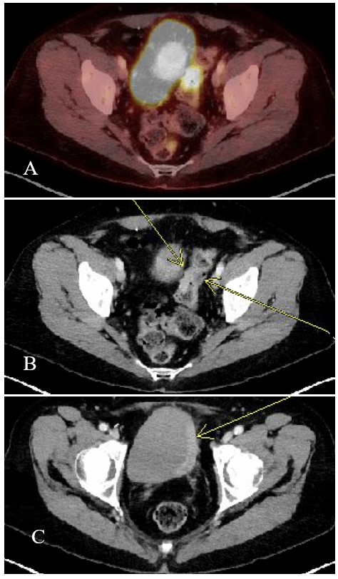
In October 2024, surveillance PET/CT noted focal intense activity in the sigmoid colon with a recommendation for endoscopic correlation. In early 2025, she developed recurrent gross hematuria, initially evaluated in the emergency department with negative urine culture, followed by hospitalization for clot retention and anemia requiring transfusion. PET/CT in February 2025 demonstrated focal FDG uptake with short-segment thickening in the mid sigmoid colon (SUVmax 10.4), a mildly hypermetabolic 0.6 cm left external iliac lymph node (SUVmax 4.6), and asymmetric left bladder wall thickening without hypermetabolic activity Figure 1.
Cystoscopy in March 2025 revealed extensive clot burden and a firm, oozing left bladder wall; the most actively bleeding area was resected, and the remaining left lateral wall was cauterized. Bladder biopsy demonstrated poorly cohesive adenocarcinoma consistent with metastatic lobular breast carcinoma. IHC showed CK20 and p63 negative tumor cells (highlighting overlying urothelium), and mammaglobin/GCDFP-15 positivity, supporting breast origin in conjunction with morphology and hormone receptor positivity (ER 70% [2+], PR 30% [3+]). HER2 amplification was negative by fluorescence in situ hybridization (HER2/CEP17 ratio 0.5, average HER2 1.8 signals/cell; Group 5 per 2023 ASCO/CAP). Hematuria resolved after fulguration, with no recurrence, and there was no documented hydronephrosis or urinary obstruction.
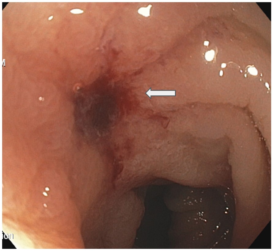
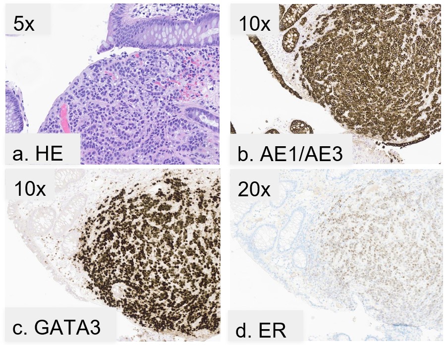
Colonoscopy in April 2025 identified an erythematous area with convergence of folds at approximately 22 cm from the anal verge Figure 2; biopsy confirmed metastatic breast carcinoma with ER 50% (1+), PR 5% (1+), HER2 IHC 2+, and negative HER2 amplification by FISH (HER2/CEP17 ratio 1.1, average HER2 2.6 signals/cell; Group 5). The Ki-67 labeling fraction on the sigmoid biopsy was 50%. IHC from the sigmoid biopsy additionally demonstrated keratin AE1/AE3 positivity and strong GATA3 positivity Figure 3. CDH1 IHC was not performed; instead, next-generation sequencing of the left bladder wall tumor identified a pathogenic CDH1 loss-of-function mutation, providing molecular confirmation of lobular phenotype. CK20 and CDX2 testing were not performed.
Comprehensive molecular profiling with next-generation sequencing (TEMPUS xT) of the left bladder wall tumor performed in April 2025 identified CDH1 loss-of-function and PIK3CA gain-of-function mutations, with a tumor mutational burden–high (28.4 mutations/Mb) and microsatellite-stable profile. Tumor markers had demonstrated a rising trend over the preceding months, with CA 15-3 increasing from 56 U/mL (June 2024) to 79 U/mL (September 2024), 146 U/mL (January 2025), and 170 U/mL (April 2025; reference range <30 U/mL). Similarly, CA 27.29 increased from 53.9 U/mL (June 2024) to 77.6 U/mL (September 2024), 159.7 U/mL (February 2025), and 157.3 U/mL (April 2025; reference range <38 U/mL). She initiated fulvestrant and everolimus in April 2025, and levels declined to 57 U/mL (June 2025) for CA 15-3 and 83.1 U/mL (June 2025) for CA 27.29. These marker trends were interpreted in conjunction with imaging and clinical findings rather than as independent evidence of treatment response.
PET/CT in July 2025 demonstrated resolution of previously FDG-avid uptake in the mid sigmoid colon and improvement in the left external iliac lymph node (decreased size to 0.4 cm and SUVmax 3.8), without new lesions. Follow-up PET/CT in October 2025 showed diminished mild uptake in the left iliac chain node (size decreased to less than 1 cm) and otherwise stable findings, with no new metastatic disease. Measurements for the bladder wall and sigmoid colon lesions could not be obtained reliably on PET/CT imaging. As of October 2025 (6 months into treatment on fulvestrant and everolimus), her disease remains stable and improved from prior imaging and lab testing. Everolimus safety monitoring included serial complete blood counts, comprehensive metabolic panels, and periodic assessments of glucose and lipids, all of which remained stable during treatment. Adverse effects were limited to grade 1 stomatitis, managed with prophylactic and therapeutic steroid mouth rinse, salt–baking soda rinses, and topical anesthetics, with complete resolution, and intermittent grade 1–2 diarrhea managed symptomatically with loperamide. No dose interruptions or reductions were required.
Discussion
Metastatic involvement of the GI tract from breast cancer is rare, particularly in the colon, where lesions may closely mimic primary colorectal malignancy. Invasive lobular carcinoma (ILC) exhibits a distinct metastatic pattern compared with invasive ductal carcinoma, with a greater propensity for spread to the GI tract, peritoneum, ovaries, and GU system [4,5,6,7,8,1,2,9,3,10]. This predilection is largely attributed to loss of CDH1, which results in discohesive tumor growth and facilitates dissemination along serosal and mucosal surfaces rather than forming discrete masses [12,6,13,10]. As a result, ILC metastases may present insidiously and remain clinically occult for prolonged periods.
The interval between initial breast cancer diagnosis and detection of colonic metastases is often prolonged, ranging from 5 to 30 years in reported series [4,6,7,8,1,14,9,15]. Clinical manifestations are frequently nonspecific, including abdominal pain, changes in bowel habits, or bleeding, and lesions may be discovered incidentally on surveillance imaging or endoscopy [5,7,8,1,2,9,10]. In the colon, metastatic ILC commonly infiltrates the submucosa and muscularis propria while sparing the mucosa, further complicating recognition and increasing the risk of misdiagnosis as primary colorectal carcinoma, inflammatory bowel disease, or ischemic colitis [5,1,2,3].
Accurate diagnosis, therefore, relies on histopathologic evaluation with panel-based IHC interpretation. Metastatic breast carcinoma typically demonstrates estrogen and progesterone receptor positivity, expression of breast-lineage markers such as GATA3 and CK7, and absence of GI markers, including CK20 and CDX2 [11]. Loss of CDH1 expression further supports lobular histology [12,13]. CA 15-3 and CA 27.29 are serum tumor markers that detect circulating MUC-1 glycoprotein and are useful for monitoring treatment response in metastatic breast cancer; serial declines correlate with disease response, while rising levels raise concern for progression [16,17]. However, these markers have limited specificity, as elevations can occur in benign conditions (e.g., liver disease) and other malignancies, and transient increases may be seen even in responding disease [18]. Therefore, CA 15-3 and CA 27.29 should always be interpreted in conjunction with imaging studies and clinical assessment rather than used in isolation to guide treatment decisions. Tissue diagnosis was essential to correctly classify the new lesions as metastatic disease rather than new primary malignancy, recognizing the limits of site-specific IHC.
Functional imaging adds another layer of diagnostic complexity. Although PET/CT is widely used in metastatic breast cancer, its sensitivity is reduced in ILC due to lower cellular density, diffuse infiltrative growth, and reduced glycolytic activity [6,10]. As a result, PET/CT may underestimate disease burden or fail to detect sites of involvement. This limitation was evident in our case, as the bladder wall lesion manifested as asymmetric wall thickening without hypermetabolic activity despite histologic confirmation of metastatic disease. These findings underscore that negative or low-uptake PET/CT results do not exclude metastatic involvement in patients with ILC and should prompt tissue evaluation when imaging abnormalities or unexplained symptoms are present, rather than reassurance based on imaging alone.
Simultaneous metastases to the colon and urinary bladder are exceedingly uncommon. In a systematic review by Malinaric et al., only 74 cases of bladder metastases from breast cancer were reported between 1950 and 2022, most of which were associated with lobular histology [19]. Bladder wall lesions in this context may mimic primary urothelial carcinoma clinically and radiographically [19]. GATA3 is highly expressed in both breast carcinoma (94% sensitivity) and urothelial carcinoma (86% sensitivity), limiting its specificity for tumor origin when used in isolation and thus demanding panel-based IHC interpretation [20]. In our case, absence of urothelial lineage markers (p63 and CK20 negativity) combined with focal mammaglobin/GCDFP-15 positivity, hormone receptor expression, and compatible morphology supported a metastatic lobular breast origin rather than a primary bladder malignancy. The diagnosis was established using the IHC panel available at our institution, and additional urothelial rule-out testing (p63, p40, uroplakin, CK5, CK6, and thrombomodulin) was not pursued, given the concordant morphologic features, hormone receptor expression, and supportive breast-lineage immunoprofile.
Management of GI and GU metastases from breast cancer is guided primarily by systemic therapy rather than surgical resection [18]. For hormone receptor–positive disease, endocrine therapy remains the cornerstone of treatment. After progression on long-term aromatase inhibition and CDK4/6 inhibitor therapy, the patient transitioned to fulvestrant plus everolimus, consistent with guideline-supported options for endocrine-resistant metastatic disease [18]. At the time of treatment selection, actionable molecular results were not yet available and therefore did not inform initial therapy choice. Subsequent molecular profiling using Tempus xT on both the sigmoid colon and bladder biopsies identified CDH1 loss-of-function and PIK3CA gain-of-function mutations, findings that may broaden future therapeutic options, including PI3K inhibitors (e.g., alpelisib) or antibody–drug conjugates (e.g., trastuzumab deruxtecan) in the setting of HER2-low disease [21]. Such findings underscore the importance of next-generation sequencing in tailoring treatment strategies for metastatic invasive lobular carcinoma.
Interpretation of HER2 status requires careful adherence to established testing and reporting standards. Although HER2 IHC varied across metastatic sites, both the adrenal and sigmoid lesions met ASCO/CAP Group 5 criteria for HER2-negative disease based on negative fluorescence in situ hybridization results [22,23]. HER2 IHC 0 versus HER2-low status appears to be unstable across patient samples, with close to 40% of cases switching between IHC 0 and IHC 1+ or 2+/ISH not-amplified results when paired primary and metastatic samples are compared [22]. While the concept of "HER2-low" has emerged in the context of antibody–drug conjugate eligibility, it is not a distinct diagnostic category in HER2 pathology reporting per the 2023 ASCO/CAP guideline update [22]. The clinical relevance of distinguishing IHC 0 from IHC 1+ is based on the DESTINY-Breast04 clinical trial entry criteria rather than the demonstration of a new predictive or prognostic threshold [22]. A clear distinction between pathologic classification and therapeutic implications is essential to avoid misinterpretation and inappropriate treatment selection.
Reported survival outcomes for metastatic ILC vary widely and are derived from heterogeneous cohorts that include patients with substantially different metastatic burdens and patterns of organ involvement [19,24,10]. These cohorts often combine patients with bone-only disease, isolated GI or GU metastases, and multi-site visceral involvement, with liver metastases consistently associated with significantly worse outcomes [24]. Published prognostic estimates for metastatic ILC may overgeneralize risk and may not apply to patients with isolated GI or GU metastases. Survival estimates should be interpreted cautiously, and the primary teaching value of the present case lies in diagnostic recognition and accurate disease classification rather than prognostic inference.
This report is limited by its single-patient design and retrospective reconstruction of a complex clinical course, which restricts generalizability and precludes causal inference. Observed radiographic and tumor marker changes represent a temporal association within an individual clinical course and should not be interpreted as evidence of treatment efficacy. Molecular profiling evolved, with next-generation sequencing performed only at recurrence, limiting correlation across earlier disease phases. Measurements of the bladder wall and sigmoid colon lesions could not be reliably obtained on PET/CT imaging, limiting standardized response assessment. Despite these limitations, the case offers important diagnostic and teaching value regarding late GI and GU metastases of ILC.
Conclusion
This case illustrates late GI and GU involvement by metastatic ILC in a patient with long-standing stage IV disease. Invasive lobular carcinoma demonstrates distinct metastatic behavior and may involve the colon or urinary bladder many years after initial diagnosis, often mimicking primary malignancies at these sites. Recognition of this pattern is critical, as misclassification may lead to unnecessary local interventions or delayed initiation of appropriate systemic therapy. This case underscores the importance of maintaining diagnostic vigilance, obtaining tissue confirmation, and applying panel-based IHC interpretation, especially when imaging findings are subtle or discordant. Accurate histopathologic classification, rather than early detection alone, is central to guiding management in patients with metastatic invasive lobular breast carcinoma.
Conflicts of Interest
The authors declare no competing interests that could have influenced the objectivity or outcome of this research.
Funding Source
This study was not supported by any sponsor or funder.
Acknowledgments
No acknowledgments.
Informed Consent
Written informed consent was obtained from the patient for publication of this case report and all accompanying images.
Large Language Model
Generative artificial intelligence tools were used in a limited and supportive role during manuscript preparation. Literature searches to identify relevant background articles were performed using OpenEvidence (accessed December 2024 and January 2025). ChatGPT (OpenAI; accessed December 2024 and January 2025) was used to assist with language refinement, grammar, clarity, and formatting of the manuscript. Neither tool was used to generate original scientific content, interpret data, perform analyses, draw conclusions, or make clinical judgments. All content was reviewed, verified, and edited by the authors, who take full responsibility for the accuracy, integrity, and originality of the manuscript. No AI tool is listed as an author, in accordance with the journal’s authorship and contributorship policies.
Authors Contribution
Conceptualization was done by NP. Data curation was carried out by NP, SMN, and VM. Writing of the original draft was done by NP, while writing, review, and editing were performed by all authors. Supervision was provided by VM, and pathology interpretation was conducted by DBM.
Data Availability
The authors confirm that all relevant clinical, imaging, pathologic, and follow-up data supporting the findings of this case report are contained within the manuscript. No publicly available dataset was generated or analyzed. Further data are not available because of patient confidentiality considerations.
References
- McLemore E. C., Pockaj B. A., Reynolds C., Gray R. J., Hernandez J. L., Grant C. S., Donohue J. H.. Breast cancer: presentation and intervention in women with gastrointestinal metastasis and carcinomatosis. Ann Surg Oncol. 2005;12(11):886-94. doi:10.1245/ASO.2005.03.030 PMID: 16177864
- Montagna E., Pirola S., Maisonneuve P., De Roberto G., Cancello G., Palazzo A., Viale G., Colleoni M.. Lobular Metastatic Breast Cancer Patients With Gastrointestinal Involvement: Features and Outcomes. Clin Breast Cancer. 2018;18(3):e401-e405. doi:10.1016/j.clbc.2017.07.003 PMID: 28778379
- Terada T.. Histopathologic study of the rectum in 1,464 consecutive rectal specimens in a single Japanese hospital: II. malignant lesions. Int J Clin Exp Pathol. 2013;6(3):385-94. PMID: 23412998 PMCID: PMC3563196
- Arif F. Z., Breese R. O., Burke R.. Breast Cancer Metastasis to Colon. Am Surg. 2023;89(9):3873-3874. doi:10.1177/00031348231173979 PMID: 37144362
- Bencardino S., Allocca M., Furfaro F., D'Amico F., Parigi T. L., Danese S., Zilli A.. Multiple Ileal and Colonic Stenoses: Is It Always Crohn's Disease?. J Crohns Colitis. 2025;19(5). doi:10.1093/ecco-jcc/jjae174 PMID: 39679549 PMCID: PMC12087567
- DiPiro P. J., Tirumani S. H., Cruz G. P., Ramaiya N. H., Lester S. C., Shinagare A. B.. Lobular breast cancer: patterns of intraabdominal metastatic spread on imaging and prognostic significance. Abdom Radiol (NY). 2019;44(1):362-369. doi:10.1007/s00261-018-1722-x PMID: 30076430
- Falco G., Mele S., Zizzo M., Di Grezia G., Cecinato P., Besutti G., Coiro S., Gatta G., Vacondio R., Ferrari G.. Colonic metastasis from breast carcinoma detection by CESM and PET/CT: A case report. Medicine (Baltimore). 2018;97(21):e10888. doi:10.1097/MD.0000000000010888 PMID: 29794798 PMCID: PMC6392653
- Inoue H., Arita T., Kuriu Y., Shimizu H., Kiuchi J., Yamamoto Y., Konishi H., Morimura R., Shiozaki A., Ikoma H., Kubota T., Fujiwara H., Okamoto K., Otsuji E.. Colonic Metastasis from Breast Cancer: A Case Report and Review of the Literature. In Vivo. 2022;36(1):522-527. doi:10.21873/invivo.12733 PMID: 34972757 PMCID: PMC8765167
- Nagtegaal I. D., Snoek J. A. A., Bult P., Tol J., Siesling S., Voorham Q. J., Hugen N.. The value of case reports in rare oncological scenarios: mixed method analysis of colorectal metastases from breast cancer. Clin Exp Metastasis. 2023;40(3):205-216. doi:10.1007/s10585-023-10207-9 PMID: 37106226 PMCID: PMC10232590
- Winn J. S., Baker M. G., Fanous I. S., Slack-Davis J. K., Atkins K. A., Dillon P. M.. Lobular Breast Cancer and Abdominal Metastases: A Retrospective Review and Impact on Survival. Oncology. 2016;91(3):135-42. doi:10.1159/000447264 PMID: 27355204
- National Comprehensive Cancer Network. Occult Primary (Cancer of Unknown Primary [CUP]). 2024.
- Bullock E., Brunton V. G.. E-Cadherin-Mediated Cell-Cell Adhesion and Invasive Lobular Breast Cancer. Adv Exp Med Biol. 2025;1464:259-275. doi:10.1007/978-3-031-70875-6_14 PMID: 39821030
- Fang C., Kang Y.. E-Cadherin: Context-Dependent Functions of a Quintessential Epithelial Marker in Metastasis. Cancer Res. 2021;81(23):5800-5802. doi:10.1158/0008-5472.CAN-21-3302 PMID: 34853039
- Mourra N., Jouret-Mourin A., Lazure T., Audard V., Albiges L., Malbois M., Bouzourene H., Duvillard P.. Metastatic tumors to the colon and rectum: a multi-institutional study. Arch Pathol Lab Med. 2012;136(11):1397-401. doi:10.5858/arpa.2011-0432-OA PMID: 23106585
- Pedersen R. N., Esen B. O., Mellemkjaer L., Christiansen P., Ejlertsen B., Lash T. L., Norgaard M., Cronin-Fenton D.. The Incidence of Breast Cancer Recurrence 10-32 Years After Primary Diagnosis. J Natl Cancer Inst. 2022;114(3):391-399. doi:10.1093/jnci/djab202 PMID: 34747484 PMCID: PMC8902439
- Lauro S., Trasatti L., Bordin F., Lanzetta G., Bria E., Gelibter A., Reale M. G., Vecchione A.. Comparison of CEA, MCA, CA 15-3 and CA 27-29 in follow-up and monitoring therapeutic response in breast cancer patients. Anticancer Res. 1999;19(4C):3511-5. PMID: 10629644
- Tampellini M., Berruti A., Bitossi R., Gorzegno G., Alabiso I., Bottini A., Farris A., Donadio M., Sarobba M. G., Manzin E., Durando A., Defabiani E., De Matteis A., Ardine M., Castiglione F., Danese S., Bertone E., Alabiso O., Massobrio M., Dogliotti L.. Prognostic significance of changes in CA 15-3 serum levels during chemotherapy in metastatic breast cancer patients. Breast Cancer Res Treat. 2006;98(3):241-8. doi:10.1007/s10549-005-9155-y PMID: 16670941
- National Comprehensive Cancer Network. NCCN Clinical Practice Guidelines in Oncology: Breast Cancer. 2025.
- Malinaric R., Balzarini F., Granelli G., Ferrari A., Trani G., Ambrosini F., Mantica G., Panarello D., De Rose A. F., Terrone C.. From women to women-hematuria during therapy for metastatic breast cancer, what to suspect and when to be alarmed; Bladder metastasis from breast cancer-our experience and a systematic literature review. Front Oncol. 2022;12:976947. doi:10.3389/fonc.2022.976947 PMID: 36248976 PMCID: PMC9557997
- Liu H., Shi J., Wilkerson M. L., Lin F.. Immunohistochemical evaluation of GATA3 expression in tumors and normal tissues: a useful immunomarker for breast and urothelial carcinomas. Am J Clin Pathol. 2012;138(1):57-64. doi:10.1309/AJCP5UAFMSA9ZQBZ PMID: 22706858
- Burstein H. J., DeMichele A., Somerfield M. R., Henry N. L., Biomarker Testing and Endocrine and Targeted Therapy in Metastatic Breast Cancer Expert Panels. Testing for ESR1 Mutations to Guide Therapy for Hormone Receptor-Positive, Human Epidermal Growth Factor Receptor 2-Negative Metastatic Breast Cancer: ASCO Guideline Rapid Recommendation Update. J Clin Oncol. 2023;41(18):3423-3425. doi:10.1200/JCO.23.00638 PMID: 37196213
- Wolff A. C., Somerfield M. R., Dowsett M., Hammond M. E. H., Hayes D. F., McShane L. M., Saphner T. J., Spears P. A., Allison K. H.. Human Epidermal Growth Factor Receptor 2 Testing in Breast Cancer: ASCO-College of American Pathologists Guideline Update. J Clin Oncol. 2023;41(22):3867-3872. doi:10.1200/JCO.22.02864 PMID: 37284804
- Woo J. W., Lee K., Chung Y. R., Jang M. H., Ahn S., Park S. Y.. The updated 2018 American Society of Clinical Oncology/College of American Pathologists guideline on human epidermal growth factor receptor 2 interpretation in breast cancer: comparison with previous guidelines and clinical significance of the proposed in situ hybridization groups. Hum Pathol. 2020;98:10-21. doi:10.1016/j.humpath.2020.01.003 PMID: 32027910
- Raghavendra A. S., Bassett R., Jr., Damodaran S., Barcenas C. H., Mouabbi J. A., Layman R., Tripathy D.. Clinical Characteristics and Survival Outcomes of Metastatic Invasive Lobular and Ductal Carcinoma. JAMA Netw Open. 2025;8(4):e251888. doi:10.1001/jamanetworkopen.2025.1888 PMID: 40293750 PMCID: PMC12038507
References
1. McLemore EC, Pockaj BA, Reynolds C, Gray RJ, Hernandez JL, Grant CS, Donohue JH. Breast cancer: presentation and intervention in women with gastrointestinal metastasis and carcinomatosis. Ann Surg Oncol. 2005;12(11):886-94. [PMID: 16177864, https://doi.org/10.1245/ASO.2005.03.030].
2. Montagna E, Pirola S, Maisonneuve P, De Roberto G, Cancello G, Palazzo A, Viale G, Colleoni M. Lobular Metastatic Breast Cancer Patients With Gastrointestinal Involvement: Features and Outcomes. Clin Breast Cancer. 2018;18(3):e401-5. [PMID: 28778379, https://doi.org/10.1016/j.clbc.2017.07.003].
3. Terada T. Histopathologic study of the rectum in 1,464 consecutive rectal specimens in a single Japanese hospital: II. malignant lesions. Int J Clin Exp Pathol. 2013;6(3):385-94. [PMID: 23412998, PMCID: PMC3563196].
4. Arif FZ, Breese RO, Burke R. Breast Cancer Metastasis to Colon. Am Surg. 2023;89(9):3873-4. [PMID: 37144362, https://doi.org/10.1177/00031348231173979].
5. Bencardino S, Allocca M, Furfaro F, D'Amico F, Parigi TL, Danese S, Zilli A. Multiple Ileal and Colonic Stenoses: Is It Always Crohn's Disease? J Crohns Colitis. 2025;19(5). [PMID: 39679549, PMCID: PMC12087567, https://doi.org/10.1093/ecco-jcc/jjae174].
6. DiPiro PJ, Tirumani SH, Cruz GP, Ramaiya NH, Lester SC, Shinagare AB. Lobular breast cancer: patterns of intraabdominal metastatic spread on imaging and prognostic significance. Abdom Radiol (NY). 2019;44(1):362-9. [PMID: 30076430, https://doi.org/10.1007/s00261-018-1722-x].
7. Falco G, Mele S, Zizzo M, Di Grezia G, Cecinato P, Besutti G, Coiro S, Gatta G, Vacondio R, Ferrari G. Colonic metastasis from breast carcinoma detection by CESM and PET/CT: A case report. Medicine (Baltimore). 2018;97(21):e10888. [PMID: 29794798, PMCID: PMC6392653, https://doi.org/10.1097/MD.0000000000010888].
8. Inoue H, Arita T, Kuriu Y, Shimizu H, Kiuchi J, Yamamoto Y, Konishi H, Morimura R, Shiozaki A, Ikoma H, Kubota T, Fujiwara H, Okamoto K, Otsuji E. Colonic Metastasis from Breast Cancer: A Case Report and Review of the Literature. In Vivo. 2022;36(1):522-7. [PMID: 34972757, PMCID: PMC8765167, https://doi.org/10.21873/invivo.12733].
9. Nagtegaal ID, Snoek JAA, Bult P, Tol J, Siesling S, Voorham QJ, Hugen N. The value of case reports in rare oncological scenarios: mixed method analysis of colorectal metastases from breast cancer. Clin Exp Metastasis. 2023;40(3):205-16. [PMID: 37106226, PMCID: PMC10232590, https://doi.org/10.1007/s10585-023-10207-9].
10. Winn JS, Baker MG, Fanous IS, Slack-Davis JK, Atkins KA, Dillon PM. Lobular Breast Cancer and Abdominal Metastases: A Retrospective Review and Impact on Survival. Oncology. 2016;91(3):135-42. [PMID: 27355204, https://doi.org/10.1159/000447264].
11. National Comprehensive Cancer Network. Occult Primary (Cancer of Unknown Primary [CUP]); 2024. [updated 2024/09/11]. Available from: https://www.nccn.org.
12. Bullock E, Brunton VG. E-Cadherin-Mediated Cell-Cell Adhesion and Invasive Lobular Breast Cancer. Adv Exp Med Biol. 2025;1464:259-75. [PMID: 39821030, https://doi.org/10.1007/978-3-031-70875-6_14].
13. Fang C, Kang Y. E-Cadherin: Context-Dependent Functions of a Quintessential Epithelial Marker in Metastasis. Cancer Res. 2021;81(23):5800-2. [PMID: 34853039, https://doi.org/10.1158/0008-5472.CAN-21-3302].
14. Mourra N, Jouret-Mourin A, Lazure T, Audard V, Albiges L, Malbois M, Bouzourene H, Duvillard P. Metastatic tumors to the colon and rectum: a multi-institutional study. Arch Pathol Lab Med. 2012;136(11):1397-401. [PMID: 23106585, https://doi.org/10.5858/arpa.2011-0432-OA].
15. Pedersen RN, Esen BO, Mellemkjaer L, Christiansen P, Ejlertsen B, Lash TL, Norgaard M, Cronin-Fenton D. The Incidence of Breast Cancer Recurrence 10-32 Years After Primary Diagnosis. J Natl Cancer Inst. 2022;114(3):391-9. [PMID: 34747484, PMCID: PMC8902439, https://doi.org/10.1093/jnci/djab202].
16. Lauro S, Trasatti L, Bordin F, Lanzetta G, Bria E, Gelibter A, Reale MG, Vecchione A. Comparison of CEA, MCA, CA 15-3 and CA 27-29 in follow-up and monitoring therapeutic response in breast cancer patients. Anticancer Res. 1999;19(4C):3511-5. [PMID: 10629644].
17. Tampellini M, Berruti A, Bitossi R, Gorzegno G, Alabiso I, Bottini A, Farris A, Donadio M, Sarobba MG, Manzin E, Durando A, Defabiani E, De Matteis A, Ardine M, Castiglione F, Danese S, Bertone E, Alabiso O, Massobrio M, Dogliotti L. Prognostic significance of changes in CA 15-3 serum levels during chemotherapy in metastatic breast cancer patients. Breast Cancer Res Treat. 2006;98(3):241-8. [PMID: 16670941, https://doi.org/10.1007/s10549-005-9155-y].
18. National Comprehensive Cancer Network. NCCN Clinical Practice Guidelines in Oncology: Breast Cancer; 2025. [updated 2025/04/17]. Available from: https://www.nccn.org/professionals/physician_gls/pdf/breast.pdf.
19. Malinaric R, Balzarini F, Granelli G, Ferrari A, Trani G, Ambrosini F, Mantica G, Panarello D, De Rose AF, Terrone C. From women to women-hematuria during therapy for metastatic breast cancer, what to suspect and when to be alarmed; Bladder metastasis from breast cancer-our experience and a systematic literature review. Front Oncol. 2022;12:976947. [PMID: 36248976, PMCID: PMC9557997, https://doi.org/10.3389/fonc.2022.976947].
20. Liu H, Shi J, Wilkerson ML, Lin F. Immunohistochemical evaluation of GATA3 expression in tumors and normal tissues: a useful immunomarker for breast and urothelial carcinomas. Am J Clin Pathol. 2012;138(1):57-64. [PMID: 22706858, https://doi.org/10.1309/AJCP5UAFMSA9ZQBZ].
21. Burstein HJ, DeMichele A, Somerfield MR, Henry NL, Biomarker T, Endocrine, Targeted Therapy in Metastatic Breast Cancer Expert P. Testing for ESR1 Mutations to Guide Therapy for Hormone Receptor-Positive, Human Epidermal Growth Factor Receptor 2-Negative Metastatic Breast Cancer: ASCO Guideline Rapid Recommendation Update. J Clin Oncol. 2023;41(18):3423-5. [PMID: 37196213, https://doi.org/10.1200/JCO.23.00638].
22. Wolff AC, Somerfield MR, Dowsett M, Hammond MEH, Hayes DF, McShane LM, Saphner TJ, Spears PA, Allison KH. Human Epidermal Growth Factor Receptor 2 Testing in Breast Cancer: ASCO-College of American Pathologists Guideline Update. J Clin Oncol. 2023;41(22):3867-72. [PMID: 37284804, https://doi.org/10.1200/JCO.22.02864].
23. Woo JW, Lee K, Chung YR, Jang MH, Ahn S, Park SY. The updated 2018 American Society of Clinical Oncology/College of American Pathologists guideline on human epidermal growth factor receptor 2 interpretation in breast cancer: comparison with previous guidelines and clinical significance of the proposed in situ hybridization groups. Hum Pathol. 2020;98:10-21. [PMID: 32027910, https://doi.org/10.1016/j.humpath.2020.01.003].
24. Raghavendra AS, Bassett RJ, Damodaran S, Barcenas CH, Mouabbi JA, Layman R, Tripathy D. Clinical Characteristics and Survival Outcomes of Metastatic Invasive Lobular and Ductal Carcinoma. JAMA Netw Open. 2025;8(4):e251888. [PMID: 40293750, PMCID: PMC12038507, https://doi.org/10.1001/jamanetworkopen.2025.1888].
Data Availability Statement
The authors confirm that all relevant clinical, imaging, pathologic, and follow-up data supporting the findings of this case report are contained within the manuscript. No publicly available dataset was generated or analyzed. Further data are not available because of patient confidentiality considerations.
License
Copyright (c) 2026 Navin Roger Prasad, Stanley M Nwabudike, David Brian Meredith, Vipin Mittal

This work is licensed under a Creative Commons Attribution 4.0 International License.
Copyright and Licensing
All articles published in ASIDE Oncology are open access and freely available online immediately upon publication, without embargo.
This journal applies the terms of the Creative Commons Attribution 4.0 International License (CC BY 4.0). Under this license, anyone may copy, redistribute, remix, transform, and build upon the material for any purpose, even commercially, provided that proper credit is given to the original author(s), a link to the license is provided, and any changes made are indicated.
-
License link: https://creativecommons.org/licenses/by/4.0/
-
Machine-readable license metadata is embedded in each article and its DOI record.
Copyright Holder
Copyright in all articles remains with the author(s). ASIDE Oncology is published and owned by PubPorta Publishing LLC. By submitting a manuscript, authors agree to grant the publisher a non-exclusive license to publish, distribute, archive, and identify itself as the original publisher of the work under the terms of the CC BY 4.0 license.
Academic oversight and scholarly guidance are provided by the American Society for Inclusion, Diversity, and Equity in Healthcare (ASIDE). Copyright and licensing arrangements are administered by the publisher.
Third-party Material
Any material (e.g., images, figures, or tables) not covered by CC BY 4.0 will be clearly indicated in the article. Users must obtain permission directly from the copyright holder for reuse of such third-party material.
Author Rights and Self-Archiving
Authors are permitted to deposit all versions of their work (preprint, author’s accepted manuscript, and version of record) in institutional or subject repositories of their choice immediately upon publication, without embargo, provided that the DOI and license information are included.

Published
Issue
Section
How to Cite
Article history
- Received
- 2 Feb 2026
- Received in revised form
- 23 Feb 2026
- Accepted
- 3 Mar 2026
- Published
- 8 Mar 2026