Acute Myeloid Leukemia Presenting as Bilateral Proptosis: A Case Report with Literature Review
Ahmed Mostafa Youssef1, Mohamad Monif AsskerⒾ2*, Farah Alkhaleefa3, Mahasin Shaheen3
- 1Department of Education, Al Qassimi Hospital, Sharjah, UAE
- 2Department of Radiology, Sheikh Khalifa Medical City, Abu Dhabi, UAE
- 3Department of Internal Medicine, Al Qassimi Hospital, Sharjah, UAE
Abstract
Introduction: Acute myeloid leukemia (AML), constituting 30% of pediatric malignancies, is the most common childhood cancer. This paper explores the rare presentation of AML with extramedullary involvement, specifically bilateral proptosis, in a 15-year-old boy.
Case report: The patient initially presented with worsening shortness of breath, palpitations, extreme fatigue, and bruising. Examination revealed bilateral proptosis, watery discharge from the right eye, and petechial rash. Blood investigations revealed low hemoglobin, severe thrombocytopenia, and high WBC count. Computed tomography (CT) revealed bilateral orbital infiltrative soft tissue lesions. Leukemia fusion gene screening identified RUNX1-RUNX1T1 later. The patient was admitted, received transfusions, and started on antibiotics. Despite initial improvement, he later developed sepsis, septic shock, and severe pancytopenia, necessitating intensive care and specific AML M2 targeting therapy.
Conclusion: Bilateral proptosis in AML, termed myeloid sarcoma (MS), is rare but responsive to chemotherapy. Orbital MS has higher responsiveness and survival rates in pediatric cases. The case highlights the importance of identifying AML subtypes, like RUNX1-RUNX1T1-positive AML, for tailored treatment strategies. This case underscores the challenges in diagnosing and treating pediatric AML with extramedullary involvement. Early recognition of AML subtypes is crucial for prognosis prediction and treatment tailoring.
Keywords: Acute Myeloid Leukemia, Myeloid Sarcoma, Bilateral proptosis, Di Guglielmo syndrome, M6 subtype, RUNX1-RUNX1T1
Article information
Introduction
Acute leukemia, constituting approximately 30% of pediatric malignancies, stands as the most prevalent cancer in children [1]. Specifically, Acute myeloid leukemia (AML), which is the second most common form of leukemia, manifests in up to 15% of the pediatric leukemic population [2]. Leukemia, defined as a hematopoietic system malignancy, results in the infiltration of the bone marrow by myeloblasts in AML. These myeloblasts disrupt the development of normal blood cells in the bone marrow and are subsequently released into the bloodstream, reaching distant extramedullary sites such as the orbit, soft tissues, and bones, forming a solid tumor known as myeloid sarcoma (MS) [3]. While extramedullary involvement is rare in AML, it is exceptionally uncommon in bone structures [4]. Myeloid Sarcomas are most prevalent in specific AML subtypes, notably M6 (Di Guglielmo syndrome), M5a (monoblastic), M5b (monocytic), M4 (myelomonocytic), and M2 (myeloblastic with maturation) [5]. The incidence of extramedullary disease is low, accounting for 2.5–9% of AML, with 60% of Myeloid sarcomas occurring in children under 15 years old [6]. Notably, the incidence appears slightly higher in Asia, the region of origin for our patient [7].
While extramedullary involvement in AML has been previously documented, the presentation of bilateral proptosis as the initial manifestation of AML with the RUNX1-RUNX1T1 fusion gene is exceptionally rare, with limited cases in the literature. This case contributes to understanding orbital involvement in AML and highlights the importance of early molecular characterization in guiding treatment decisions. In this report, we describe a case of AML in a 15-year-old boy who presented primarily to the Emergency Department with the chief complaint of fatigue, dyspnea, and bilateral proptosis.
Case Presentation
History
A 15-year-old male patient from South Asia presented with a chief complaint of worsening shortness of breath and palpitations on minimal exertion and extreme fatigue that was associated with back pain for the past 15 days. The patient has been feeling progressively weak for the past week, with no recent fever. Additionally, he reported associated bruising without any history of trauma or family history of similar conditions. The surgical history and medical history are unremarkable. There’s no recent travel history for three years and no known allergies. The patient had been attending the gym for the past few months, intentionally reducing weight from 98 to 92 kg. No chest pain, abdominal pain, nausea, vomiting, changes in urine or bowel habits, melena, or blood in stool were reported.
Examination
On examination, the patient was vitally stable with a temperature of 37.1 °C, heart rate of 110, respiratory rate of 20, blood pressure of 106/60, and maintaining SpO2 at 99% on room air. The patient has a patent airway, bilaterally equal, and clear entry with no added sounds. The patient is tachycardic, with the Glasgow Coma Scale at 15; however, he appears extremely pale. As shown in Figure 1, bilateral proptosis of the eyes (right more than left) led to a change in facial appearance that has worsened over the past 15 days. There is a watery discharge from the right eye with lower lid retraction and reduced hearing bilaterally without pain and normal eye movements. No visual disturbances or ocular pain were reported. He exhibited a petechial rash, mainly noted on the lower lip.
Investigations
The patient’s blood workup revealed critical values: low hemoglobin of (Normal range: ), severe thrombocytopenia of (Normal range: ). High WBC count of (Normal range: ). Further investigations showed CRP: 13, MCV: 97, retic count: 0.97, LDH: 213, Haptoglobin: (Normal range: ), INR: 1, and creatinine: 86. Liver function tests were within normal limits.
Blood film results indicated predominantly normocytic normochromic RBCs and leukocytosis with the following differential: Neutrophils 3%, Lymphocytes 9%, Monocytes 2%, Blasts + promyelocytes 86%. Blasts displayed characteristics such as Auer rods, Buttock cells, large size, scant granular cytoplasm, high N: C ratio, irregular nuclear contours, immature chromatin, and prominent 1–2 nucleoli.
The leukemia fusion gene report Table 1 detected positive gene fusion mRNA of RUNX1-RUNX1T1 that correlates with the translocation in t(8;21) (q22;q22), which has been associated with both de novo and therapy-related AML and has a favorable prognosis.
| Translocation | Gene Fusion | Result |
|---|---|---|
| t(9;11) (p22;q23) | KMT2A-MLLT3 | Fusion mRNA Not Detected |
| t(15;17) (q24;q21) | PML-RARA | Fusion mRNA Not Detected |
| t(8;21) (q22;q22) | RUNX1-RUNX1T1 | Fusion mRNA of RUNX1-RUNX1T1 Detected |
| t(4;11) (q21;q23) | KMT2A-AFF1 | Fusion mRNA Not Detected |
| t(12;21) (p13;q22) | ETV6-RUNX1 | Fusion mRNA Not Detected |
| t(1;19) (q23;p13) | TCF3-PBX1 | Fusion mRNA Not Detected |
| t(11;19) (q23;p13.3) | KMT2A-MLLT1 | Fusion mRNA Not Detected |
| t(9;22) (q34;q11) | BCR-ABL1 | Fusion mRNA Not Detected |
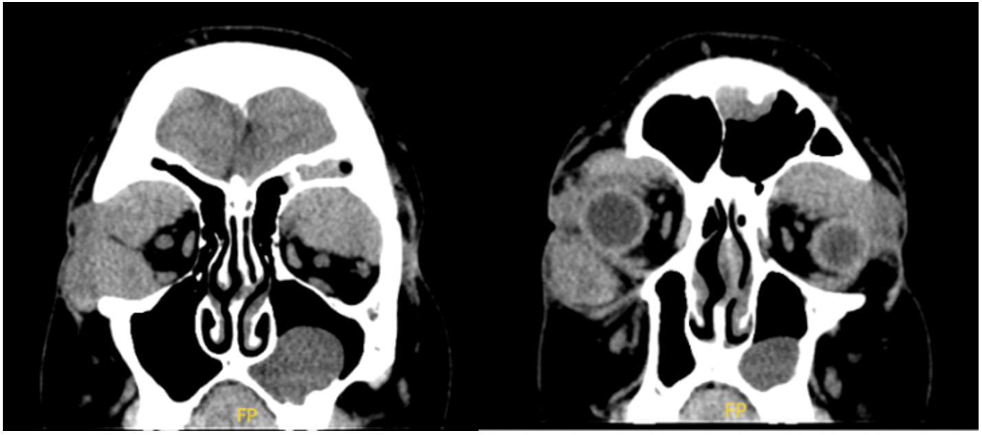
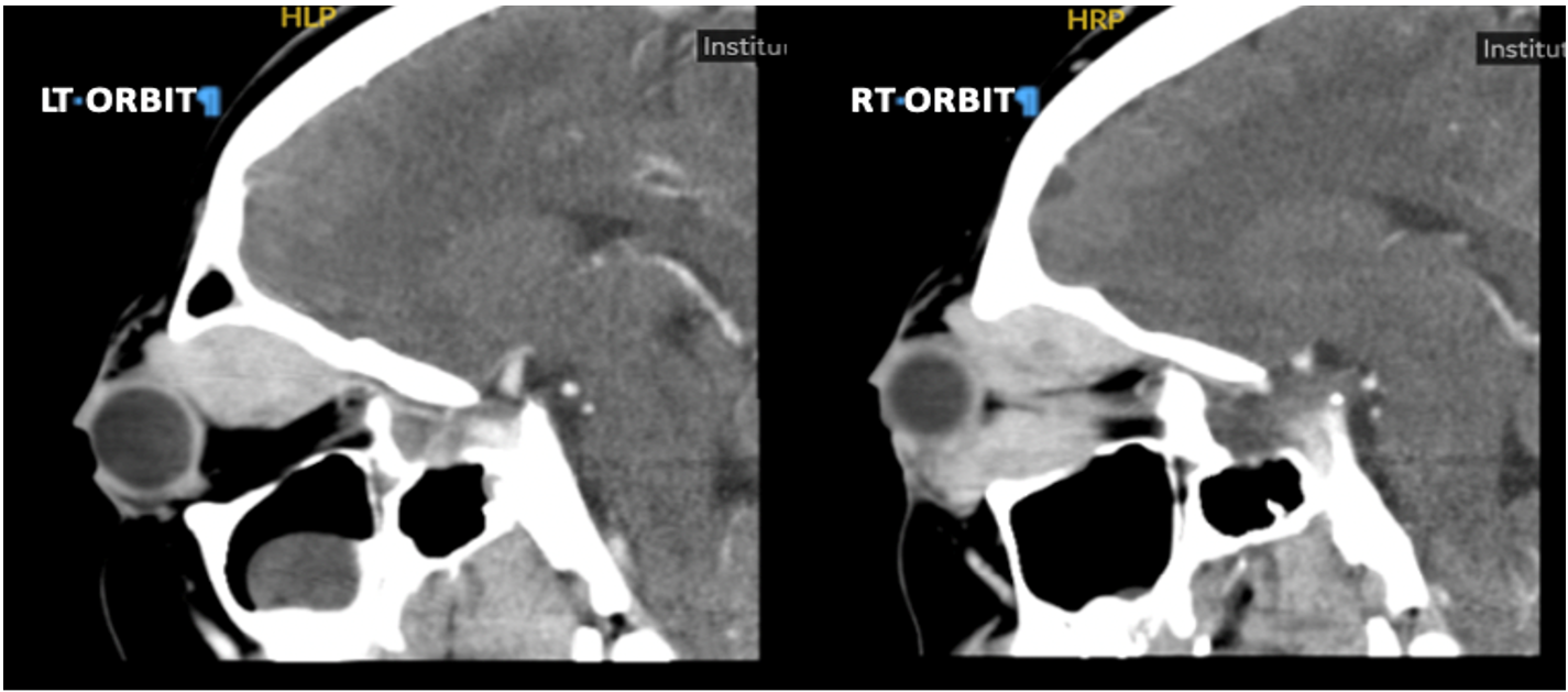
FLT-3: negative, Flow cytometry post-Induction I: 3% CD34 positive, CD117 positive blasts. The Computed tomography (CT) Orbit Sella with Contrast Figure 2 and Figure 3 displayed bilateral, almost symmetrical enhancing extra-conchal soft tissue lesions observed at both orbits’ superior and lateral aspects. A biopsy was not obtained to confirm the diagnosis of myeloid sarcoma.

Treatment
The patient is admitted to the High Dependency Unit (HDU). Anemia workup, TFT, occult blood, and blood film were sent. Autoimmune and viral screens were sent. An urgent type of cross for four units of PRBC/4 FFP/6 platelets was requested. Two units of PRBCs and six units of platelets were transfused, after which his HB improved to 6.5 and platelets improved to 30,000. Pan cultures were sent, and empirical ceftriaxone was started. Ophthalmology advised tobramycin-dexamethasone eye drops and a pan CT of the orbital region with contrast once the patient is stable vitally. Blood pressure is monitored, and MAP is kept above 65.
The patient was started on Intravenous hydration along with an Intravenous antibiotic given the blood workup, and the blood film showed the possibility of acute myeloid Leukemia for further workup. The next day, the patient’s right eye showed more protrusion and redness with dryness compared to yesterday. The left eye was showing a new sub-scleral hemorrhage but no ophthalmoplegia. Eye movements were preserved. The patient was accepted to be transferred to a specialized hospital where an Oncology service is available for Acute Myeloid Leukemia chemotherapy.
Outcomes and Follow-up
After 1 month after admission, the patient was brought by ambulance as a case of altered level of consciousness, lethargy, and loss of appetite and presented with chief complaints of fever, SOB, vomiting, and generalized weakness for one day. He vomited twice, but no coffee ground vomitus, no blood vomiting, no cough, no chest pain, and no bleeding from any site. On examination, the patient is vitally stable with HR: 78 bpm, BP 88/22 mm Hg, RR: 16 bpm. SpO2: 96%. He was alert, conscious, oriented, chest clear, and his abdomen was soft, not tender. The patient developed sepsis and septic shock, acute kidney injury, and severe pancytopenia. Blood culture was positive for gram-negative rods identified as Klebsiella pneumoniae pan-sensitive. The patient required inotropic support and Intensive Care Unit admission, as well as being managed accordingly with cefepime, teicoplanin, and voriconazole.
WBC 0.03 x10(3)/mcL, HB 5.80 g/dL, platelet 9.00 x10(3)/mcL, he was started on filgrastim 300 mcg SC OD till absolute neutrophile 0.5 and managed with platelets and packed RBCs transfusion. Abdominal US showed mild hepatomegaly.
One month later, the patient was treated according to the Tawam AML protocol, which consists of standard induction with cytarabine (100 mg/m² continuous infusion for 7 days) and daunorubicin (60 mg/m² for 3 days), followed by four cycles of high-dose cytarabine consolidation therapy, tailored explicitly for RUNX1-RUNX1T1-positive AML M2 subtype. His chemotherapy was complicated by septic shock with gram-negative rods, which he recovered from successfully after prolonged admission and PICU stay. Fungal infection of the lungs, with Chest CT suggestive of fungal infection, was managed with Voriconazole. He had bilateral Retinal detachment and is currently followed by ophthalmology.
Discussion
Bilateral proptosis, being the main presentation in an AML patient, has been previously reported in some reports across the globe. Albeit far less common than their counterparts’ presentations, unilaterally, Leukemic cellular infiltration of the retro-orbital space is the main pathophysiologic phenomenon reported behind proptosis in AML cases [8,9,10,11,12,13]. In our case, bilateral proptosis was observed as the initial presentation of the disease. This aligns with several reports in the literature that describe orbital myeloid sarcoma as an initial manifestation of AML, where orbital involvement precedes the diagnosis of the underlying hematologic malignancy [14,15].
Other contributory reasons include retrobulbar hemorrhages, obstructed venous drainage, and extraocular muscle infiltrates within the orbital space [16]. This specific extramedullary manifestation of AML was recognized by Allen Burns in 1811 as the green tumor and subsequently termed granulocytic sarcoma (GS) [17]. The green coloration due to myeloperoxidase concentration is variable, however, which leads to the broader nomenclature of orbital leukemic infiltrates by the term myeloid sarcoma (MS), or more commonly, chloroma [18].
Our patient, a 15-year-old male, presented with bilateral proptosis, which, although consistent with orbital involvement, falls slightly outside the most affected age group reported in the literature. Orbital MS is most frequently seen in children, with a mean age of presentation around 7 years [19]. Moreover, studies highlight a predilection for the 3–10-year-old age range, making it important to recognize that such presentations can also occur in older adolescents. More importantly, orbital MS was more responsive to Children’s Cancer Group (CCG) chemotherapy protocols, 96% of which achieved complete remission at the end of the second treatment course compared to other non-CNS MS (78%) and non-MS (78%) AML patients undergoing similar protocols. Event-free survival (76%) and overall survival (92%) were also markedly higher in orbital MS compared to other extramedullary variants [20]. Independent survival rates, orbital MS was proven to be more likely associated with the M2 morphology of AML and t(8;21), which were noncontributory to the significance of survival difference compared to other AML morphologies. These characteristics of orbital MS were in consensus with another study conducted in Soweto, South Africa, but prognostic data remain conflicted with another study in Turkey on a similar population and is still a topic for further investigation [21,22].
The significance of identifying the type of AML lies in the varying clinical picture it presents. The M2 subtype has the lowest mean hemoglobin levels among all other subtypes, reaching as low as 3g/dl, while having the highest platelet counts, reaching as much as platelets /L [23]. Immunophenotyping variation was also significant in AML M2, as CD7 positivity was most predominant, guiding toward the use of promising targeted chemotherapeutic agents like the recent autologous CD7 CAR T-cell therapy [24].
The CT of the orbits shows poor definition of the superior and lateral recti muscles in both orbits, more prominently on the right side. This contrasts with previous studies that demonstrated diffuse infiltration and enlargement of all extraocular muscles without an associated mass [11,25].
AML is a heterogeneous disease with various subtypes, each characterized by distinct genetic abnormalities [26]. Two of these subtypes are RUNX1-RUNX1T1 (previously known as AML1-ETO) and CBFB-MYH11-positive AML [27]. RUNX1-RUNX1T1 is one of the most common genetic abnormalities found in the AML M2 subtype and is among the initial fusion genes employed for the surveillance of minimal residual disease [28]. It results from a translocation and fusion between chromosomes 8 and 21. Patients with RUNX1-RUNX1T1-positive AML typically have a more favorable prognosis than many other AML subtypes. This subtype is often associated with younger age, a higher likelihood of achieving complete remission (CR) with treatment, and special morphological features such as lower white blood cell counts, sizable blasts characterized by ample basophilic cytoplasm filled with numerous azurophilic granules, and Auer rods. There have also been instances of blasts with exceptionally large granules, possibly suggesting a fusion of these cells [29]. Treatment response in this specific subtype is primarily reliant on anthracyclines and cytarabine, coupled with subsequent 2-4 rounds of cytarabine; treatment can be enhanced through the incorporation of gemtuzumab-ozogamicin (GO), an antibody targeting CD33 [30]. However, long-term survival can vary, and additional factors, such as the presence of other mutations, are taken into consideration [31].
Another AML variant belonging to the favorable risk subtypes is CBFB-MYH11-positive AML, which is associated with the AML-M4Eo subtype and results from the inv (16) (p13q22) translocation. This subtype has a higher likelihood of achieving CR and prolonged disease-free survival [32]. Patients with CBFB-MYH11-positive AML tend to be younger and have other distinctive morphologic features, including an abundance of monocytes and a distinctive, atypical eosinophil element that defines this particular AML subgroup, which is why it is often referred to as M4Eo AML [33]. However, like RUNX1-RUNX1T1-positive AML, the presence of additional genetic mutations can influence prognosis. Response of this specific subtype was specifically proven to be sensitive to high-dose cytarabine-based consolidation regimens, thereby guiding treatment modalities and options towards better and more specific regimens [34,35]. However, it is worth noting that relapses affect nearly 50% of adult patients, and the survival rate beyond five years is merely around 50% [36,37,38].
AML characterized by t(8;21) or inv(16) is typically grouped under the term "core binding factor AML (CBF-AML) [39]." CBF-AML comprises about 25% of pediatric and 15% of adult patients with newly diagnosed AML, making it the most prevalent cytogenetic subtype of AML [40]. Survival outcomes for pediatric CBF-AML patients, when compared to AML with typical cytogenetics, show a slight improvement. However, a subset with a less favorable prognosis exists within this patient population. This suggests that there is diversity among these patients, and it is likely that additional mutational changes can impact the development of the disease [41].
Given the rarity of extramedullary AML involvement, especially in the context of bone structures, additional diagnostic workup may be necessary to identify the subtype accurately. This may involve cytogenetic and molecular genetic testing to detect specific translocations, mutations, and fusion genes associated with AML subtypes. Management of AML in pediatric patients typically involves intensive chemotherapy regimens aimed at achieving CR. For patients with RUNX1-RUNX1T1 and CBFB–MYH11–positive AML, standard induction chemotherapy, often including anthracycline -based regimens, is the first-line treatment. Hematopoietic stem cell transplantation (HSCT) may be considered for eligible patients, particularly in cases of high-risk AML or relapsed disease. HSCT can offer a chance for long-term remission and potential cure, but it comes with its own set of risks and complications.
The patient developed bilateral retinal detachment, a rare but serious complication in leukemic patients, often linked to leukemic infiltration or hemorrhage. According to the updated Retina Society Classification for proliferative vitreoretinopathy (PVR), retinal changes are graded from A to CA/CP based on severity and location. Grade A includes mild findings like vitreous haze, while more advanced PVR is classified as posterior (CP) or anterior (CA), depending on whether the proliferative changes and retinal folds are located behind or in front of the equator [42]. Given the risk of relapses and the potential for long-term ophthalmologic complications such as retinal detachment, close and coordinated follow-up is essential. The patient will require regular hematological monitoring of visual outcomes and management of any late sequelae of orbital involvement or treatment-related complications.
Although 15-year-olds may fall at the intersection of pediatric and adult care, emerging evidence supports the use of pediatric protocols for adolescents with AML. Studies have shown that adolescents benefit more from pediatric AML protocols than adult ones, with studies showing improved remission and survival rates compared to those treated with adult protocols [27,43]. The use of the pediatric-based Tawam AML protocol, in this case, aligns with current recommendations, especially given the favorable RUNX1-RUNX1T1 cytogenetic profile. In this case, the patient’s clinical condition and response to initial treatment would be critical in determining the appropriate course of action. The development of sepsis and septic shock, along with severe pancytopenia, indicates a complex clinical scenario that may require intensive supportive care in addition to disease-specific AML M2 targeting therapy. Regular monitoring, including minimal residual disease assessment, is crucial for evaluating treatment response and guiding further therapeutic decisions.
Conclusions
This case report sheds light on the intricacies of diagnosing and treating Acute Myeloid Leukemia (AML) in a pediatric patient presenting with extramedullary involvement and bilateral proptosis. Recognizing the specific AML subtype, such as RUNX1-RUNX1T1 and CBFB-MYH11–positive AML, is pivotal in predicting prognosis and tailoring the treatment strategy. Early and precise diagnosis, coupled with a comprehensive multidisciplinary approach to care, can significantly enhance the outcomes for pediatric AML patients, positively impacting their overall well-being and recovery.
Conflicts of Interest
The authors declare no competing interests that could have influenced the objectivity or outcome of this research.
Funding Source
This research received no specific grant from funding agencies in the public, commercial, or not-for-profit sectors.
Acknowledgments
None
Informed Consent
Informed consent was obtained from the patient, and verbal witness informed consent was obtained from parents, anonymizing identifiable information.
Large Language Model
None
Authors Contribution
AMY conceived and designed the study. AMY and MMA provided research materials and collected and organized data. AMY, MMA, and FA wrote the initial and final drafts of the article. MS has critically reviewed and approved the final draft. MS supervised the conduction of the research.
Data Availability
This case report is based on a single patient’s clinical presentation, diagnostic findings, and treatment course. All relevant data are included within the article. No new datasets were generated or analyzed beyond the information documented in the patient’s medical records, which are not publicly available due to privacy and confidentiality considerations.
References
- Al-Mujaini A., Al-Shaaibi M., Al-Mughaizwi T., Wali Y., Ganesh A.. Unilateral Proptosis: A Rare Presenting Sign of Acute Myeloid Leukemia. Oman Med J. 2022;37(4):e400. doi:10.5001/omj.2022.10 PMID: 35915765 PMCID: PMC9260739
- Stein-Wexler R., Wootton-Gorges S. L., West D. C.. Orbital granulocytic sarcoma: an unusual presentation of acute myelocytic leukemia. Pediatr Radiol. 2003;33(2):136-9. doi:10.1007/s00247-002-0834-0 PMID: 12557072
- Huang Y. C., Wang S. C., Chen S. N., Jou J. R.. Bilateral Acute Proptosis as Initial Manifestation of Acute Myeloid Leukemia. Orbit. 2015;34(5):248-52. doi:10.3109/01676830.2015.1049371 PMID: 26186043
- Geetha N., Sreelesh K. P., Priya M. J., Lali V. S., Rekha N.. Osteolytic Bone Lesions - A Rare Presentation of AML M6. Mediterr J Hematol Infect Dis. 2015;7(1):e2015017. doi:10.4084/MJHID.2015.017 PMID: 25745544 PMCID: PMC4344168
- Byrd J. C., Edenfield W. J., Shields D. J., Dawson N. A.. Extramedullary myeloid cell tumors in acute nonlymphocytic leukemia: a clinical review. J Clin Oncol. 1995;13(7):1800-16. doi:10.1200/JCO.1995.13.7.1800 PMID: 7602369
- Athukuri P., Khan A. B., Gadot R., Haque M., Lee S., Gallagher K. K., Mims M. P., Rivero G. A., Barbieri A., Patel A. J., Jalali A.. Myeloid sarcoma of the skull base: A case report and systematic literature review. Surg Neurol Int. 2022;13:220. doi:10.25259/SNI_255_2022 PMID: 35673665 PMCID: PMC9168333
- Jaiprakash M. P., Antia P. K., Shetty P. A.. Extramedullary deposits in acute myelogenous leukaemia. Indian J Cancer. 1981;18(1):51-3. PMID: 6943124
- Almalki A. M. J., Alotaibi F. A., Jabr H. F., Mastan A. R.. Unilateral Proptosis As An Initial Sign Of Acute Myeloid Leukemia In A Child: A Case Report. Int Med Case Rep J. 2019;12:319-323. doi:10.2147/IMCRJ.S206596 PMID: 31695517 PMCID: PMC6817491
- Ansari S., Rauniyar R. K., Dhungel K., Sah P. L., Ahmad K., Gupta M. K., Agrawal M.. Acute myeloid leukemia presenting as bilateral proptosis and right temporal swelling. Oman J Ophthalmol. 2014;7(1):35-7. doi:10.4103/0974-620X.127927 PMID: 24799802 PMCID: PMC4008900
- Chaudhry I. A., Alaraj A. M., Alkatan H. M.. Unilateral eyelid swelling, proptosis and diplopia as initial manifestation of acute myeloid leukemia. Saudi J Ophthalmol. 2012;26(2):241-4. doi:10.1016/j.sjopt.2012.03.004 PMID: 23960999 PMCID: PMC3729485
- Chen E., Morrison D. G., Donahue S. P.. Acute myeloid leukemia presenting as bilateral proptosis from diffuse extraocular muscle infiltration. Am J Ophthalmol. 2004;137(5):948-50. doi:10.1016/j.ajo.2003.10.050 PMID: 15126169
- Shahriari M., & Saleh, F. A case of unilateral proptosis: what is your diagnosis?. Middle East Journal of Cancer. 2015.
- Sune Pradeep, Sune Mona. Bilateral proptosis as initial sign of acute myeloid leukemia: case report and review of literature. International Journal of Contemporary Pediatrics. 2014;1(2). doi:10.5455/2349-3291.ijcp20140807
- Chandra Tejasvini, Khan Perwez, Khan Lubna, Gupta Anshika. Bilateral Proptosis - Initial and Rare Presentation: Aml. Indian Journal of Applied Research. 2021:17-18. doi:10.36106/ijar/0610699
- Pecorella Irene, Manna Nunzia, Calbi Valeria, Omona Venice, Okello Tom Richard. Bilateral proptosis as an early manifestation of juvenile myelomonocitic leukemia in an African child. Indian Journal of Pathology and Oncology. 2021;8(1):152-154. doi:10.18231/j.ijpo.2021.028
- Bidar M., Wilson M. W., Laquis S. J., Wilson T. D., Fleming J. C., Wesley R. E., Ribeiro R. C., Haik B. G.. Clinical and imaging characteristics of orbital leukemic tumors. Ophthalmic Plast Reconstr Surg. 2007;23(2):87-93. doi:10.1097/IOP.0b013e3180333a85 PMID: 17413619
- Bhat V. K., Naseeruddin K., Narayanaswamy G. N.. Sino-orbital chloroma presenting as unilateral proptosis in a boy. Int J Pediatr Otorhinolaryngol. 2005;69(11):1595-8. doi:10.1016/j.ijporl.2005.03.044 PMID: 15939484
- Faderl Stefan, Kantarjian Hagop M.. Clinical Manifestations and Treatment of Acute Myeloid Leukemia. Hematology. 2018:924-943. doi:10.1016/b978-0-323-35762-3.00059-7
- Tanveer Shafiq, Khilji Maryum, Hassan Rao, Tanveer Safina, Khilji Ayesha. Approach to a Child with Bilateral Proptosis as a Rare Presentation of Acute Myeloid Leukemia. American Journal of Health, Medicine and Nursing Practice. 2023;9(2):42-53. doi:10.47672/ajhmn.1600
- Johnston D. L., Alonzo T. A., Gerbing R. B., Lange B. J., Woods W. G.. Superior outcome of pediatric acute myeloid leukemia patients with orbital and CNS myeloid sarcoma: a report from the Children's Oncology Group. Pediatr Blood Cancer. 2012;58(4):519-24. doi:10.1002/pbc.23201 PMID: 21618422 PMCID: PMC3165066
- Gozdasoglu S., Yavuz G., Unal E., Tacyldz N., Cavdar A. O.. Orbital granulocytic sarcoma and AML with poor prognosis in Turkish children. Leukemia. 2002;16(5):962; author reply 963. doi:10.1038/sj.leu.2402449 PMID: 11986967
- Schwyzer R., Sherman G. G., Cohn R. J., Poole J. E., Willem P.. Granulocytic sarcoma in children with acute myeloblastic leukemia and t(8;21). Med Pediatr Oncol. 1998;31(3):144-9. doi:10.1002/(sici)1096-911x(199809)31:3<144::aid-mpo3>3.0.co;2-b PMID: 9722895
- Basharat M., Khan S. A., Din N. U., Ahmed D.. Immunophenotypic characterisation of morphologically diagnosed cases of Acute Myeloid Leukaemia (AML). Pak J Med Sci. 2019;35(2):470-476. doi:10.12669/pjms.35.2.614 PMID: 31086535 PMCID: PMC6500804
- Cao X., Dai H., Cui Q., Li Z., Shen W., Pan J., Shen H., Ma Q., Li M., Chen S., Chen J., Zhu X., Meng H., Yang L., Wu D., Tang X.. CD7-directed CAR T-cell therapy: a potential immunotherapy strategy for relapsed/refractory acute myeloid leukemia. Exp Hematol Oncol. 2022;11(1):67. doi:10.1186/s40164-022-00318-6 PMID: 36175988 PMCID: PMC9523980
- Nagarajan Shastikaa, Ramesh Prasanna V., Rangasami Sathiskumar, Azad Ashik. Behind the bulging eyes: A rare case of acute myeloid leukemia M2 presenting as bilateral proptosis. Indian Journal of Ophthalmology - Case Reports. 2025;5(1):131-133. doi:10.4103/ijo.Ijo_2143_24
- Dohner H., Weisdorf D. J., Bloomfield C. D.. Acute Myeloid Leukemia. N Engl J Med. 2015;373(12):1136-52. doi:10.1056/NEJMra1406184 PMID: 26376137
- Creutzig U., Kutny M. A., Barr R., Schlenk R. F., Ribeiro R. C.. Acute myelogenous leukemia in adolescents and young adults. Pediatr Blood Cancer. 2018;65(9):e27089. doi:10.1002/pbc.27089 PMID: 29667722 PMCID: PMC6105504
- Allan J. M.. Genetic susceptibility to breast cancer in lymphoma survivors. Blood. 2019;133(10):1004-1006. doi:10.1182/blood-2019-01-894279 PMID: 30846507
- Harrison C. J., Hills R. K., Moorman A. V., Grimwade D. J., Hann I., Webb D. K., Wheatley K., de Graaf S. S., van den Berg E., Burnett A. K., Gibson B. E.. Cytogenetics of childhood acute myeloid leukemia: United Kingdom Medical Research Council Treatment trials AML 10 and 12. J Clin Oncol. 2010;28(16):2674-81. doi:10.1200/JCO.2009.24.8997 PMID: 20439644
- Al-Harbi S., Aljurf M., Mohty M., Almohareb F., Ahmed S. O. A.. An update on the molecular pathogenesis and potential therapeutic targeting of AML with t(8;21)(q22;q22.1);RUNX1-RUNX1T1. Blood Adv. 2020;4(1):229-238. doi:10.1182/bloodadvances.2019000168 PMID: 31935293 PMCID: PMC6960481
- Mishra S. R., Rawal L., Othman M. A. K., Thatai A., Sarkar A., Lal V., Bhattacharya S. K.. Complex rearrangement in acute myeloid leukemia M2 with RUNX1/RUNX1T1 fusion involving chromosomes 8, 17 and 21. Mol Cytogenet. 2021;14(1):28. doi:10.1186/s13039-021-00541-6 PMID: 34020686 PMCID: PMC8140419
- Talami A., Bettelli F., Pioli V., Giusti D., Gilioli A., Colasante C., Galassi L., Giubbolini R., Catellani H., Donatelli F., Maffei R., Martinelli S., Barozzi P., Potenza L., Marasca R., Trenti T., Tagliafico E., Comoli P., Luppi M., Forghieri F.. How to Improve Prognostication in Acute Myeloid Leukemia with CBFB-MYH11 Fusion Transcript: Focus on the Role of Molecular Measurable Residual Disease (MRD) Monitoring. Biomedicines. 2021;9(8). doi:10.3390/biomedicines9080953 PMID: 34440157 PMCID: PMC8391269
- Sethapati V. R., Jabr R., Shune L., El Atrouni W., Gonzales P. R., Cui W., Golem S.. De Novo Acute Myeloid Leukemia with Combined CBFB-MYH11 and BCR-ABL1 Gene Rearrangements: A Case Report and Review of Literature. Case Rep Hematol. 2020;2020:8822670. doi:10.1155/2020/8822670 PMID: 33489389 PMCID: PMC7787850
- Bloomfield C. D., Lawrence D., Byrd J. C., Carroll A., Pettenati M. J., Tantravahi R., Patil S. R., Davey F. R., Berg D. T., Schiffer C. A., Arthur D. C., Mayer R. J.. Frequency of prolonged remission duration after high-dose cytarabine intensification in acute myeloid leukemia varies by cytogenetic subtype. Cancer Res. 1998;58(18):4173-9. PMID: 9751631
- Dohner H., Estey E. H., Amadori S., Appelbaum F. R., Buchner T., Burnett A. K., Dombret H., Fenaux P., Grimwade D., Larson R. A., Lo-Coco F., Naoe T., Niederwieser D., Ossenkoppele G. J., Sanz M. A., Sierra J., Tallman M. S., Lowenberg B., Bloomfield C. D., European LeukemiaNet. Diagnosis and management of acute myeloid leukemia in adults: recommendations from an international expert panel, on behalf of the European LeukemiaNet. Blood. 2010;115(3):453-74. doi:10.1182/blood-2009-07-235358 PMID: 19880497
- Marcucci G., Mrozek K., Ruppert A. S., Maharry K., Kolitz J. E., Moore J. O., Mayer R. J., Pettenati M. J., Powell B. L., Edwards C. G., Sterling L. J., Vardiman J. W., Schiffer C. A., Carroll A. J., Larson R. A., Bloomfield C. D.. Prognostic factors and outcome of core binding factor acute myeloid leukemia patients with t(8;21) differ from those of patients with inv(16): a Cancer and Leukemia Group B study. J Clin Oncol. 2005;23(24):5705-17. doi:10.1200/JCO.2005.15.610 PMID: 16110030
- Prebet T., Boissel N., Reutenauer S., Thomas X., Delaunay J., Cahn J. Y., Pigneux A., Quesnel B., Witz F., Thepot S., Ugo V., Terre C., Recher C., Tavernier E., Hunault M., Esterni B., Castaigne S., Guilhot F., Dombret H., Vey N., Acute Leukemia French Association, Groupe Ouest-Est des leucemies et autres maladies du sang, Core Binding Factor Acute Myeloid Leukemia intergroup. Acute myeloid leukemia with translocation (8;21) or inversion (16) in elderly patients treated with conventional chemotherapy: a collaborative study of the French CBF-AML intergroup. J Clin Oncol. 2009;27(28):4747-53. doi:10.1200/JCO.2008.21.0674 PMID: 19720919
- Schlenk R. F., Benner A., Krauter J., Buchner T., Sauerland C., Ehninger G., Schaich M., Mohr B., Niederwieser D., Krahl R., Pasold R., Dohner K., Ganser A., Dohner H., Heil G.. Individual patient data-based meta-analysis of patients aged 16 to 60 years with core binding factor acute myeloid leukemia: a survey of the German Acute Myeloid Leukemia Intergroup. J Clin Oncol. 2004;22(18):3741-50. doi:10.1200/JCO.2004.03.012 PMID: 15289486
- Duployez N., Marceau-Renaut A., Boissel N., Petit A., Bucci M., Geffroy S., Lapillonne H., Renneville A., Ragu C., Figeac M., Celli-Lebras K., Lacombe C., Micol J. B., Abdel-Wahab O., Cornillet P., Ifrah N., Dombret H., Leverger G., Jourdan E., Preudhomme C.. Comprehensive mutational profiling of core binding factor acute myeloid leukemia. Blood. 2016;127(20):2451-9. doi:10.1182/blood-2015-12-688705 PMID: 26980726 PMCID: PMC5457131
- Schoch C., Kern W., Schnittger S., Buchner T., Hiddemann W., Haferlach T.. The influence of age on prognosis of de novo acute myeloid leukemia differs according to cytogenetic subgroups. Haematologica. 2004;89(9):1082-90. PMID: 15377469
- Satelite Symposium V, Meet-the-Professor Sessions I and II, Main Sessions I-IX. Annals of Hematology. 2004;83(S1):S59-S137. doi:10.1007/s00277-004-0850-2
- Di Lauro S., Kadhim M. R., Charteris D. G., Pastor J. C.. Classifications for Proliferative Vitreoretinopathy (PVR): An Analysis of Their Use in Publications over the Last 15 Years. J Ophthalmol. 2016;2016:7807596. doi:10.1155/2016/7807596 PMID: 27429798 PMCID: PMC4939352
- Nasir S. S., Giri S., Nunnery S., Martin M. G.. Outcome of Adolescents and Young Adults Compared With Pediatric Patients With Acute Myeloid and Promyelocytic Leukemia. Clin Lymphoma Myeloma Leuk. 2017;17(2):126-132 e1. doi:10.1016/j.clml.2016.09.011 PMID: 27836483