Real-World Epidemiological Analysis of Gastrointestinal Neuroendocrine Carcinomas: A TriNetX-Based Study
Bahaaeldin BarakaⒾ1, Mahmoud NassarⒾ2, Ahmed Y. AzzamⒾ3, Mahmoud M. MorsyⒾ4, Eyad IbrahimⒾ5, Jin Wu6, Ahmad GhorabⒾ7*
- 1Nottingham University Hospitals NHS Trust, Nottingham, United Kingdom
- 2Department of Medicine, Division of Endocrinology, Diabetes and Metabolism, Jacobs School of Medicine and Biomedical Sciences, University at Buffalo, New York, USA
- 3SNU Medical Big Data Research Center, Seoul National University, Seoul, South Korea
- 4American Society for Inclusion, Diversity, and Health Equity (ASIDE), Delaware (DE), USA
- 5University of Houston, Houston, TX, USA
- 6National Institute of Neurological Disorders and Stroke, Bethesda, MD, USA
- 7Department of Medicine, Division of Hematology and Oncology, Dan L Duncan Comprehensive Cancer Center, Baylor College of Medicine, Houston, Texas, USA
Abstract
Introduction: Gastrointestinal neuroendocrine carcinomas (GI-NECs) are a diverse group of aggressive tumors with variable clinical outcomes. Although progress has been made in classifying and treating these cancers, detailed real-world data on their anatomical distribution and survival rates are scant. This study utilizes a large database to explore the epidemiological and anatomical distribution patterns and to assess the survival outcomes of GI-NECs.
Methods:We accessed the TriNetX global health research network, comprising about 197 million patient records from 160 healthcare organizations, to perform a retrospective analysis of GI-NEC cases through November 2024. Patients were identified via the ICD-O-3 morphology code 8246/3. We analyzed TNM staging and survival rates across various GI locations.
Results: We identified 4,515 cases of NECs with a nearly equal gender distribution (47.27% male, 47.35% female) and an average age of 71 years. Unknown primary sites were the most common (n=692) followed by Small intestinal NECs (n=682) and others. The least common were liver and intrahepatic biliary NECs (n=71). Survival varied significantly by site, from a high of 37.5% in small intestinal NECs to just 11.4% in hepatic/biliary NECs, highlighting notable differences even within the same organ, such as between appendiceal and cecal NECs (44.8% vs. 26.4%).
Conclusions: This study highlights the necessity for site-specific treatment and improved diagnostic strategies, especially for the worst-prognosis NECs found in hepatic and biliary locations. Our findings are vital for developing targeted therapies and refining prognostic tools based on anatomical sites.
Keywords: Neuroendocrine Carcinoma, Gastrointestinal, Cancer, Epidemiology, Real-World Evidence, Real-World Data
Article information
Introduction
The landscape of gastrointestinal neuroendocrine carcinomas (GI-NECs) has undergone significant transformations in recent decades, marked by evolving epidemiological patterns and refined classification systems. Emerging epidemiological data has enhanced our understanding of these complex neoplasms, revealing notable geographic incidence variations. In Norway, GI-NECs have exhibited a remarkable 200% increase from 1993 to 2021, particularly those originating from the gastrointestinal tract [1]. Similarly, England has suffered from growth, with age-standardized incidence of neuroendocrine neoplasms (NENs) reaching 9 per 100,000 in 2016 [2]. This trend contrasts with data from Switzerland, where despite a steady increase in gastroenteropancreatic neuroendocrine tumors (GEP-NETs) incidence (1.7% annually in men and 1.3% in women), GEP-NEC rates remained relatively stable from 1976 to 2016 [3]. The complexity of these neoplasms is further illustrated by their diverse biological behavior and histopathological characteristics. The World Health Organization’s refined classification system has been instrumental in delineating crucial distinctions between well-differentiated neuroendocrine tumors (NETs) and poorly differentiated neuroendocrine carcinomas (NECs) [4]. This classification emphasizes the critical role of proliferation indices and differentiation status, where NETs span grades G1 through G3 based on mitotic counts and Ki-67 indices, while NECs consistently manifest as high-grade (G3) neoplasms [5]. The introduction of high-grade well-differentiated tumors (NET G3) as a distinct entity has particularly revolutionized our approach to diagnosis and treatment strategies [6].
Our study aims to address several gaps in the current understanding of GI-NECs. While previous studies have documented increasing incidence rates across different geographical regions, real-world data on site-specific survival patterns and TNM staging distributions remain scarce. Existing studies have primarily focused on single-institution experiences or specific anatomical sites, lacking the breadth needed to establish comparative outcomes across different GI locations. Furthermore, while the prognostic implications of tumor grade and differentiation are well-established, the relationship between anatomical location and survival outcomes remains inadequately characterized. By analyzing a large, multi-institutional cohort through TriNetX, our study provides significant insights into site-specific staging patterns, subsite variations in survival, and the prognostic implications of anatomical location. Our paper aims to analyze real-world epidemiological data using the TriNetX platform. We aim to bridge critical knowledge gaps in understanding the contemporary landscape of GI-NECs. This investigation is particularly pertinent given the reported poor prognosis of metastatic cases [7] and the imperative need for precise classification to guide treatment decisions [8].
Methods
Study Design and Data Source:
In this retrospective cohort study, we utilized the data from the TriNetX Research Network, which includes around 197 million electronic health records to date from about 160 healthcare organizations around the world, mainly in the United States [9], but also including around a total of 21 countries from all over the world (https://trinetx.com/solutions/live-platform/). The dataset provides rich patient-level information, including demographics, diagnoses, treatments, procedures, and outcomes, coded using standard medical classification systems such as ICD-10 and CPT. Our analysis focused on extracting comprehensive data related to GI-NECs across multiple anatomical sites within the gastrointestinal tract until November 2024.
Study Population and Cohort Definition:
We systematically identified patients with histologically confirmed NECs using the International Classification of Diseases for Oncology, Third Edition (ICD-O-3) morphology code 8246/3. Our study encompassed primary GI-NECs originating from five distinct anatomical locations: stomach, pancreas, liver and intrahepatic biliary ducts, small intestine, and large intestine, as defined by the International Classification of Diseases, Tenth Revision, Procedure Coding System (ICD-10-PCS). To maintain diagnostic precision and cohort homogeneity, we explicitly excluded cases of well-differentiated neuroendocrine tumors (ICD-O 8240/3) and neuroendocrine neoplasms originating from sites outside our predefined anatomical regions of interest.
data Extraction and Variables:
Through the TriNetX explore cohort tool, we extracted comprehensive demographic characteristics for each anatomical site-specific cohort, including age at diagnosis, sex distribution, and racial demographics. We utilized the TriNetX oncology module to identify and classify cases according to the TNM staging system to ensure accurate staging information. This approach allowed us to stratify cases based on tumor extent (T), lymph node involvement (N), and presence of distant metastasis (M), providing crucial insights into disease presentation and progression patterns.
Survival Analysis:
We employed the Cox proportional hazards model after verifying the proportional hazards assumption using Schoenfeld residuals for survival analysis. The model included the following covariates: age, gender, race, anatomical site, TNM stage, and presence of metastasis. The proportionality assumption was tested globally and for each covariate. Time-dependent covariates were created and tested when the proportional hazards assumption was violated.
Statistical Considerations:
The TriNetX statistical analysis framework is designed to handle potential confounding factors and ensure robust comparative analyses across different anatomical sites and patient subgroups. We utilized the built-in statistical capabilities of the TriNetX platform, which automatically adjusts for demographic variations and accounts for missing data patterns in the real-world dataset. The platform handled missing data automatically without any action from our side.
Results
Our analysis encompassed 4,515 patients, who confirmed all bodies from various sites across different anatomical sites. The demographic profile revealed a relatively balanced gender distribution (47.27% male, 47.35% female) with a mean age of 71 ± 14 years (18-90 years). The cohort was predominantly comprised of White patients (52.71%), followed by Black or African American (12.56%) and Asian (1%) populations, with 33.73% categorized as Other/Unknown race. Among those with documented ethnicity, 55.13% were identified as Not Hispanic or Latino, while 3.81% were Hispanic or Latino.
Anatomical Distribution and Staging:
The Unknown primary sites were the most common (n=692), followed by Small intestinal NECs (n=682), large intestine (n=269), pancreas (n=252), and stomach (n=193). Liver and intrahepatic biliary ducts represented the least common primary site (n=71), as listed in Table 1. TNM staging analysis revealed variable patterns across anatomical locations Table 2. For T-staging, 23% of all cases were classified as TX, indicating challenges in primary tumor assessment. Among staged tumors, T1 (16%) and T2 (15%) were most prevalent, followed by T3 (13%) and T4 (9%). Notably, the small intestine demonstrated a higher proportion of T3 tumors (21%) than other sites.
Nodal involvement analysis showed that 31% of cases were N0, while 17% were N1, with significant variations across sites. The small intestine exhibited the highest rate of N1 disease (39%), whereas liver and intrahepatic biliary tract cases showed predominantly early or undetermined nodal status. Metastatic disease (M1) was present in 27% of all cases, with the highest proportion observed in small intestinal NECs (32%) and liver/biliary NECs (13% of documented cases).
Survival Analysis:
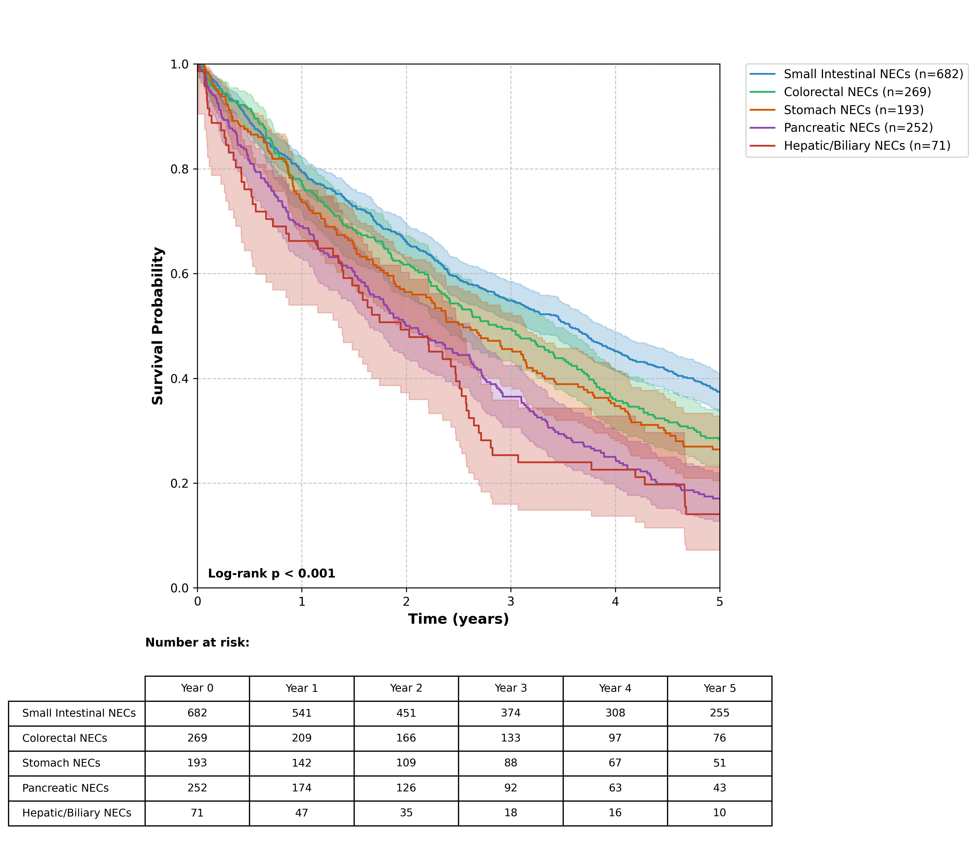
Five-year overall survival rates demonstrated marked variations across anatomical sites and subsites Table 3. Small intestinal NECs showed the most favorable prognosis with a 37.5% five-year survival rate, varying by specific location (duodenum 32.8%, jejunum 36.9%, ileum 38.4%). Colorectal NECs demonstrated the second-best survival outcomes at 31.8%, with notable variations between subsites - ranging from 26.4% in cecal NECs to 44.8% in appendiceal NECs. Gastric NECs showed intermediate survival rates (23.7%), with some variation between cardia (19.4%) and body/fundus (24.2%) locations Figure 1. Pancreatic NECs demonstrated poorer outcomes with a 15.9% five-year survival rate, showing modest variations based on anatomical location within the pancreas (head 13.8%, body 16.2%, tail 17.1%). Hepatic and biliary NECs exhibited the poorest prognosis with an 11.4% five-year survival rate, with intrahepatic lesions showing particularly poor outcomes (8.9%). These survival patterns correlate with the staging distributions observed across different anatomical sites, reflecting the impact of disease extent on patient outcomes.
| Characteristic | All Sites* | Small Intestine | Unknown Sites | Large Intestine | Pancreas | Stomach | Liver and Intra-Hepatic Ducts |
|---|---|---|---|---|---|---|---|
| Number of Patients | 4515 | 682 | 692 | 269 | 252 | 193 | 71 |
| Age (mean ± SD) | 71 ± 14 | 70 ± 12 | 73 ± 13 | 67 ± 16 | 68 ± 14 | 68 ± 15 | 70 ± 12 |
| Age range (years) | 18-90 | 29-90 | 31-90 | 18-90 | 24-90 | 20-90 | 32-90 |
| Male (%) | 52.63 | 52.66 | 51.14 | 46.45 | 53.47 | 55.19 | 49.25 |
| Female (%) | 47.37 | 47.34 | 48.86 | 53.55 | 46.53 | 44.81 | 50.75 |
| White (%) | 80.77 | 87.89 | 78.42 | 84.45 | 83.65 | 75.77 | 75.61 |
| Black or African American (%) | 19.23 | 12.11 | 21.58 | 15.55 | 16.35 | 24.23 | 24.39 |
| Hispanic or Latino (%) | 6.47 | 4.81 | 6.15 | 9.48 | 8 | 10.53 | 25.64 |
| Not Hispanic or Latino (%) | 93.53 | 95.19 | 93.85 | 90.52 | 92 | 89.47 | 74.36 |
| Category | Stage | All (n=2892)* | Small Intestine (n=493) | Large Intestine (n=210) | Stomach (n=145) | Pancreas (n=160) | Liver and Intra-Hepatic Biliary Ducts (n=142) |
|---|---|---|---|---|---|---|---|
| T Stage | TX | 1042 (23%) | 172 (25%) | 98 (36%) | 54 (28%) | 58 (23%) | 94 (14%) |
| T0 | 18 (0%) | 10 (1%) | 10 (4%) | 10 (5%) | 10 (4%) | 10 (1%) | |
| T1 | 719 (16%) | 69 (10%) | 48 (18%) | 46 (24%) | 37 (15%) | 24 (3%) | |
| T2 | 672 (15%) | 97 (14%) | 35 (13%) | 26 (13%) | 30 (12%) | 14 (2%) | |
| T3 | 565 (13%) | 146 (21%) | 50 (19%) | 16 (8%) | 29 (12%) | 12 (2%) | |
| T4 | 407 (9%) | 93 (14%) | 31 (12%) | 22 (11%) | 13 (5%) | 15 (2%) | |
| N Stage | N0 | 1397 (31%) | 164 (24%) | 110 (41%) | 79 (41%) | 83 (33%) | 46 (7%) |
| N1 | 747 (17%) | 268 (39%) | 75 (28%) | 35 (18%) | 33 (13%) | 17 (2%) | |
| N2 | 284 (6%) | 21 (3%) | 12 (4%) | 10 (5%) | 10 (4%) | 10 (1%) | |
| N3 | 125 (3%) | 10 (1%) | 0 (0%) | 10 (5%) | 0 (0%) | 10 (1%) | |
| NX | 883 (20%) | 131 (19%) | 61 (23%) | 40 (21%) | 56 (22%) | 88 (13%) | |
| M Stage | M0 | 1468 (33%) | 246 (36%) | 109 (41%) | 87 (45%) | 68 (27%) | 46 (7%) |
| M1 | 1234 (27%) | 216 (32%) | 84 (31%) | 55 (28%) | 73 (29%) | 88 (13%) |
Site-Specific Epidemiological Patterns and TNM Distributions:
Stomach:
Among gastric NECs (n=193), the cardia accounted for 24 cases, while the body/fundus comprised 27 cases. Tumor staging indicated that 28% of cases were classified as TX, reflecting incomplete tumor assessment. The proportions of T1 and T2 cases were 24% and 13%, respectively, with notable staging challenges in the cardia.
Pancreas:
Pancreatic NECs (n=252) displayed distinct anatomical subsites, with the head of the pancreas (n=213) showing the highest proportion of T3 lesions (23%). The tail of the pancreas demonstrated a more favorable distribution with a significant proportion of T2 cases (32%) compared to the head (22%).
Small Intestine:
The small intestine (n=682) demonstrated significant variation between its subsites: duodenum (n=120), jejunum (n=19), and ileum (n=207). Notably, T3 staging was more prevalent in the ileum (27%) and jejunum (53%), suggesting a pattern of advanced local invasion in these subsites. Nodal involvement was highest in ileal NECs.
Liver and Intrahepatic Biliary Ducts:
Liver and intrahepatic biliary NECs (n=71) exhibited the poorest TNM profile, with 15% of cases classified as TX and a significant proportion of patients presenting with metastatic disease (M1). This aligns with the aggressive nature of NECs in this anatomical site.
Unknown Primary Sites:
A substantial cohort (n=692) had NECs of unknown primary origin. These cases exhibited the highest mean age at diagnosis (73 ± 13 years) and significant staging ambiguity, with 14% categorized as TX. The high proportion of M1 staging (29%).
Comparative Insights Across Sites:
A comprehensive analysis revealed that the proportion of advanced-stage (T3/T4) disease was highest in the small intestine and pancreas, with lower stages more common in stomach NECs. Lymph node involvement (N1) was most frequent in small intestine NECs (39%), with markedly lower rates in liver and biliary tract cases.
Discussion
Our analysis of GI-NECs using the TriNetX database uncovers valuable insights into epidemiological trends, staging distributions, and survival outcomes across various anatomical sites. The balanced gender distribution and predominant occurrence in older adults (mean age: 71 ± 14 years) align with previous epidemiological studies. However, our cohort demonstrated a slightly higher mean age compared to the median age of 65 years reported in the Norwegian registry data from 1993 to 2010 [10]. The racial distribution in our study, with a predominance of White patients (52.71%), reflects similar patterns observed in other large-scale epidemiological studies. However, our cohort demonstrated a higher proportion of Black or African American patients (12.56%) compared to previous reports.
This variation might reflect advancements in diagnostic capabilities for small intestinal NECs, as highlighted by Koffas et al. (2023), or real geographical differences in disease distribution [11]. Our finding of a substantial proportion of cases with unknown primary sites (n=692) underscores the diagnostic challenges emphasized by Koffas et al. (2023), further reinforcing the need for enhanced diagnostic strategies, such as PET/CT imaging and circulating tumor cell analysis [11]. The TNM staging descriptive data revealed important patterns that impact clinical management. The high proportion of TX classification (23%) across all sites in our study underscores persistent challenges in primary tumor assessment, particularly in anatomically challenging locations. These findings align with Merola et al. (2020), who highlighted the complexities of achieving accurate histopathological diagnoses in GEP-NENs, particularly in non-specialist settings [12]. Similarly, the higher proportion of T3 tumors in small intestinal NECs (21%) compared to other sites suggests a potential delay in diagnosis, likely due to the anatomical location and nonspecific presenting symptoms, as noted by Lee et al. (2019), who emphasized the advanced presentation of small intestinal NECs due to diagnostic difficulties [13].
| Anatomical Site | 5-Year Overall Survival (%) |
|---|---|
| Stomach NECs (All) | 23.7 |
| Stomach Cardia | 19.4 |
| Stomach Body/Fundus | 24.2 |
| Pancreatic NECs (All) | 15.9 |
| Head of Pancreas | 13.8 |
| Body of Pancreas | 16.2 |
| Tail of Pancreas | 17.1 |
| Hepatic and Biliary NECs (All) | 11.4 |
| Intrahepatic | 8.9 |
| Colorectal NECs (All) | 31.8 |
| Cecum | 26.4 |
| Appendix | 44.8 |
| Ascending Colon | 27.9 |
| Sigmoid Colon | 32.4 |
| Rectum | 36.7 |
| Small Intestinal NECs (All) | 37.5 |
| Duodenum | 32.8 |
| Jejunum | 36.9 |
| Ileum | 38.4 |
Survival outcomes in our study demonstrated marked variations across anatomical sites, with small intestinal NECs showing the most favorable five-year survival rate (37.5%). This finding contrasts with earlier studies, such as Alese et al. (2019), which reported poor survival outcomes for high-grade GI NECs overall, emphasizing the aggressive nature of these tumors [14]. The particularly poor prognosis observed in hepatic and biliary NECs (11.4% five-year survival) is consistent with the aggressive nature of high-grade NECs described by Venizelos et al. (2021), who highlighted their molecular complexity and limited treatment options [15]. The variation in survival rates between different subsites within the same organ system (e.g., appendiceal NECs at 44.8% versus cecal NECs at 26.4%) highlights the prognostic influence of anatomical location. While our findings may relate to differences in lymphatic drainage patterns and detection timing, site-specific molecular heterogeneity, as discussed by Venizelos et al. (2021), could also play a role [15].
Our observation of higher nodal involvement in small intestinal NECs (39% N1 disease) emphasizes the aggressive nature of these tumors. While Burkart et al. (2018) primarily explored molecular targets like BRAF mutations, their findings highlight the metastatic potential of gastrointestinal NECs, which may correlate with lymphotropic behavior [16]. The presence of metastatic disease in 27% of all cases at diagnosis underscores the aggressive nature of GI-NECs and aligns with observations by Chen et al. (2021) regarding late-stage presentation [17]. The particularly poor outcomes in hepatic and biliary NECs (8.9% five-year survival for intrahepatic lesions) reflect the challenges in managing these anatomically complex tumors. Mestre-Alagarda et al. (2023) highlighted the molecular heterogeneity and poor prognosis associated with aggressive NETs and NECs, which likely contribute to the difficulties in treatment [18]. The significant variation in survival outcomes between different anatomical subsites highlights the importance of site-specific approaches to management. For instance, the relatively better outcomes in appendiceal NECs (44.8% five-year survival) than other colorectal sites suggest potential biological differences warrant further investigation. The gradual deterioration in survival rates from proximal to distal pancreatic NECs (tail 17.1% vs. head 13.8%) may reflect differences in presentation timing and surgical accessibility.
Our study has several important limitations that warrant consideration. First, the predominant representation of U.S. healthcare organizations in the TriNetX database may introduce geographic and demographic biases. While our cohort showed diversity in racial distribution (52.71% White, 12.56% Black or African American), these proportions may not accurately reflect global population demographics, potentially limiting generalizability to other geographic regions, particularly Asia and South America, where GI-NEC epidemiology may differ substantially. Second, our analysis is subject to several potential biases inherent to retrospective database studies. Coding inaccuracies and misclassification errors may exist, particularly in distinguishing between well-differentiated NETs and poorly differentiated NECs, as this distinction often requires detailed histopathological review. The high proportion of unknown primary sites (n=692) and TX classification (23%) might reflect both genuine diagnostic challenges and documentation limitations within the database. Third, our study’s temporal scope may not fully capture recent advances in diagnostic techniques and therapeutic approaches. The rapid evolution of molecular profiling and targeted therapies in NECs means that some patients in our cohort may have received different standard-of-care treatments depending on their diagnosis date. Advanced molecular techniques, such as the use of liquid biopsies to complement solid tumor analyses, as suggested by Knappskog et al. (2023), could improve the identification of targetable mutations and enhance biomarker assessment, particularly in patients with limited tumor tissue availability [19].
The applicability of our findings across different healthcare settings requires careful consideration. While our results demonstrate clear anatomical site-specific survival patterns, these outcomes may vary in healthcare systems with different diagnostic capabilities and treatment accessibility. For instance, the superior survival rates observed in small intestinal NECs (37.5%) may reflect earlier detection in well-resourced healthcare settings, and these outcomes might not be reproducible in regions with limited access to advanced imaging or surgical expertise. The demographic characteristics of our cohort, particularly the mean age of 71 years and racial distribution should be considered when applying these findings to different populations. Healthcare systems serving younger populations or different ethnic compositions may observe varying patterns of disease presentation and outcomes. Additionally, the treatment patterns and survival outcomes observed in our U.S.-predominant cohort may not directly translate to healthcare systems with different organizational structures or resource availability.
Despite these limitations, our study’s large sample size and detailed anatomical analysis provide valuable insights for clinical practice. The observed survival differences between subsites within organs (e.g., appendiceal versus cecal NECs) remain relevant across different healthcare settings, as they likely reflect underlying biological differences rather than treatment variations. Furthermore, our findings regarding the poor prognosis of hepatic and biliary NECs (11.4% survival) highlight a universal need for improved therapeutic strategies for these anatomical locations, regardless of geographic setting.
Conclusions
Our large-scale analysis of GI-NECs through the TriNetX database reveals critical patterns that significantly impact patient care and outcomes. The marked variations in survival rates across anatomical sites, ranging from 44.8% in appendiceal NECs to 8.9% in intrahepatic lesions, emphasize the necessity for site-specific treatment approaches rather than a one-size-fits-all strategy. The high proportion of advanced-stage disease at diagnosis, particularly in small intestinal NECs, with 39% showing N1 disease, underscores the urgent need for improved early detection methods. The high number of cases with unknown primary sites (n=692) and high TX classification rates (23%) highlights a critical gap in current diagnostic capabilities. This finding suggests the potential value of implementing standardized diagnostic algorithms incorporating advanced imaging techniques and molecular profiling. Furthermore, the notably poor outcomes in hepatic and biliary NECs (11.4% five-year survival) identify a specific patient subgroup requiring innovative therapeutic strategies. Our findings directly impact clinical practice, supporting the development of anatomical site-specific treatment protocols and suggesting the need for more aggressive surveillance in high-risk anatomical locations. The significant survival differences between subsites within the same organ system, such as the variance between appendiceal and cecal NECs, indicate that tumor location should be a key consideration in prognostication and treatment planning. Our epidemiological results raise an important concern in targeted therapies according to anatomical regions and biomolecular profiles, particularly in investigating the biological basis for site-specific outcome variations and developing targeted therapeutic approaches. Integrating these findings with emerging molecular and genetic data could further refine our understanding of GI-NECs and lead to more effective, personalized treatment strategies.
Conflicts of Interest
The authors declare no competing interests that could have influenced the objectivity or outcome of this research.
Funding Source
None
Acknowledgments
None
Institutional Review Board (IRB)
The IRB Board at the Jacobs School of Medicine and Biomedical Sciences, University at Buffalo, NY, USA, reviewed the study and determined it was exempt from IRB review as it does not constitute human subjects research, given the study design and type.
Large Language Model
We have employed an advanced large language model (LLM) to enhance and refine English-language writing. This process focused solely on improving the text’s clarity and style without generating or adding any new information to the content.
Authors Contribution
BB conceptualized the study. MN and AYA developed the methodology and performed the statistical analysis. MMM and EI drafted the manuscript. JW and AG reviewed the manuscript and supervised the work. All authors reviewed and approved the final version of the manuscript.
Data Availability
Available on TriNetX Database Based on Institutional Collaborations.
References
- Thiis-Evensen E., Boyar Cetinkaya R.. Incidence and prevalence of neuroendocrine neoplasms in Norway 1993-2021. J Neuroendocrinol. 2023;35(4):e13264. doi:10.1111/jne.13264 PMID: 36988112
- White B. E., Rous B., Chandrakumaran K., Wong K., Bouvier C., Van Hemelrijck M., George G., Russell B., Srirajaskanthan R., Ramage J. K.. Incidence and survival of neuroendocrine neoplasia in England 1995-2018: A retrospective, population-based study. Lancet Reg Health Eur. 2022;23:100510. doi:10.1016/j.lanepe.2022.100510 PMID: 36176500 PMCID: PMC9513765
- Alwan H., La Rosa S., Andreas Kopp P., Germann S., Maspoli-Conconi M., Sempoux C., Bulliard J. L.. Incidence trends of lung and gastroenteropancreatic neuroendocrine neoplasms in Switzerland. Cancer Med. 2020;9(24):9454-9461. doi:10.1002/cam4.3524 PMID: 33078908 PMCID: PMC7774736
- Rindi G., Klimstra D. S., Abedi-Ardekani B., Asa S. L., Bosman F. T., Brambilla E., Busam K. J., de Krijger R. R., Dietel M., El-Naggar A. K., Fernandez-Cuesta L., Kloppel G., McCluggage W. G., Moch H., Ohgaki H., Rakha E. A., Reed N. S., Rous B. A., Sasano H., Scarpa A., Scoazec J. Y., Travis W. D., Tallini G., Trouillas J., van Krieken J. H., Cree I. A.. A common classification framework for neuroendocrine neoplasms: an International Agency for Research on Cancer (IARC) and World Health Organization (WHO) expert consensus proposal. Mod Pathol. 2018;31(12):1770-1786. doi:10.1038/s41379-018-0110-y PMID: 30140036 PMCID: PMC6265262
- Gill A. J.. Why did they change that? Practical implications of the evolving classification of neuroendocrine tumours of the gastrointestinal tract. Histopathology. 2021;78(1):162-170. doi:10.1111/his.14172 PMID: 33382490
- Inzani F., Petrone G., Rindi G.. The New World Health Organization Classification for Pancreatic Neuroendocrine Neoplasia. Endocrinol Metab Clin North Am. 2018;47(3):463-470. doi:10.1016/j.ecl.2018.04.008 PMID: 30098710
- Escobar K. M., Vicente-Villardon J. L., Villacis Gonzalez R. E., Castillo Cordova P. H., Sanchez Rodriguez J. M., De la Cruz-Velez M., Siteneski A.. Neuroendocrine Tumors: An Analysis of Prevalence, Incidence, and Survival in a Hospital-Based Study in Ecuador. Healthcare (Basel). 2022;10(8). doi:10.3390/healthcare10081569 PMID: 36011226 PMCID: PMC9408119
- Helderman N. C., Suerink M., Kilinc G., van den Berg J. G., Nielsen M., Tesselaar M. E. T.. Relation between WHO Classification and Location- and Functionality-Based Classifications of Neuroendocrine Neoplasms of the Digestive Tract. Neuroendocrinology. 2024;114(2):120-133. doi:10.1159/000534035 PMID: 37690447 PMCID: PMC10836754
- Palchuk M. B., London J. W., Perez-Rey D., Drebert Z. J., Winer-Jones J. P., Thompson C. N., Esposito J., Claerhout B.. A global federated real-world data and analytics platform for research. JAMIA Open. 2023;6(2):ooad035. doi:10.1093/jamiaopen/ooad035 PMID: 37193038 PMCID: PMC10182857
- Boyar Cetinkaya R., Aagnes B., Thiis-Evensen E., Tretli S., Bergestuen D. S., Hansen S.. Trends in Incidence of Neuroendocrine Neoplasms in Norway: A Report of 16,075 Cases from 1993 through 2010. Neuroendocrinology. 2017;104(1):1-10. doi:10.1159/000442207 PMID: 26562558
- Koffas A., Giakoustidis A., Papaefthymiou A., Bangeas P., Giakoustidis D., Papadopoulos V. N., Toumpanakis C.. Diagnostic work-up and advancement in the diagnosis of gastroenteropancreatic neuroendocrine neoplasms. Front Surg. 2023;10:1064145. doi:10.3389/fsurg.2023.1064145 PMID: 36950054 PMCID: PMC10025557
- Merola E., Zandee W., de Mestier L., Klumpen H. J., Makulik K., Geboes K., van Velthuysen M. L., Couvelard A., Cros J., van Eeden S., Hoorens A., Stephenson T., Zajecki W., de Herder W., Munir A.. Histopathological Revision for Gastroenteropancreatic Neuroendocrine Neoplasms in Expert Centers: Does It Make the Difference?. Neuroendocrinology. 2021;111(1-2):170-177. doi:10.1159/000507082 PMID: 32155627
- Lee J. E., Hong S. H., Jung H. I., Son M. W., Ahn T. S., Han S. W., Cho J. H.. Small-cell neuroendocrine carcinoma of the ileum: case report and literature review. BMC Surg. 2019;19(1):135. doi:10.1186/s12893-019-0591-8 PMID: 31510991 PMCID: PMC6739994
- Alese O. B., Jiang R., Shaib W., Wu C., Akce M., Behera M., El-Rayes B. F.. High-Grade Gastrointestinal Neuroendocrine Carcinoma Management and Outcomes: A National Cancer Database Study. Oncologist. 2019;24(7):911-920. doi:10.1634/theoncologist.2018-0382 PMID: 30482824 PMCID: PMC6656466
- Venizelos A., Elvebakken H., Perren A., Nikolaienko O., Deng W., Lothe I. M. B., Couvelard A., Hjortland G. O., Sundlov A., Svensson J., Garresori H., Kersten C., Hofsli E., Detlefsen S., Krogh M., Sorbye H., Knappskog S.. The molecular characteristics of high-grade gastroenteropancreatic neuroendocrine neoplasms. Endocr Relat Cancer. 2021;29(1):1-14. doi:10.1530/ERC-21-0152 PMID: 34647903 PMCID: PMC8630776
- Burkart J., Owen D., Shah M. H., Abdel-Misih S. R. Z., Roychowdhury S., Wesolowski R., Haraldsdottir S., Reeser J. W., Samorodnitsky E., Smith A., Konda B.. Targeting BRAF Mutations in High-Grade Neuroendocrine Carcinoma of the Colon. J Natl Compr Canc Netw. 2018;16(9):1035-1040. doi:10.6004/jnccn.2018.7043 PMID: 30181415
- Chen D., Bao X., Zhang R., Ding Y., Zhang M., Li B., Zhang H., Li X., Tong Z., Liu L., Zhou X., Wang S., Cheng X., Zheng Y., Ruan J., Fang W., Zhao P.. Depiction of the genomic and genetic landscape identifies CCL5 as a protective factor in colorectal neuroendocrine carcinoma. Br J Cancer. 2021;125(7):994-1002. doi:10.1038/s41416-021-01501-y PMID: 34331023 PMCID: PMC8476633
- Mestre-Alagarda C., Srirajaskanthan R., Zen Y., Giwa M., Howard M., Ooft M. L.. Genetic and epigenetic prognosticators of neuroendocrine tumours of the GI tract, liver, biliary tract and pancreas: A systematic review and meta-analysis. Histopathology. 2024;84(2):255-265. doi:10.1111/his.15025 PMID: 37565289
- Knappskog S., Grob T., Venizelos A., Amstutz U., Hjortland G. O., Lothe I. M., Kersten C., Hofsli E., Sundlov A., Elvebakken H., Garresori H., Couvelard A., Svensson J., Sorbye H., Perren A.. Mutation Spectrum in Liquid Versus Solid Biopsies From Patients With Advanced Gastroenteropancreatic Neuroendocrine Carcinoma. JCO Precis Oncol. 2023;7:e2200336. doi:10.1200/PO.22.00336 PMID: 36753687 PMCID: PMC9928986