Benign Pheochromocytoma Coming Back with Bony Metastasis: A Case Report and Literature Review
Tarek ZahoⒾ1, Ezzeldin AbdellatefⒾ2, Shueb MohamedⒾ3, Angad Singh GillⒾ4, FNU ManasⒾ5*
- 1Research Fellowship, Wake Forest University, Winston-Salem, NC, USA
- 2Research Fellowship, Duke University, Durham, NC, USA
- 3Medical Residency, Wayne State University, Detroit, MI, USA
- 4Endocrinology, Diabetes, and Metabolism Fellowship, University at Buffalo, Plattsburgh, NY, USA
- 5Endocrinology, Diabetes, and Metabolism Fellowship, Henry Ford Health University, Detroit, MI, USA
Abstract
Pheochromocytoma is an uncommon adrenal medulla tumor that secretes catecholamines. It is usually a benign condition and is generally treated with adrenalectomy; in a small proportion of cases, however, it might return or spread. Pheochromocytoma is uncommon to recur or metastasize, meaning every reported case is clinically relevant.
A 46-year-old Caucasian woman initially presented with conventional catecholamine excess symptoms—refractory hypertension, palpitations, diaphoresis, and headaches. Imaging showed a sizable right adrenal mass; biochemical testing indicated very high plasma metanephrines. A medical decision was made to perform a right adrenalectomy; pathology confirmed a benign pheochromocytoma. After surgery, her symptoms went away, but she was missed for follow-up. Four years later, she came back with comparable symptoms; biochemical screening found increased metanephrines once more, and a computed tomography (CT) scan revealed local recurrence in the right adrenal bed. Notably, the CT also disclosed a lytic lesion in a lumbar vertebra and evidence of bone metastases from the pheochromocytoma. This instance highlights a rare situation in which a pheochromocytoma, initially thought to be innocuous, reoccurred with distant skeletal involvement.
In conclusion, a very rare, recurrent pheochromocytoma with bone metastases underscores the importance of continuous monitoring even after complete tumor removal. Early recognition of metastasis or relapse can significantly impact therapy and outcomes. This case highlights the necessity of ongoing follow-up in patients with pheochromocytoma and alerts individuals to potential late metastatic symptoms.
Keywords: Pheochromocytoma, Bone metastasis, Adrenal tumor, Catecholamine excess, Adrenalectomy
Article information
Introduction
Pheochromocytomas are rare neuroendocrine malignancies originating from chromaffin cells of the adrenal medulla. Usually benign, but 10% are approximately malignant; the incidence of malignancy is 2.5%-13% [1]. No clear histopathological standards differentiate benign from malignant neoplasms at the time of initial diagnosis. Malignant pheochromocytomas usually metastasize to the bone, kidney, lymph nodes, and lung and rarely to the brain, prostate, skin, and urinary bladder. Although bone is the commonest site for metastases, vertebral involvement is rare [2] . Pheochromocytomas exhibit symptoms of catecholamine excess, including headaches, paroxysmal hypertension, palpitations, flushing, and sweating. However, a small percentage show malignant behavior, determined by local recurrence or distal metastases; many times, these tumors are benign and surgically excised; that is, adrenalectomy is curable. Though rare, estimated in approximately 10–14% of patients, pheochromocytoma recurs years or even decades after the first therapy following curative surgery. Long-term follow-up is necessary because of the possibility of recurrence or metastasis, even in tumors initially deemed "benign. " This case report has clinical importance since it describes a patient with a pheochromocytoma first handled as a benign tumor who later showed recurrence with bone metastases. Such a presentation highlights gaps in our ability to accurately forecast tumor behavior, which is uncommon.
Case presentation
Patient Details: A 46-year-old Caucasian female with long-standing insulin-dependent diabetes mellitus and a history of poorly controlled hypertension on several antihypertensive drugs including Hydrochlorthiazide 25 mg daily (started as first line at the time of diagnosis), Losartan 100 mg daily (added on when diagnosed with diabetes), Amlodipine 10 mg daily, and Hydralazine 50 mg tid. Presented to the emergency department with extreme weariness, ongoing upper abdominal discomfort, and undesired weight loss. She also noted sporadic heart pounding, chest heaviness, profuse perspiration, face flushing, and severe headaches over the past few weeks. Her vital signs during presentation were as follows: blood pressure, 186/104 mmHg; heart rate, 110 beats per minute; respiratory rate, 18 breaths per minute; temperature, 36.8°C; and oxygen saturation, 98%. No known family history of pheochromocytoma or other endocrine tumors existed. Physical examination showed persistent hypertension and facial flushing but was otherwise normal for the initial appearance and diagnostic work-up. Differential diagnosis considered for these signs and symptoms included: hypertensive urgency, acute coronary syndrome, perimenopause, hyperthyroidism. Abdominal CT scan was ordered to investigate the cause of abdominal pain, and it revealed an adrenal mass. Subsequently, a diagnosis of pheochromocytoma was considered.
Biochemical testing confirmed a hypercatecholaminergic state by revealing remarkably high plasma-free metanephrine levels (>13,000 pg/mL; normal <200 pg/mL). TSH hormone level was ordered to exclude hyperthyroidism and was within normal range. Her set of symptoms led one to consider a catecholamine-secreting tumor.
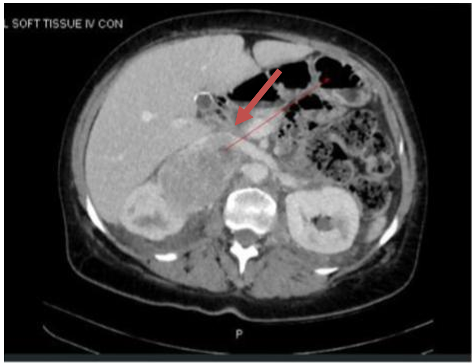
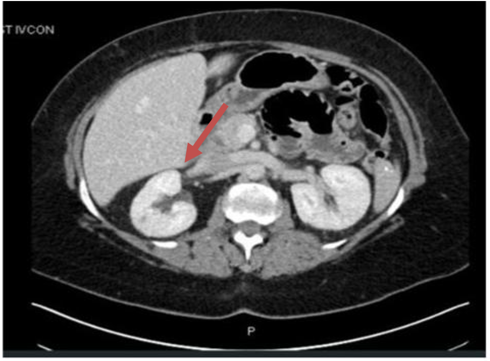
An abdominal CT scan was done for tumor localization; it showed a vast right adrenal mass measuring around 12 cm in size. On preliminary CT abdomen, the big right adrenal tumor compressing nearby structures Figure 1 shows no evident metastatic lesions. Based on the imaging, biochemical evidence, and clinical picture, a right adrenal pheochromocytoma was diagnosed. Initial therapy and results: The patient underwent an open right adrenalectomy Figure 2 following suitable preoperative Doxazosin initially started at 1 mg bid three weeks prior to surgery and titrated up to 8 mg bid, and then metoprolol 50 mg bid started 3 days before surgery. Post-surgery antihypertensive Hydralazine was stopped immediately. Histopathology confirmed a pheochromocytoma restricted to the adrenal gland with no cancerous characteristics discovered; the resected tumor was well-encapsulated. Surgical borders were well-defined, with no infiltration into adjacent tissues. At the time, no adjuvant therapy was advised. Postoperative follow-up biochemical tests revealed normalized plasma metanephrines, and the patient’s symptoms resolved completely; her blood pressure also improved, allowing a reduction in antihypertensive medication. Though, unfortunately, she was lost to follow-up following surgery and did not have scheduled monitoring, she was counseled on continuous monitoring with periodic biochemical testing and imaging.
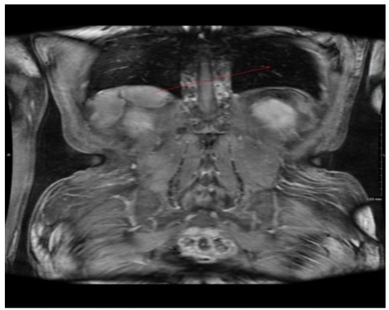
Around four years following the first adrenalectomy, the patient reappeared with a recurrence of symptoms. Reminiscent of her original presentation, she reported new episodes of palpitations, sweating, facial flushing, and elevated blood pressure levels. Clinicians swiftly requested further biochemical testing and imaging, knowing the possibility of tumor recurrence. Biochemical test results showed that ( 3,500 pg/mL) were plasma metanephrine levels. A contrast-enhanced CT scan of the stomach strongly indicated loco-regional recurrence of pheochromocytoma in the adrenal bed since it revealed several irregular soft tissue masses (approximately 2x4 cm each) in the right retroperitoneum close to the site of the previous adrenalectomy. Notably, the CT scan also found a solitary lytic lesion in the body of the L2 vertebra, thereby generating suspicion of a metastatic deposit from the pheochromocytoma Figure 3. CT scan at recurrence showed a recurrent tumor mass in the right adrenal bed and a lytic destructive lesion in the L2 vertebral body consistent with metastatic spread. Magnetic Resonance Imaging revealed no apparent liver, lungs, or other organ lesions. Later, a whole-body radionuclide investigation, metaiodobenzylguanidine scan, or PET scan was planned to confirm the adrenergic character of the spinal lesion. In this instance, the clinical setting indicated that the vertebral lesion was a metastasis of the pheochromocytoma; therefore, the tumor was classified as malignant. The patient’s situation was addressed at a multidisciplinary cancer board. Given the loco-regional recurrence and solitary bone metastasis, a combined treatment approach was intended. The patient was put under alpha- and beta-adrenergic blockage to help her stabilize her cardiovascular condition. She then had surgical excision of the recurring retroperitoneal tumor masses. Options for treating vertebral metastasis included surgical stabilization, radiation treatment, or targeted radionuclide therapy (e.g., I-131 MIBG or peptide receptor radionuclide therapy). Ultimately, localized radiation therapy was performed on the L2 vertebra to control the metastatic lesion and avoid spinal cord compression. Biochemical metanephrine levels after treatment revealed a notable decrease, and the patient’s symptoms improved. The patient was set for close long-term follow-up every six to twelve months with intermittent biochemical monitoring and imaging.
Discussion
Pheochromocytoma is a rare tumor that arises from the chromaffin cells of the adrenal gland. The annual incidence of pheochromocytoma is less than 1 per 100,000 people [3] and the prevalence is 1 in 6500 [4]. The usual peak age of diagnosis is the third to fifth decade of life, with equal incidence in men and women [5]. Most pheochromocytomas are sporadic, but about 40% of the pheochromocytomas are familial, associated with genetic mutations [4,6]. VHL (von Hippel-Lindau, vHL), NF1 (Neurofibromatosis Type 1, NF1), and RET (Multiple Endocrine Neoplasia Type 2, MEN 2) are the common mutations associated with pheochromocytoma [7]. Approximately 10% of all pheochromocytomas are malignant [3]. About 50% of the patients with metastatic disease have germline mutations [8]. The five-year survival rate is about 90% in benign pheochromocytoma and about 40% in malignant pheochromocytoma [3]. Mortality and morbidity are related to the mass effect and the high catecholamine burden leading to hypertension, arrhythmias, cardiomyopathy, stroke, and even death [7]. Male sex is associated with higher mortality [9]. Because of their broad and nonspecific clinical symptoms, pheochromocytomas are often referred to in medicine as "the great mimicker. " Although pheochromocytomas can sometimes present atypically or even be found incidentally, this patient’s initial presentation—hypertension, headaches, perspiration, palpitations—is typical. Orthostatic hypotension and syncope are seen in some patients [10]. Diagnosis begins with biochemical confirmation of catecholamine excess [11] (increased plasma or urinary metanephrines), then imaging to locate the tumor. Genetic testing should be considered in all patients [12].
Complete surgical resection (adrenalectomy) is the ultimate therapy for localized disease and usually results in a cure for benign tumors. Minimally invasive adrenalectomy is preferred in most patients, while open resection is reserved for patients with large (>6cm) or invasive tumors to prevent tumor rupture and local recurrence [12]. The tumor should be pathologically confirmed for the absence of malignancy, even though histopathological features do not reliably distinguish between benign and malignant pheochromocytomas [13]. Post-operatively, biochemical testing should be done regularly to confirm complete resection [14].
Initial care for our patient followed these principles and appeared effective, as evidenced by normalized laboratory results post-surgery and symptom resolution. Though the best first treatment is in place, a small number of patients can experience tumor recurrence. Reported recurrence rates of pheochromocytoma following adrenalectomy range between 6% and 16% in several series. One long-term study found recurrence in up to 10–14% [14] of cases. Recurrence in our patient four years after this window falls. In this instance, a lack of routine follow-up postponed the detection of recurrence until it became symptomatic. This highlights a major difficulty: patients might become non-adherent to follow-up after the first tumor is eliminated and symptoms subside, therefore missing an early diagnosis of recurrence.
Especially for those with bigger tumors, hereditary syndromes, or any other risk factors, lifelong surveillance with yearly or bi-yearly biochemical testing and frequent imaging is often advised to detect recurrence at a treatable level. The malignant potential of pheochromocytomas is another important point demonstrated by this case. There are no predictive criteria for the recurrence of pheochromocytoma. As per one study, patients with primary tumor size >5cm and average Ki-67 counts >3% are at higher risk of recurrence [15].
Recurrence is associated with multiple node involvement and distant metastasis and, therefore, is difficult to treat with surgery alone [15]. The recurrence time is variable, ranging from 1 year after surgery to decades later, but is usually observed after 2-3 years. Therefore, lifelong follow-up is recommended to detect recurrence or metastasis [16]. Annual follow-up should be preferred in patients with familial, large, or bilateral tumors, while biennial follow-up might be adequate for low-risk risk [17]. Malignancy in pheochromocytoma is characterized by metastases to areas where chromaffin tissue is typically absent (e.g., bones, lungs, liver, and distant lymph nodes). Malicious behavior is observed in approximately 10% of pheochromocytomas (or slightly higher in some recent studies). No completely trustworthy histological standards predict malignancy: characteristics like cellular atypia or further adrenal extension do not definitively indicate that a pheochromocytoma will metastasize.
In reality, the only certain indicator of cancer is the documentation of metastatic spread. Though the subsequent appearance of a bone lesion showed its cancerous nature, in our patient’s first operation, the tumor was reported as "benign," indicating no invasion or metastasis was apparent at that moment. This case emphasizes the diagnostic difficulty that even a tumor designated benign may have malignant potential that is only discovered over time. Although some research has attempted to identify predictors of aggressive behavior (such as tumor size exceeding 5 cm, a high cellular proliferation index (like Ki-67) greater than 3%, or specific genetic mutations), these findings are inconclusive.
In pheochromocytomas, genetic testing is crucial; up to 40% are associated with germline mutations (e.g., VHL, RET, NF1, SDHB/D). Malignant cases are more likely to have underlying mutations (about half of metastatic pheochromocytomas show hereditary mutations). Although no genetic testing was initially performed in our case, it would be wise to check for changes like SDHB (which is frequently associated with aggressive, metastatic pheochromocytoma), given the tumor’s behavior. Bone metastasis in pheochromocytoma is a noteworthy finding, as shown in this patient. Among metastatic pheochromocytomas, bone is indeed one of the most common locations of dissemination. Research indicates that bone involvement occurs in approximately 60–70% of malignant cases. Patients with bone metastases may show bone pain, pathological fractures, or neurological symptoms if the spine is affected [18].
The discovery of a single bone metastasis presented significant management issues. While loco-regional recurrence can usually be managed with multiple surgeries, disseminated disease may require systemic treatment. For the recurrent adrenal lesion, this patient’s treatment comprised surgery and radiation for the bony metastasis. Other or supplemental treatments for metastatic pheochromocytoma include targeted radiotherapy using radioactive iodine-labeled MIBG [19] and systemic chemotherapy (including cyclophosphamide-based regimens [20]. Every strategy must be tailored to the patient’s tumor biology and the extent of the disease. Our case highlights how even "benign" pheochromocytomas can behave aggressively and underscores the importance of a multidisciplinary approach when they do.
Comparing this case to the current literature, it fits with other studies stressing the unpredictability of Pheochromocytoma recurrence. Recurrence has been noted several years after resection; a few pheochromocytomas were initially non-metastatic, but then developed distant metastases, including skeletal lesions. The clinical result can vary: while some patients respond well to additional therapies and second surgeries, others may have a more aggressive course. Prognostically, benign pheochromocytomas have a high 5-year survival ( 90%), whereas malignant pheochromocytomas have a poorer prognosis (5-year survival often around 40–50%, heavily dependent on the extent of metastasis and effectiveness of therapy). Early detection of recurrence, as in the case of our patient, once she sought medical care, likely increases the chances of a good outcome by enabling prompt action. This case helps to advance the field of knowledge by highlighting a rare instance of repeated pheochromocytoma with bone metastasis. This case report emphasizes three important ideas: the need for ongoing follow-up, the potential for metastases to develop even after curative surgery, and the importance of physicians being alert for signs of recurrence. It also emphasizes the need for more studies on predictive indicators of malignancy and relapse in pheochromocytoma, which might finally inform early interventions and surveillance strategies for high-risk individuals.
Conclusion
Though rare, recurrent pheochromocytoma with far-off metastases is a vital clinical entity. This case illustrates the importance of lifetime monitoring, demonstrating how pheochromocytomas, initially treated as benign tumors, can recur years later with malignant characteristics. For doctors, it is important to keep a high index of suspicion for recurrence in any patient with a history of pheochromocytoma exhibiting suspicious symptoms, even many years following surgical resection. Early detection of recurrence or metastasis enables quick, aggressive treatment that can improve patient outcomes. To find late recurrences or metastases, thorough long-term follow-up is required. A multidisciplinary approach is recommended to manage these uncommon but life-threatening incidents; even "benign" pheochromocytomas are not entirely benign.
Conflicts of Interest
The authors declare that they have no competing interests.
Funding Source
No funding was received for the conduct of this study or the preparation of this manuscript.
Acknowledgments
This case was previously presented at the Endocrine Society Annual Meeting 2022 (ENDO 2022) as abstract PSUN15: “Recurrence of Benign Pheochromocytoma.” The abstract was published in the Journal of the Endocrine Society, Volume 6, Issue Supplement_1, November-December 2022, Page A122. https://doi.org/10.1210/jendso/bvac150.249
Informed Consent
Consent for publication was obtained from the patient involved in this case report.
Large Language Model
None
Authors Contribution
FM analyzed and interpreted the patient data and provided critical revisions to the manuscript. AG reviewed and provided critical revisions to the manuscript. TZ was a major contributor to writing the manuscript. EA contributed to writing the manuscript. SM contributed to writing the manuscript.
Data Availability
Patient data related to this study are not publicly available but can be obtained upon request from the corresponding author.
References
- Yurt A., Arda M. N., Vardar E.. Metastatic pheochromocytoma of the thoracic spinal extradural space. Case report and review of the literature. Kobe J Med Sci. 2005;51(3-4):49-53. PMID: 16444096
- Kasliwal M. K., Sharma M. S., Vaishya S., Sharma B. S.. Metachronous pheochromocytoma metastasis to the upper dorsal spine-6-year survival. Spine J. 2008;8(5):845-8. doi:10.1016/j.spinee.2007.06.004 PMID: 18024223
- Liu S., Song A., Zhou X., Kong X., Li W. A., Wang Y., Liu Y.. Malignant pheochromocytoma with multiple vertebral metastases causing acute incomplete paralysis during pregnancy: Literature review with one case report. Medicine (Baltimore). 2017;96(44):e8535. doi:10.1097/MD.0000000000008535 PMID: 29095319 PMCID: PMC5682838
- Chen H., Sippel R. S., O'Dorisio M. S., Vinik A. I., Lloyd R. V., Pacak K., North American Neuroendocrine Tumor Society. The North American Neuroendocrine Tumor Society consensus guideline for the diagnosis and management of neuroendocrine tumors: pheochromocytoma, paraganglioma, and medullary thyroid cancer. Pancreas. 2010;39(6):775-83. doi:10.1097/MPA.0b013e3181ebb4f0 PMID: 20664475 PMCID: PMC3419007
- Guerrero M. A., Schreinemakers J. M., Vriens M. R., Suh I., Hwang J., Shen W. T., Gosnell J., Clark O. H., Duh Q. Y.. Clinical spectrum of pheochromocytoma. J Am Coll Surg. 2009;209(6):727-32. doi:10.1016/j.jamcollsurg.2009.09.022 PMID: 19959041
- Neumann H. P. H., Young W. F., Jr., Eng C.. Pheochromocytoma and Paraganglioma. N Engl J Med. 2019;381(6):552-565. doi:10.1056/NEJMra1806651 PMID: 31390501
- Fishbein L., Merrill S., Fraker D. L., Cohen D. L., Nathanson K. L.. Inherited mutations in pheochromocytoma and paraganglioma: why all patients should be offered genetic testing. Ann Surg Oncol. 2013;20(5):1444-50. doi:10.1245/s10434-013-2942-5 PMID: 23512077 PMCID: PMC4291281
- Hamidi O., Young W. F., Jr., Iniguez-Ariza N. M., Kittah N. E., Gruber L., Bancos C., Tamhane S., Bancos I.. Malignant Pheochromocytoma and Paraganglioma: 272 Patients Over 55 Years. J Clin Endocrinol Metab. 2017;102(9):3296-3305. doi:10.1210/jc.2017-00992 PMID: 28605453 PMCID: PMC5587061
- Hamidi O., Young W. F., Jr., Gruber L., Smestad J., Yan Q., Ponce O. J., Prokop L., Murad M. H., Bancos I.. Outcomes of patients with metastatic phaeochromocytoma and paraganglioma: A systematic review and meta-analysis. Clin Endocrinol (Oxf). 2017;87(5):440-450. doi:10.1111/cen.13434 PMID: 28746746 PMCID: PMC5854189
- Ueda T., Oka N., Matsumoto A., Miyazaki H., Ohmura H., Kikuchi T., Nakayama M., Kato S., Imaizumi T.. Pheochromocytoma presenting as recurrent hypotension and syncope. Intern Med. 2005;44(3):222-7. doi:10.2169/internalmedicine.44.222 PMID: 15805711
- Bravo E. L.. Pheochromocytoma: new concepts and future trends. Kidney Int. 1991;40(3):544-56. doi:10.1038/ki.1991.244 PMID: 1787652
- Lenders J. W., Duh Q. Y., Eisenhofer G., Gimenez-Roqueplo A. P., Grebe S. K., Murad M. H., Naruse M., Pacak K., Young W. F., Jr., Endocrine Society. Pheochromocytoma and paraganglioma: an endocrine society clinical practice guideline. J Clin Endocrinol Metab. 2014;99(6):1915-42. doi:10.1210/jc.2014-1498 PMID: 24893135
- Clarke M. R., Weyant R. J., Watson C. G., Carty S. E.. Prognostic markers in pheochromocytoma. Hum Pathol. 1998;29(5):522-6. doi:10.1016/s0046-8177(98)90070-3 PMID: 9596278
- Darr R., Lenders J. W., Hofbauer L. C., Naumann B., Bornstein S. R., Eisenhofer G.. Pheochromocytoma - update on disease management. Ther Adv Endocrinol Metab. 2012;3(1):11-26. doi:10.1177/2042018812437356 PMID: 23148191 PMCID: PMC3474647
- Cui Y., Ma X., Gao Y., Chang X., Chen S., Lu L., Tong A.. Local-Regional Recurrence of Pheochromocytoma/Paraganglioma: Characteristics, Risk Factors and Outcomes. Front Endocrinol (Lausanne). 2021;12:762548. doi:10.3389/fendo.2021.762548 PMID: 34899602 PMCID: PMC8660112
- Amar L., Servais A., Gimenez-Roqueplo A. P., Zinzindohoue F., Chatellier G., Plouin P. F.. Year of diagnosis, features at presentation, and risk of recurrence in patients with pheochromocytoma or secreting paraganglioma. J Clin Endocrinol Metab. 2005;90(4):2110-6. doi:10.1210/jc.2004-1398 PMID: 15644401
- Plouin P. F., Chatellier G., Fofol I., Corvol P.. Tumor recurrence and hypertension persistence after successful pheochromocytoma operation. Hypertension. 1997;29(5):1133-9. doi:10.1161/01.hyp.29.5.1133 PMID: 9149678
- Ayala-Ramirez M., Palmer J. L., Hofmann M. C., de la Cruz M., Moon B. S., Waguespack S. G., Habra M. A., Jimenez C.. Bone metastases and skeletal-related events in patients with malignant pheochromocytoma and sympathetic paraganglioma. J Clin Endocrinol Metab. 2013;98(4):1492-7. doi:10.1210/jc.2012-4231 PMID: 23436918 PMCID: PMC5393459
- Dillon J. S., Bushnell D., Laux D. E.. High-specific-activity (131)iodine-metaiodobenzylguanidine for therapy of unresectable pheochromocytoma and paraganglioma. Future Oncol. 2021;17(10):1131-1141. doi:10.2217/fon-2020-0625 PMID: 33506713 PMCID: PMC7927907
- Tong A., Li M., Cui Y., Ma X., Wang H., Li Y.. Temozolomide Is a Potential Therapeutic Tool for Patients With Metastatic Pheochromocytoma/Paraganglioma-Case Report and Review of the Literature. Front Endocrinol (Lausanne). 2020;11:61. doi:10.3389/fendo.2020.00061 PMID: 32132978 PMCID: PMC7040234