Selective Attenuation of Acetaminophen-induced Hepatobiliary Dysfunction by Pseudoelephantopus spicatus in Sprague-Dawley Rats
Uzzal ChondraⒾ1*, Palash C. RoyⒾ2, Khaled H. RiadⒾ2, Masud A. SumonⒾ2, Md. Zahirul IslamⒾ2, S.M. Nazmul IslamⒾ2, Md. Jahid Hasan2, Pabittra K. ShillⒾ2, Mithun SikderⒾ3, Shawon S. JoyⒾ4, Annesha MajumderⒾ2
- 1Chikitsa Foundation Bangladesh (CF-BD), Dhaka, Bangladesh
- 2Department of Pharmacy, Gono Bishwabidyalay, Dhaka, Bangladesh
- 3Bangladesh Rural Advancement Committee (BRAC), Dhaka, Bangladesh
- 4Department of Physiotherapy, Gono Bishwabidyalay, Dhaka, Bangladesh
Abstract
Background: Pseudoelephantopus spicatus (PS) is used in ethnomedicine for hepatic disorders; however, its effects on biochemical markers of drug-induced liver injury remain incompletely characterized. This study evaluated the effects of the ethanolic root extract of PS on serum biochemical indices in an acetaminophen-induced acute hepatotoxicity model.
Methods: Sprague-Dawley rats (150-200 g) were allocated into five groups (n = 6/group). PS extract (500 and 1000 mg/kg) and silymarin (100 mg/kg) were administered orally for 10 days. On day 10, hepatotoxicity was induced by a single oral dose of acetaminophen (700 mg/kg) after a 12 h fast. Serum ALT, AST, ALP, total bilirubin, total protein, albumin, and globulin were quantified 24 h post-administration.
Results: Acetaminophen produced marked elevations in ALT, AST, ALP, and total bilirubin, with reduced serum protein fractions. PS treatment did not significantly reduce ALT or AST levels. However, PS significantly decreased ALP activity at both 500 and 1000 mg/kg (p < 0.001) and reduced total bilirubin dose-dependently (p < 0.01-p < 0.001 vs. acetaminophen control). At 1000 mg/kg, PS significantly restored total protein (p < 0.001) and increased albumin and globulin levels (p < 0.05), indicating partial recovery of hepatic synthetic function.
Conclusion: PS ethanolic root extract produced selective biochemical attenuation of acetaminophen-induced hepatobiliary dysfunction, significantly modulating ALP, total bilirubin, and circulating proteins without significant transaminase normalization. These findings indicate partial functional improvement rather than comprehensive hepatocellular protection. Further studies incorporating histopathology, oxidative stress, and mechanistic analyses are required to establish structural and molecular correlates.
Keywords: Acetaminophen, Hepatobiliary biomarkers, Liver enzymes, Phytotherapy, Pseudoelephantopus spicatus, Rat model
Article information
Introduction
The liver is the primary site for the metabolism and elimination of xenobiotics and endogenous compounds, rendering it particularly vulnerable to chemically induced injury [1]. Drug-induced liver injury represents a major cause of acute hepatic failure, with acetaminophen overdose being one of the most extensively studied and clinically relevant models of hepatotoxicity [2]. Under therapeutic conditions, acetaminophen is predominantly metabolized through glucuronidation and sulfation pathways; however, excessive dosing leads to cytochrome P450-mediated bioactivation to the highly reactive metabolite N-acetyl-p-benzoquinone imine (NAPQI) [3]. Accumulation of NAPQI results in depletion of intracellular glutathione reserves, covalent binding to hepatic proteins, mitochondrial dysfunction, enhanced generation of reactive oxygen species, and subsequent hepatocellular necrosis [4]. Acetaminophen-induced liver damage is commonly associated with altered ALT, AST, ALP, bilirubin, and protein levels, which are used as biochemical indicators of liver function [5].
Currently available hepatoprotective therapies are limited in efficacy, necessitating the exploration of alternative agents capable of modulating oxidative, inflammatory, and metabolic pathways involved in liver damage [6]. While oxidative stress and inflammation are recognized contributors to acetaminophen-induced hepatotoxicity, this study focused specifically on routine serum biochemical markers-ALT, AST, ALP, bilirubin, and circulating protein fractions-as primary endpoints. These biomarkers provide reliable, accessible measures of hepatocellular and hepatobiliary function, reflecting functional impairment and recovery without requiring direct assessment of oxidative or inflammatory pathways [7,8]. In this context, medicinal plants constitute a valuable source of structurally diverse bioactive compounds with potential hepatoprotective properties [9].
P. spicatus (Juss.) Rohr. (Asteraceae) is a perennial herb traditionally used to manage inflammatory disorders, infectious diseases, and hepatic dysfunction [10]. Phytochemical investigations have revealed the presence of sesquiterpene lactones, triterpenoids, sterols, and related secondary metabolites, several of which exhibit antioxidant, anti-inflammatory, and cytoprotective activities [11]. Experimental studies have reported moderate hepatoprotective effects of P. spicatus extracts against chemically induced liver injury, including toxin and drug-mediated models [12]; however, systematic evaluation of organ-specific extracts, particularly roots, in acetaminophen-induced hepatotoxicity remains limited.
Accordingly, the present study was designed to evaluate the effects of the ethanolic root extract of P. spicatus on serum biochemical markers of hepatocellular and hepatobiliary injury in a rat model of acetaminophen-induced acute hepatotoxicity. By focusing on these routine endpoints, the study aims to establish the functional hepatoprotective potential of P. spicatus before mechanistic or histopathological investigations.
Methods
Plant Collection and Extraction
The plant of P. spicatus was collected from Dhamrai, Dhaka, Bangladesh, and identified by the Bangladesh National Herbarium (BNH) and accession number (DACB87366). Thoroughly washed, shade-dried at 35-40 °C for 7 days, pulverized to a fine powder, and subjected to Soxhlet extraction using 96% ethanol (900 g powder; 30 h extraction). The combined ethanolic extract was concentrated under reduced pressure using a rotary evaporator (BUCHI Rota Vapor R-114, Switzerland), further dried on a water bath at room temperature to yield a solid extract (100 g), which was stored at 4 °C until use. Although the plant material was taxonomically authenticated and a voucher specimen deposited, the ethanolic extract was not standardized using a quantified chemical marker or chromatographic fingerprint, and variation in phytochemical composition may therefore influence reproducibility.
Chemicals and Reagents
Acetaminophen (APAP) was procured from Gonoshasthaya Pharmaceuticals Ltd. aspartate aminotransferase (AST), alanine aminotransferase (ALT), alkaline phosphatase (ALP), and total bilirubin (TB) were obtained from HUMAN GmbH (Germany), while total protein and albumin were quantified using colorimetric diagnostic kits supplied by Erba Mannheim (Transasia Bio-Medicals Ltd., India); all other reagents used in the study were of analytical grade.
Experimental Animals
Sprague-Dawley rats of either sex (4-8 weeks old) were housed three per polypropylene cage with standard bedding and environmental enrichment materials under standard hygienic conditions at the animal house of the Department of Pharmacy, Jahangirnagar University. Environmental conditions were maintained at 27 ± 2 °C with a 12 h light/12 h dark cycle, and animals had free access to a standard pellet diet and water. All animals allocated to the experimental groups (n = 6 per group) were included in the final biochemical evaluation, and no animals were excluded from analysis. All procedures were conducted in accordance with institutional and international ethical guidelines. Animals of both sexes were included; however, the allocation of males and females within individual experimental groups was not recorded during the study. Consequently, sex-balanced group assignment and sex-specific analyses could not be performed.
Acute Oral Toxicity Study
A preliminary acute oral toxicity study was conducted in Swiss albino female mice (25-30 g, n = 4 animals per dose step) as an exploratory dose-tolerability assessment using a stepwise design broadly based on the acute toxic class approach described in OECD Test Guideline 423, in which animals received a single oral dose of ethanolic P. spicatus root extract and were continuously monitored for 4 h, periodically over 24 h, and daily up to 14 days for clinical signs, including locomotor activity, grooming, respiration, food and water intake, tremors, and mortality, with body weights recorded on days 0, 7, and 14. No mortality or treatment-related clinical signs were observed at doses up to 1000 mg/kg. Based on these observations, 500 and 1000 mg/kg were selected for subsequent biochemical evaluation in Sprague-Dawley rats [13,10].
Experimental Design and Assessment of Liver Function
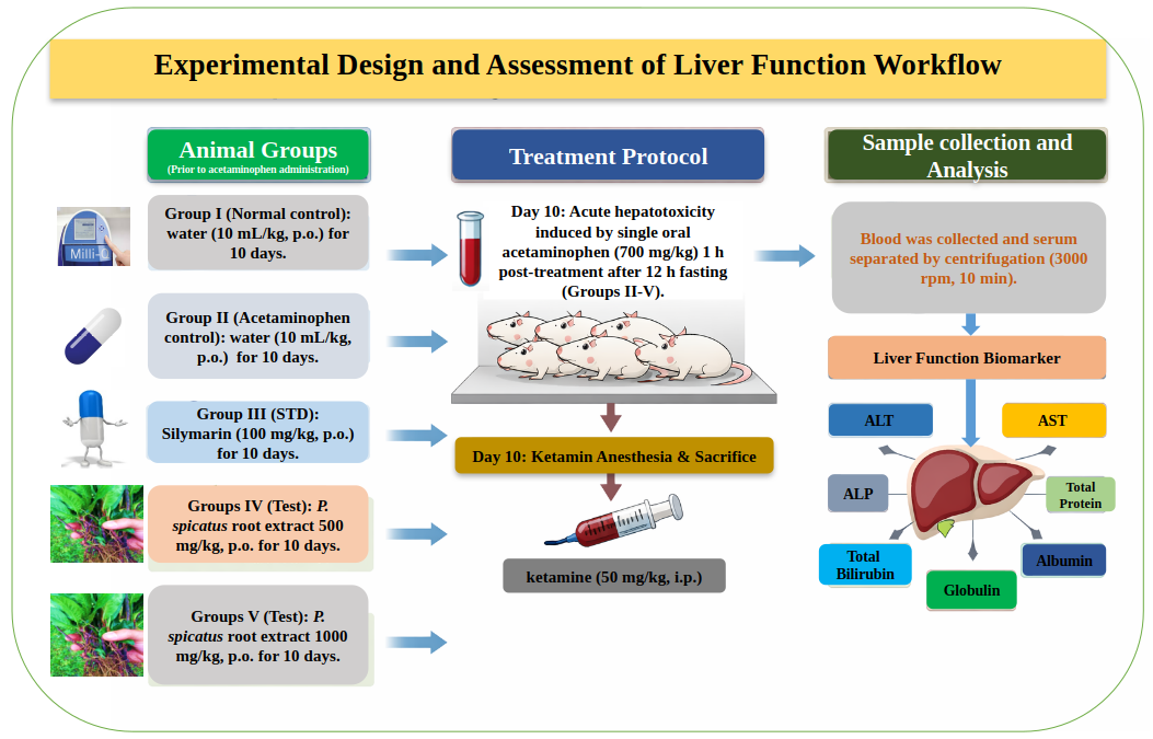
The in vivo study was conducted at the Pharmacology Laboratory, Department of Pharmacy, Jahangirnagar University (Savar, Dhaka), using adult Sprague-Dawley rats of either sex (150-200 g) housed in polypropylene cages under controlled environmental conditions (27 ± 2 °C, standard pellet diet, ad libitum access to water) following a minimum 2-week acclimatization period. All protocols for the animal experiment were approved by the Research Ethical Committee under the Center for Multidisciplinary Research of Chikitsa Foundation Bangladesh (Approval No. CF-BD/EC/023). Animals were monitored daily for clinical signs of toxicity or distress, including changes in behavior, locomotion, grooming, and food and water intake, and humane endpoints were applied in accordance with approved ethical guidelines. Before acetaminophen administration, animals received their respective treatments once daily for 10 consecutive days. Rats were randomly assigned to five groups (n = 6 per group) after body weight measurement to ensure comparable mean body weights across groups. Group allocation was performed using a simple randomization method based on a random number sequence.
Group I received distilled water (10 mL/kg, p.o.) for 10 days and served as the normal control.
Group II received distilled water (10 mL/kg, p.o.) for 10 days, followed by a single oral dose of acetaminophen (700 mg/kg).
Group III received silymarin (100 mg/kg, p.o.) for 10 days, followed by a single oral dose of acetaminophen (700 mg/kg).
Groups IV and V received P. spicatus root extract (500 and 1000 mg/kg, p.o., respectively) for 10 days, followed by a single oral dose of acetaminophen (700 mg/kg).
Acute hepatotoxicity was induced by a single oral administration of acetaminophen (700 mg/kg) on day 10, administered 1 h after the final dose of the respective treatments, following 12 h of overnight fasting [14,15]. Food was withheld for 12 h before acetaminophen administration, while water was provided ad libitum. The animals were observed for 24 h after dosing. The dried extract yield was 11.1% (w/w) relative to the starting plant material. The extract was suspended in distilled water and administered orally at a dosing volume of 10 mL/kg body weight. The formulation was freshly prepared, stored at 25 °C protected from light, and used within 2 h to ensure stability. Following treatment, animals were anesthetized with ketamine (50 mg/kg, intraperitoneal) [16,17] to achieve deep anesthesia before blood collection. Animals were sacrificed under anesthesia in accordance with institutional ethical approval. Blood samples were collected and centrifuged (3000 rpm, 10 min) to obtain serum, and liver function biomarkers, including ALT, AST, ALP, and total bilirubin [18], total protein, and albumin were determined using standard enzymatic colorimetric assays Figure 1 [19]. Serum biochemical assays and statistical analyses were conducted using coded samples so that investigators performing the analyses were unaware of the treatment group allocation.
Statistical Analysis
Data were analyzed using one-way ANOVA followed by Bonferroni’s post hoc test in SPSS v16.0. Results are presented as mean ± SEM, with statistical significance was defined as *p < 0.05, **p < 0.01, and ***p < 0.001, where the normal control group (Group I) was compared with the acetaminophen control (Group II) to confirm hepatotoxicity, and each treatment group-PS 500 mg/kg (Group IV), PS 1000 mg/kg (Group V), and silymarin 100 mg/kg (Group III)-was statistically compared exclusively against the acetaminophen control (Group II) without intergroup comparisons or reference to the normal control.
Results
Acetaminophen-induced hepatic injury and effects on serum transaminases and ALP by P. spicatus
Acetaminophen administration resulted in elevated serum ALT, AST, and ALP levels compared with the normal control group, consistent with the expected biochemical pattern of hepatic injury in this experimental model. Relative to the acetaminophen control group, treatment with P. spicatus (PS) at 500 mg/kg did not produce statistically significant reductions in ALT or AST levels; however, ALP activity was significantly decreased (p < 0.001). Similarly, PS at 1000 mg/kg did not significantly alter ALT or AST levels compared with the acetaminophen control group, while ALP levels were significantly reduced (p < 0.001), suggesting selective modulation of hepatobiliary enzyme activity. In contrast, silymarin (100 mg/kg) significantly decreased ALT, AST, and ALP levels (p < 0.001) relative to the acetaminophen control group. Collectively, PS treatment was associated with a significant reduction in ALP but not in transaminase levels, whereas silymarin significantly reduced all measured hepatic enzyme parameters Table 1 and Figure 2.
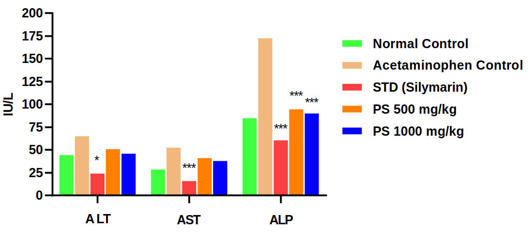
| Group | ALT (IU/L) | AST (IU/L) | ALP (IU/L) | Total Bilirubin (mg/dl) |
|---|---|---|---|---|
| Normal Control | 44.0 ± 2.25 | 28.33 ± 3.72 | 84.50 ± 9.62 | 0.78 ± 0.08 |
| Acetaminophen Control | 64.67 ± 10.80 | 52.17 ± 5.78 | 172.33 ± 13.36 | 1.92 ± 0.15 |
| STD (Silymarin 100 mg/kg) | 23.83 ± 8.85* | 15.50 ± 1.67*** | 60.33 ± 4.09*** | 0.55 ± 0.12*** |
| PS 500 mg/kg | 50.50 ± 7.31 | 40.67 ± 2.09 | 94.33 ± 9.83*** | 1.07 ± 0.18** |
| PS 1000 mg/kg | 45.50 ± 12.71 | 37.50 ± 4.91 | 89.83 ± 3.69*** | 0.65 ± 0.06*** |
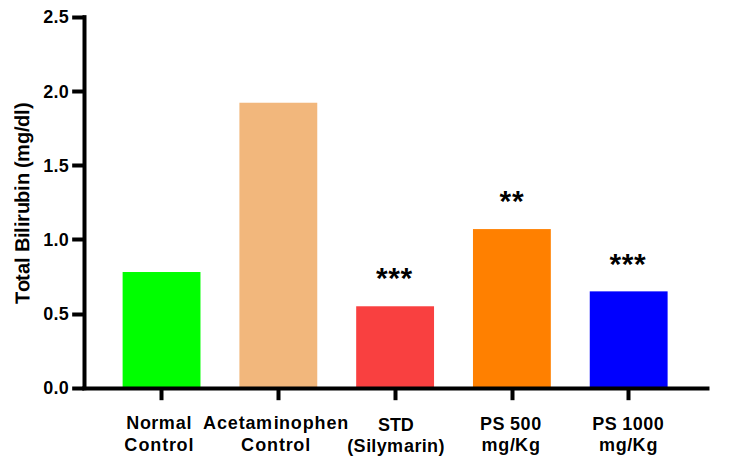
Data are expressed as mean ± SEM (n = 6) and were analyzed by one-way ANOVA followed by Bonferroni’s post hoc test; p* < 0.05, p** < 0.01, and p*** < 0.001 indicate statistically significant, highly significant, and very highly significant differences, respectively, where the normal control group (Group I) was compared with the acetaminophen control (Group II) to confirm hepatotoxicity, and each treatment group—PS 500 mg/kg (Group IV), PS 1000 mg/kg (Group V), and silymarin 100 mg/kg (Group III)—was statistically compared exclusively against the acetaminophen control (Group II) without intergroup comparisons or reference to the normal control.
Acetaminophen-induced alterations in serum total bilirubin and effects of P. spicatus
Acetaminophen administration resulted in elevated serum total bilirubin levels compared with the normal control group. Relative to the acetaminophen control group, treatment with P. spicatus (PS) at 500 mg/kg significantly reduced serum total bilirubin levels (p < 0.01). Administration of PS at 1000 mg/kg produced a further significant reduction in bilirubin levels (p < 0.001) compared with the acetaminophen control group. Similarly, silymarin (100 mg/kg) significantly decreased serum total bilirubin levels (p < 0.001) relative to the acetaminophen control group. Collectively, PS treatment was associated with a dose-dependent reduction in serum bilirubin levels compared with acetaminophen-induced elevation Table 1 and Figure 3.
| Group | Total Protein (g/dl) | Albumin (g/dl) | Globulin (g/dl) |
|---|---|---|---|
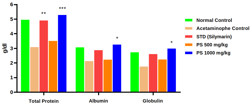
| Normal Control | 4.95 ± 0.36 | 3.06 ± 0.11 | 2.72 ± 0.35 |
| Acetaminophen Control | 3.08 ± 0.33 | 2.12 ± 0.27 | 1.75 ± 0.16 |
| STD (Silymarin 100 mg/kg) | 4.90 ± 0.20** | 2.87 ± 0.31 | 2.60 ± 0.36 |
| PS 500 mg/kg | 3.50 ± 0.20 | 2.22 ± 0.12 | 2.23 ± 0.20 |
| PS 1000 mg/kg | 5.28 ± 0.16*** | 3.25 ± 0.23* | 2.98 ± 0.14* |
Data are expressed as mean ± SEM (n = 6) and were analyzed by one-way ANOVA followed by Bonferroni’s post hoc test; p* < 0.05, p** < 0.01, and p*** < 0.001 indicate statistically significant, highly significant, and very highly significant differences, respectively, where the normal control group (Group I) was compared with the acetaminophen control (Group II) to confirm hepatotoxicity, and each treatment group—PS 500 mg/kg (Group IV), PS 1000 mg/kg (Group V), and silymarin 100 mg/kg (Group III)—was statistically compared exclusively against the acetaminophen control (Group II) without intergroup comparisons or reference to the normal control.
Acetaminophen-induced alterations in serum total protein, albumin, and globulin, and effects of P. spicatus
Acetaminophen administration resulted in marked reductions in serum total protein, albumin, and globulin levels compared with those in the normal control group. Treatment with P. spicatus (PS) at 500 mg/kg did not produce statistically significant changes in any of these parameters relative to the acetaminophen control group. In contrast, PS at 1000 mg/kg significantly increased serum total protein levels (p < 0.001) and elevated albumin and globulin concentrations (p < 0.05) compared with the acetaminophen control group. Silymarin (100 mg/kg) significantly increased serum total protein levels (p < 0.01) relative to the acetaminophen control group, whereas albumin and globulin showed no significant changes Table 2 and Figure 4. Mean ± SEM values for serum ALT, AST, ALP, total bilirubin, total protein, albumin, and globulin across all experimental groups, together with corresponding statistical significance levels, are provided in (Table 1, Table 2) to ensure full transparency of inter-animal variability and the dispersion of biochemical responses.
Discussion
Acetaminophen (APAP) is widely used as an analgesic and antipyretic, yet overdose results in severe hepatotoxicity due to metabolic activation and oxidative stress [20]. Under therapeutic conditions, acetaminophen is primarily detoxified via glucuronidation and sulfation pathways; however, a minor fraction is bioactivated by cytochrome P450 enzymes to the reactive intermediate N-acetyl-p-benzoquinone imine (NAPQI), which is normally neutralized through conjugation with reduced glutathione (GSH) [21]. Following overdose, saturation of conjugation pathways leads to excessive NAPQI formation, hepatic GSH depletion, and covalent binding of NAPQI to cellular macromolecules, triggering lipid peroxidation, calcium dysregulation, mitochondrial dysfunction, and hepatocellular necrosis [22,23]. Hepatocellular damage disrupts membrane integrity, resulting in leakage of cytosolic enzymes into the circulation, making serum transaminases and alkaline phosphatase reliable biomarkers of liver injury [24,25]. These biomarkers reflect both hepatocellular membrane integrity and hepatobiliary functional status, and differential modulation of these parameters may indicate selective biochemical effects rather than uniform cytoprotection.
In the present study, acetaminophen administration was associated with elevated serum ALT, AST, ALP, and bilirubin levels, confirming hepatocellular and hepatobiliary dysfunction. Treatment with P. spicatus (PS) root extract attenuated these alterations, with significant reductions observed in ALP at both doses and in bilirubin in a dose-dependent manner. However, ALT and AST levels were not significantly reduced at either dose, indicating that the extract did not demonstrate statistically significant protection against hepatocellular enzyme leakage under the present experimental conditions. The higher dose produced marked reductions in ALP and bilirubin levels, suggesting a possible modulatory effect on hepatobiliary parameters rather than comprehensive hepatocellular protection [26]. Because histopathological assessment and additional cholestatic markers (e.g., GGT or bile acids) were not evaluated, any inference regarding selective canalicular or biliary membrane protection should be interpreted cautiously and considered hypothesis-generating rather than definitive. Accordingly, the observed reductions in ALP and bilirubin, in the absence of significant normalization of ALT and AST, suggest selective modulation of hepatobiliary biochemical parameters rather than comprehensive hepatocellular protection under the present experimental conditions [26]. Similar patterns of partial biochemical modulation, characterized by improvement in cholestatic markers with limited transaminase normalization, have been reported in certain plant-derived hepatoprotective studies, suggesting that phytochemical extracts may differentially influence hepatobiliary and hepatocellular components depending on dose, composition, and injury severity [27,28].
In addition to enzymatic changes, PS treatment at 1000 mg/kg significantly restored serum total protein, albumin, and globulin levels, indicating improved circulating protein levels. Notably, serum protein fractions in the 1000 mg/kg group exceeded values observed in the normal control group. Such supranormal elevations may reflect biological variability in circulating proteins after acute injury and recovery, adaptive compensatory responses of the liver, acute phase reactant changes, or transient shifts in hydration and plasma volume rather than definitive supraphysiological protein synthesis. Without histopathological correlation and comprehensive biomarker profiling, these elevated protein levels should be interpreted cautiously as exploratory observations rather than conclusive evidence of enhanced synthetic capacity [29,30]. Since the liver is the primary site of plasma protein synthesis [31], reductions in these parameters are indicative of impaired hepatocellular function in acute hepatic injury [32]. Restoration of protein levels following PS administration suggests enhancement of hepatocyte functional integrity and biosynthetic activity [33].
Nevertheless, because transaminase levels were not significantly normalized and structural assessment was not performed, this improvement should be interpreted as functional modulation rather than confirmed cytoprotective regeneration. Thus, the improvements in circulating protein fractions likely reflect partial restoration of hepatic synthetic function rather than confirmed structural regeneration of hepatocytes.
Overall, the findings demonstrate that P. spicatus root extract ameliorated selected biochemical alterations induced by acetaminophen, particularly ALP and bilirubin elevation, and improved hepatic synthetic parameters at the higher dose. These effects indicate selective biochemical improvement and partial functional restoration, but do not establish comprehensive hepatocellular protection. In addition to statistical significance, the magnitude of change in ALP and bilirubin relative to the acetaminophen control group indicates a biologically meaningful attenuation of hepatobiliary dysfunction. However, effect size estimation would further strengthen interpretation in future studies [34,35]. Although oxidative stress is central to acetaminophen-induced hepatotoxicity, oxidative stress markers, antioxidant enzyme activity, and histopathological validation were not assessed in this study; therefore, attribution of antioxidant or membrane-stabilizing mechanisms would be premature [36]. Therefore, any mechanistic interpretation regarding antioxidant, anti-inflammatory, or membrane-stabilizing activity should be considered speculative and requires direct biochemical and structural validation [37]. Future studies should incorporate oxidative stress profiling, molecular pathway analyses, and liver histology to clarify the mechanistic basis of the observed effects. Additionally, the relatively high doses employed (500-1000 mg/kg crude extract) necessitate careful consideration of dose optimization, extract standardization, pharmacokinetic characterization, and safety evaluation before translational extrapolation can be justified [38]. Inclusion of extract-only control groups in future investigations would further help delineate intrinsic hepatic effects independent of acetaminophen-induced injury. Importantly, because an extract-only control group was not included in the present study, the extract’s independent biochemical effects in the absence of an acetaminophen challenge cannot be excluded. Another limitation of the present study is that the crude ethanolic extract was not chemically standardized using chromatographic fingerprinting or quantified marker compounds; therefore, potential variability in phytochemical composition may influence reproducibility. In addition, because the sex distribution within individual groups was not recorded, it was not possible to assess whether males and females responded differently to treatment. This remains a potential source of biological variability that could not be addressed in the present study. This represents a design limitation that should be addressed in future investigations.
Conclusion
In this acetaminophen-induced acute liver injury model, the ethanolic root extract of P. spicatus produced selective biochemical attenuation of hepatobiliary dysfunction, demonstrated by significant reductions in ALP and total bilirubin, as well as restoration of circulating protein fractions at 1000 mg/kg. The absence of significant normalization of ALT and AST indicates that comprehensive hepatocellular protection was not achieved under the present experimental conditions. These findings indicate partial biochemical improvement in hepatobiliary functional markers. Definitive validation will require histological examination, assessment of oxidative stress and inflammatory markers, inclusion of extract-only controls, and standardized pharmacological characterization before translational extrapolation can be made.
Conflicts of Interest
The authors declare no competing interests that could have influenced the objectivity or outcome of this research.
Funding Source
The authors declare that no specific grant or funding was received for this research from any public, commercial, or not-for-profit funding agency.
Acknowledgments
We gratefully acknowledge Professor Masum Shahriar (Department of Pharmacy, Jahangirnagar University, Savar, Dhaka, Bangladesh) and Professor Dr. Md. Iqbal Hossain, Dean, Faculty of Health Sciences, Gono Bishwabidyalay, Savar, Dhaka, Bangladesh, for their academic guidance and institutional support of this work.
Institutional Review Board (IRB)
All animal experimental procedures were approved by the Research Ethical Committee under the Center for Multidisciplinary Research, Chikitsa Foundation Bangladesh (Approval No. CF-BD/EC/023), and conducted in accordance with institutional and international guidelines for laboratory animal care.
Large Language Model
None.
Authors Contribution
KHR, MAS, MZI, SMNI, and MJH conducted the experiments and acquired the data. PKS contributed to the study design and experimental strategy. MS, SSJ, PCR, and AM contributed to manuscript editing. UC conceived the study, oversaw the overall workflow, and finalized the manuscript.
Data Availability
All analyzed data are presented in the manuscript as group-level summary statistics (mean ± SEM) for each experimental endpoint.
References
- Wang L., Shao Z., Wang X., Lu W., Sun H.. Xenobiotic-induced liver injury: Molecular mechanisms and disease progression. Ecotoxicol Environ Saf. 2025;303:118854. doi:10.1016/j.ecoenv.2025.118854 PMID: 40845633
- Jaeschke H., Xie Y., McGill M. R.. Acetaminophen-induced Liver Injury: from Animal Models to Humans. J Clin Transl Hepatol. 2014;2(3):153-61. doi:10.14218/JCTH.2014.00014 PMID: 26355817 PMCID: PMC4521247
- Mazaleuskaya L. L., Sangkuhl K., Thorn C. F., FitzGerald G. A., Altman R. B., Klein T. E.. PharmGKB summary: pathways of acetaminophen metabolism at the therapeutic versus toxic doses. Pharmacogenet Genomics. 2015;25(8):416-26. doi:10.1097/FPC.0000000000000150 PMID: 26049587 PMCID: PMC4498995
- Ramachandran A., Jaeschke H.. Acetaminophen hepatotoxicity: A mitochondrial perspective. Adv Pharmacol. 2019;85:195-219. doi:10.1016/bs.apha.2019.01.007 PMID: 31307587 PMCID: PMC7350903
- Maharaja P, Sengottuvel T, Aarthi A, Gopalasatheeskumar K. Review on Antioxidant and Hepatoprotective activity of Medicinal plants against Paracetamol Induced animal model. Res. J. Pharmacognosy and Phytochem. 2020;12(2):114-119. doi:10.5958/0975-4385.2020.00020.55
- Wang C., Feng X., Li W., Chen L., Wang X., Lan Y., Tang R., Jiang T., Zheng L., Liu G.. Apigenin as an emerging hepatoprotective agent: current status and future perspectives. Front Pharmacol. 2024;15:1508060. doi:10.3389/fphar.2024.1508060 PMID: 39749193 PMCID: PMC11693974
- Kaplowitz N.. Drug-induced liver injury. Clin Infect Dis. 2004;38 Suppl 2:S44-8. doi:10.1086/381446 PMID: 14986274
- Maddrey Willis C.. Hepatotoxicity: The adverse effects of drugs and other chemicals on the liver. Gastroenterology. 2000;118(5):984-985. doi:10.1016/s0016-5085(00)70192-2
- El-Saadony M. T., Saad A. M., Mohammed D. M., Korma S. A., Alshahrani M. Y., Ahmed A. E., Ibrahim E. H., Salem H. M., Alkafaas S. S., Saif A. M., Elkafas S. S., Fahmy M. A., Abd El-Mageed T. A., Abady M. M., Assal H. Y., El-Tarabily M. K., Mathew B. T., AbuQamar S. F., El-Tarabily K. A., Ibrahim S. A.. Medicinal plants: bioactive compounds, biological activities, combating multidrug-resistant microorganisms, and human health benefits - a comprehensive review. Front Immunol. 2025;16:1491777. doi:10.3389/fimmu.2025.1491777 PMID: 40375989 PMCID: PMC12079674
- Ali Md. Hasan, Islam Shariful, Hasan Mohammad Mahmudul, Rahman Md. Hasanur, Rahman Asheka, Taher Mohammad Abdullah, Muslim Tanvir. Studies of phytochemical analysis, in vitro and in vivo evaluation of the local Pseudoelephantopus spicatus plant. African Journal of Pharmacy and Pharmacology. 2024;18(7):114-126. doi:10.5897/ajpp2023.5354
- Jimenez Usuga Nora del Socorro, Malafronte Nicola, Durango Edison Javier Osorio, Braca Alessandra, De Tommasi Nunziatina. Phytochemical investigation of Pseudelephantopus spiralis (Less.) Cronquist. Phytochemistry Letters. 2016;15:256-259. doi:10.1016/j.phytol.2016.02.013
- Lin C. C., Yen M. H., Chiu H. F.. The pharmacological and pathological studies on Taiwan folk medicine (VI): The effects of Elephantopus scaber subsp. oblanceolata, E. mollis and Pseudoelephantopus spicatus. Am J Chin Med. 1991;19(1):41-50. doi:10.1142/S0192415X91000065 PMID: 1897489
- Jonsson M., Jestoi M., Nathanail A. V., Kokkonen U. M., Anttila M., Koivisto P., Karhunen P., Peltonen K.. Application of OECD Guideline 423 in assessing the acute oral toxicity of moniliformin. Food Chem Toxicol. 2013;53:27-32. doi:10.1016/j.fct.2012.11.023 PMID: 23201451
- Mitchell J. R., Jollow D. J., Potter W. Z., Davis D. C., Gillette J. R., Brodie B. B.. Acetaminophen-induced hepatic necrosis. I. Role of drug metabolism. J Pharmacol Exp Ther. 1973;187(1):185-94. PMID: 4746326
- Mossanen J. C., Tacke F.. Acetaminophen-induced acute liver injury in mice. Lab Anim. 2015;49(1 Suppl):30-6. doi:10.1177/0023677215570992 PMID: 25835736
- Li C., Peng J., Hu R., Yan J., Sun Y., Zhang L., Liu W., Jiang H.. Safety and Efficacy of Ketamine Versus Ketamine-Fentanyl-Dexmedetomidine Combination for Anesthesia and Analgesia in Rats. Dose Response. 2019;17(1):1559325819825902. doi:10.1177/1559325819825902 PMID: 30792614 PMCID: PMC6376518
- Yang J., Li W., Duan M., Zhou Z., Lin N., Wang Z., Sun J., Xu J.. Large dose ketamine inhibits lipopolysaccharide-induced acute lung injury in rats. Inflamm Res. 2005;54(3):133-7. doi:10.1007/s00011-004-1334-5 PMID: 15883747
- Idoh K., Agbonon A., Potchoo Y., Gbeassor M.. Toxicological assessment of the hydroethanolic leaf extract of clerodendrum capitatum in Wistar rats. Pan Afr Med J. 2016;24:66. doi:10.11604/pamj.2016.24.66.8771 PMID: 27642406 PMCID: PMC5012807
- Anjum Rabia, Raza Chand. Hepatoprotective effect of D-carvone against rotenone-induced liver toxicity in Swiss Albino mice. The Journal of Basic and Applied Zoology. 2026;87(1):4. doi:10.1186/s41936-026-00543-z
- Ramachandran A., Jaeschke H.. Mechanisms of acetaminophen hepatotoxicity and their translation to the human pathophysiology. J Clin Transl Res. 2017;3(Suppl 1):157-169. doi:10.18053/jctres.03.2017S1.002 PMID: 28670625 PMCID: PMC5489132
- Dahlin D. C., Miwa G. T., Lu A. Y., Nelson S. D.. N-acetyl-p-benzoquinone imine: a cytochrome P-450-mediated oxidation product of acetaminophen. Proc Natl Acad Sci U S A. 1984;81(5):1327-31. doi:10.1073/pnas.81.5.1327 PMID: 6424115 PMCID: PMC344826
- Jollow D. J., Mitchell J. R., Potter W. Z., Davis D. C., Gillette J. R., Brodie B. B.. Acetaminophen-induced hepatic necrosis. II. Role of covalent binding in vivo. J Pharmacol Exp Ther. 1973;187(1):195-202. PMID: 4746327
- Moyer A. M., Fridley B. L., Jenkins G. D., Batzler A. J., Pelleymounter L. L., Kalari K. R., Ji Y., Chai Y., Nordgren K. K., Weinshilboum R. M.. Acetaminophen-NAPQI hepatotoxicity: a cell line model system genome-wide association study. Toxicol Sci. 2011;120(1):33-41. doi:10.1093/toxsci/kfq375 PMID: 21177773 PMCID: PMC3044203
- West M., Zimmerman H. J.. Serum enzymes in hepatic disease. Med Clin North Am. 1959;43(2):371-400. doi:10.1016/s0025-7125(16)34158-x PMID: 13643126
- Zimmerman H. J.. Hepatotoxicity. Dis Mon. 1993;39(10):675-787. PMID: 8223092
- Giannini E. G., Testa R., Savarino V.. Liver enzyme alteration: a guide for clinicians. CMAJ. 2005;172(3):367-79. doi:10.1503/cmaj.1040752 PMID: 15684121 PMCID: PMC545762
- Amresh G., Reddy G. D., Rao Ch V., Singh P. N.. Evaluation of anti-inflammatory activity of Cissampelos pareira root in rats. J Ethnopharmacol. 2007;110(3):526-31. doi:10.1016/j.jep.2006.10.009 PMID: 17097249
- Sahreen S., Khan M. R., Khan R. A.. Hepatoprotective effects of methanol extract of Carissa opaca leaves on CCl4-induced damage in rat. BMC Complement Altern Med. 2011;11(1):48. doi:10.1186/1472-6882-11-48 PMID: 21699742 PMCID: PMC3141600
- Fu L., Guldiken N., Remih K., Karl A. S., Preisinger C., Strnad P.. Serum/Plasma Proteome in Non-Malignant Liver Disease. Int J Mol Sci. 2024;25(4). doi:10.3390/ijms25042008 PMID: 38396688 PMCID: PMC10889128
- Wang S., Zhao M., Su Z., Yang D., Mu R.. Annual biological variation and personalized reference intervals of 8 serum liver enzymes and 3 noninvasive tests in fatty liver patients. Clin Chim Acta. 2025;569:120156. doi:10.1016/j.cca.2025.120156 PMID: 39870293
- Carvalho J. R., Verdelho Machado M.. New Insights About Albumin and Liver Disease. Ann Hepatol. 2018;17(4):547-560. doi:10.5604/01.3001.0012.0916 PMID: 29893696
- Tavill A. S.. The synthesis and degradation of liver-produced proteins. Gut. 1972;13(3):225-41. doi:10.1136/gut.13.3.225 PMID: 4114563 PMCID: PMC1412132
- Badylak S. F., Van Vleet J. F.. Sequential morphologic and clinicopathologic alterations in dogs with experimentally induced glucocorticoid hepatopathy. Am J Vet Res. 1981;42(8):1310-8. PMID: 6117238
- Nakagawa S., Cuthill I. C.. Effect size, confidence interval and statistical significance: a practical guide for biologists. Biol Rev Camb Philos Soc. 2007;82(4):591-605. doi:10.1111/j.1469-185X.2007.00027.x PMID: 17944619
- Wasserstein Ronald L., Lazar Nicole A.. The ASA Statement on p -Values: Context, Process, and Purpose. The American Statistician. 2016;70(2):129-133. doi:10.1080/00031305.2016.1154108
- Jaeschke H., McGill M. R., Ramachandran A.. Oxidant stress, mitochondria, and cell death mechanisms in drug-induced liver injury: lessons learned from acetaminophen hepatotoxicity. Drug Metab Rev. 2012;44(1):88-106. doi:10.3109/03602532.2011.602688 PMID: 22229890 PMCID: PMC5319847
- Ramaiah S. K.. A toxicologist guide to the diagnostic interpretation of hepatic biochemical parameters. Food Chem Toxicol. 2007;45(9):1551-7. doi:10.1016/j.fct.2007.06.007 PMID: 17658209
- Heinrich M., Jalil B., Abdel-Tawab M., Echeverria J., Kulic Z., McGaw L. J., Pezzuto J. M., Potterat O., Wang J. B.. Best Practice in the chemical characterisation of extracts used in pharmacological and toxicological research-The ConPhyMP-Guidelines. Front Pharmacol. 2022;13:953205. doi:10.3389/fphar.2022.953205 PMID: 36176427 PMCID: PMC9514875