Hypoxemia After Inferior Atrial Septal Defect Repair Due to Iatrogenic Diversion of Inferior Vena Cava to Left Atrium: A Case Report
Amit Kumar PaliwalⒾ1*, RK Patel1, Debraj SenⒾ1, Rachit Sharma1, Saurabh Maheshwari1
- 1Department of Radiodiagnosis & Imaging Army Hospital (Research & Referral) Delhi, India
Abstract
Iatrogenic diversion of the inferior vena cava (IVC) into the left atrium (LA) is a rare but serious complication of surgical atrial septal defect (ASD) repair, particularly in inferior or low-lying defects. When echocardiography is inconclusive, cross-sectional imaging can be critical for diagnosis. A 17-year-old girl with a history of surgical closure of a low-lying ASD at 15 years of age presented with exertional dyspnea and mild oxygen desaturation of 90%-94%. Transthoracic echocardiography showed an intact atrial septal patch. Still, it could not clearly demonstrate the relationship of the inferior vena cava to the right atrium or assess the inferior margin of the repair. Contrast-enhanced CT angiography demonstrated direct drainage of the IVC into the LA with exclusion of the IVC orifice from the right atrium by the atrial septal patch, confirming iatrogenic diversion of the IVC. The patient was referred for surgical re-exploration and corrective re-repair. However, the details of the revision surgery are not available. Iatrogenic diversion of the IVC should be suspected in postoperative ASD patients presenting with unexplained desaturation when echocardiography is inconclusive. CT angiography provides definitive delineation of caval drainage and patch anatomy, enabling accurate diagnosis. This case also underscores the importance of careful intraoperative identification of the inferior rim and confirmation of IVC drainage during repair of low-lying ASDs to prevent this complication.
Keywords: Atrial septal defect, Inferior vena cava, Iatrogenic diversion, Postoperative hypoxemia, Computed tomography, Pediatric cardiology
Article information
Introduction
Surgical closure of atrial septal defect (ASD) is a commonly performed and generally safe procedure. Rarely, iatrogenic diversion of the inferior vena cava (IVC) into the left atrium (LA) may occur, particularly when the defect involves the inferior portion of the atrial septum near the inferior vena cava (IVC) orifice, particularly in inferiorly located secundum ASDs with a deficient inferior rim or in IVC-type sinus venosus defects [1,2]. Although this complication was more frequent in the pre – cardiopulmonary bypass era due to limited visualisation and misidentification of the inferior septal margin – often confusing the Eustachian valve for the true rim – it continues to be reported in contemporary practice [3].
We report a case of delayed adolescent presentation of iatrogenic diversion of the IVC into the LA following repair of an inferior ASD, in which transthoracic echocardiography (TTE) was inconclusive and contrast-enhanced CT angiography provided a definitive anatomical diagnosis. This case highlights an important diagnostic pitfall and emphasizes the role of cross-sectional imaging in evaluating unexplained hypoxemia following repair of an inferior ASD.
Case Presentation
A 17-year-old girl presented with intermittent exertional dyspnea for two years. At 15 years of age, she had undergone surgical closure of a low-lying (inferior) ASD at another institution. Operative records described a secundum/low-ostium ASD extending toward the IVC, closed with a pericardial patch. No documentation of intraoperative transesophageal echocardiography (TEE) assessing caval drainage was available.
On examination, oxygen saturation on room air ranged from 90% to 94%. There was no history of cyanosis, clubbing, stroke, or paradoxical embolism. Growth and development were appropriate for the age. Cardiac examination revealed normal heart sounds without murmurs, and there were no signs of heart failure.
Routine laboratory investigations showed a hemoglobin level of 13.8 g/dL, without evidence of significant polycythemia. Electrocardiography demonstrated a normal sinus rhythm. Chest radiography showed clear lung fields, a normal cardiothoracic ratio, and no pulmonary vascular abnormality. TTE revealed an intact atrial septal patch, normal chamber dimensions, no demonstrable atrial-level shunt, and normal right-sided pressures. Visualization of the IVC – RA junction was suboptimal due to limited acoustic windows.
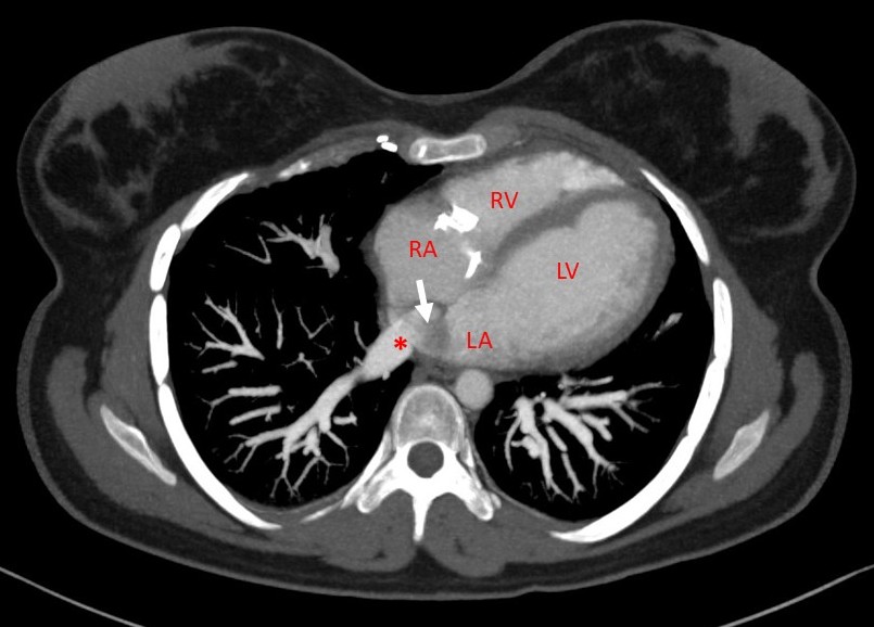
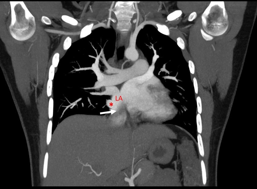
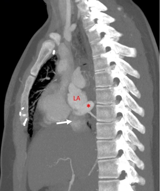
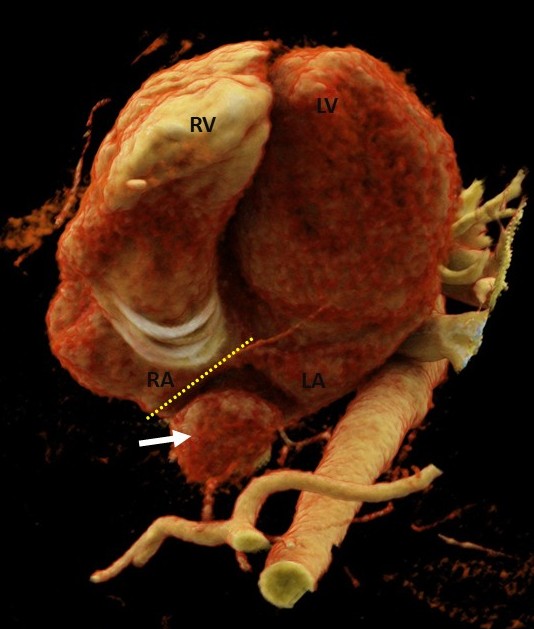
Given persistent hypoxemia and the history of prior inferior ASD repair, CT angiography was performed to evaluate postoperative atrial anatomy and caval connections. CT demonstrated IVC is coursing posteriorly and draining directly into the LA. The atrial septal patch excluded the IVC orifice from the RA, effectively diverting systemic venous return into the LA. Pulmonary venous drainage was normal, and no additional vascular anomalies were identified. These findings were diagnostic of iatrogenic diversion of the IVC to the LA, resulting in a right-to-left shunt following surgical ASD closure (Figure 1-4). Arterial blood gas analysis, contrast echocardiography, and formal exercise-induced desaturation testing were not performed prior to cross-sectional imaging.
The patient was referred for surgical re-exploration to allow direct visualization and definitive correction of the defect. Surgical revision was preferred over transcatheter intervention because the abnormality involved postoperative anatomical distortion requiring patch repositioning/baffle reconstruction, which cannot be reliably addressed with catheter-based techniques.
Also, while awaiting definitive surgical correction, the patient was counselled regarding the risk of paradoxical embolic events resulting from systemic venous admixture into the left atrium. Preventive measures included avoidance of strenuous exertion and dehydration, careful aspiration of air during intravenous access, using air filters when feasible, and close clinical monitoring for neurological or cardiopulmonary symptoms. The patient was maintained under regular follow-up until surgical re-exploration was undertaken.
Discussion
Iatrogenic diversion of the IVC, although rare in the contemporary surgical era, sporadic cases continue to be reported. The mechanism usually involves misidentification of the inferior rim of the defect, particularly in inferiorly located secundum ASDs or IVC-type sinus venosus defects, where a prominent Eustachian valve may be mistaken for the true septal margin [1,2]. Clinical presentation is variable, ranging from early postoperative cyanosis to delayed manifestations such as exertional dyspnea, hypoxemia, or paradoxical embolism years later [3,4,5].
Echocardiography is the first-line imaging modality, but may be limited in assessing the IVC – RA junction. In such cases, CT angiography provides definitive, non-invasive delineation of venous anatomy and is invaluable for diagnosis and surgical planning [2,5]. Prevention of this complication relies on meticulous intraoperative assessment in patients with inferior or low-lying atrial septal defects. Particular attention should be paid to identifying the true inferior margin of the defect and distinguishing it from the Eustachian valve, which may otherwise be mistakenly incorporated into the patch. Before completing patch closure, the surgeon should confirm that the inferior vena cava orifice remains unobstructed and drains freely into the right atrium. In addition, intraoperative transesophageal echocardiography or direct inspection after patch placement can help verify appropriate caval drainage before separation from cardiopulmonary bypass, thereby minimizing the risk of inadvertent IVC diversion [3,2].
| S No | Study | Age | Patient / Lesion Type | Timing of Presentation | Presenting Feature | Imaging Modality | Management | Distinctive Feature |
|---|---|---|---|---|---|---|---|---|
| 1 | Jain et al., 2012 | Child (NR) | Surgically repaired ASD | Early postoperative | Severe cyanosis immediately after repair | TTE (with Doppler and contrast echocardiography) | Reoperation redirecting IVC to RA | Immediate recognition after surgery |
| 2 | Tayeh et al., 2019 | 5 Y | Secundum ASD (child) | Early postoperative | Persistent cyanosis, desaturation | TTE ± TEE, CT angiography | Surgical revision | Early postoperative pediatric presentation |
| 3 | Kim et al., 2021 | Adult | After ASD closure | Late presentation | Dyspnea, systemic desaturation | TTE, CT angiography | Surgical septal revision | Delayed diagnosis of R‑L shunt |
| 4 | Darwazah et al., 2022 | Adult woman | Prior ASD repair | Delayed (years later) | Recurrent fetal loss, chronic hypoxemia | TEE, CT angiography | Surgical redirection of IVC | Rare obstetric presentation |
| 5 | Zhang et al., 2025 | Mixed ages | Post‑ASD repair (case series) | Early postoperative | Persistent hypoxemia | TTE, CT angiography | Surgical correction | Series highlighting early detection |
| 6 | Present case | 17 Y | Low‑lying inferior ASD | Early postoperative | Unexplained desaturation | TTE, CT angiography | Corrective surgical revision planned | Highlights need to confirm inferior rim and caval drainage before weaning from bypass |
ASD, atrial septal defect; IVC, inferior vena cava; TTE, transthoracic echocardiography; TEE, transesophageal echocardiography; CT, computed tomography; RA, right atrium; NR, not reported.
Table 1 summarizes the published Reports of Iatrogenic Diversion of IVC to the Left atrium following ASD closure compared with the present case. Previously reported cases span both pediatric and adult populations, with presentations ranging from immediate postoperative cyanosis to delayed manifestations years after ASD repair [3,1,4,2,5]. The present case adds to the literature by emphasizing the technical challenges of repairing low-lying inferior ASDs and the need for intraoperative verification of the true inferior margin and appropriate caval drainage before weaning from cardiopulmonary bypass.
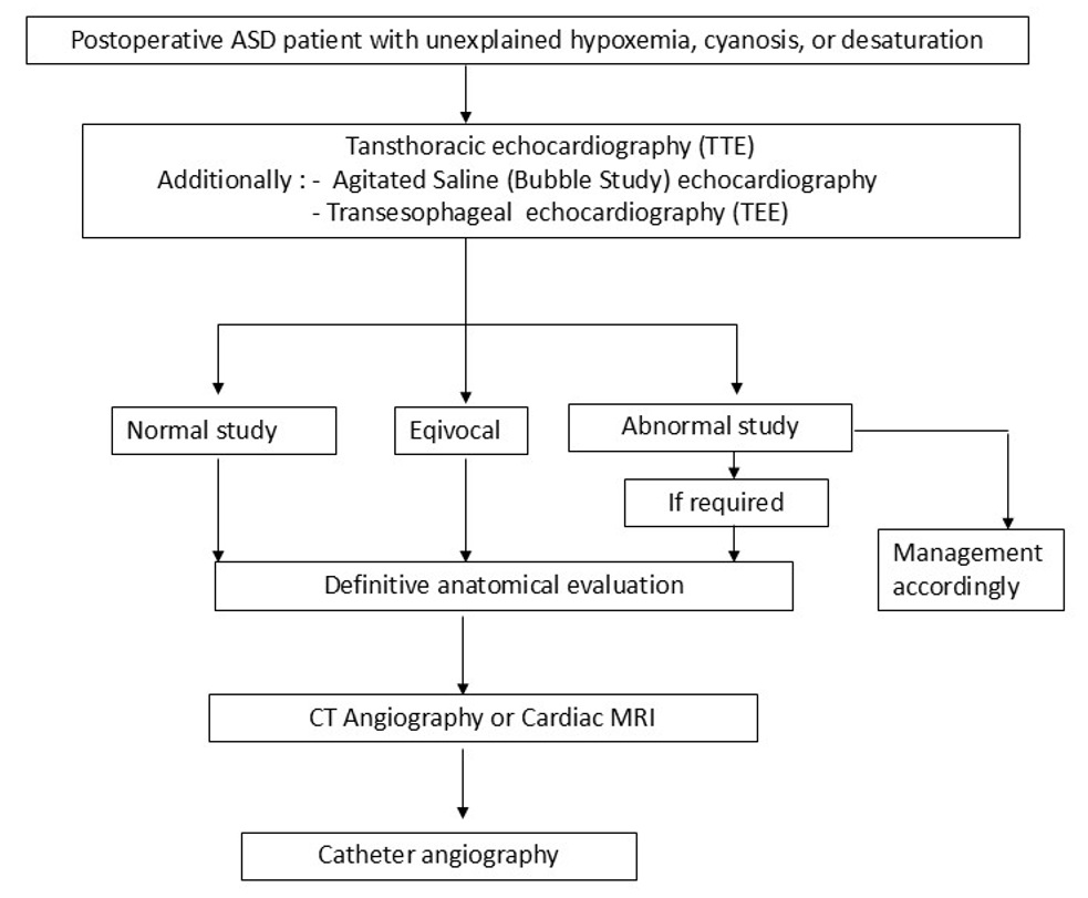
A practical diagnostic approach may be considered in postoperative patients presenting with unexplained desaturation following repair of an inferior or low-lying atrial septal defect. Initial evaluation typically includes TTE; however, visualization of the inferior vena cava – right atrial junction and postoperative atrial septal anatomy may be limited. Targeted visualization using TEE may be performed, providing superior, close-up acoustic windows to inspect the interatrial septum, evaluate the integrity of the surgical patch or closure device, and examine the proximal pulmonary veins. Further, physiologic confirmation may be obtained with an echocardiographic study using agitated saline (bubble), which may demonstrate shunting. Also, definitive anatomical evaluation can be performed with cross-sectional imaging, such as CT angiography or cardiac MRI, to delineate the IVC pathway, patch/baffle position, and atrial connections, thereby confirming inadvertent diversion and guiding surgical correction. Cardiac MRI is preferred when flow quantification, shunt physiology (Qp/Qs), and ventricular functional assessment are required in addition to anatomical evaluation. Catheter angiography is reserved for cases where hemodynamic assessment, confirmation of shunt physiology, or potential transcatheter intervention is required, or when noninvasive imaging findings remain inconclusive [6]. A diagnostic algorithm in a postoperative ASD patient with unexplained hypoxemia, cyanosis, or desaturation is suggested in the flowchart Figure 5.
Despite the clear anatomical diversion of the inferior vena cava into the left atrium, the patient demonstrated only mild resting desaturation. Similar variable or delayed desaturation in IVC-to-LA diversion has been reported earlier [3,1]. This may be explained by physiologic factors such as preferential streaming of oxygenated pulmonary venous blood toward the left ventricular inflow and partial mixing within the atrium, which can limit systemic desaturation at rest. In addition, the inferior vena cava contributes a smaller proportion of total venous return compared with the superior vena cava under resting conditions [7]. However, during exertion, increased venous return from the lower body may augment right-to-left shunting, thereby explaining the patient’s predominant exertional dyspnea and desaturation [8].
This report has certain limitations. The exact intraoperative mechanism of the diversion cannot be confirmed because original intraoperative imaging and transesophageal echocardiography documentation from the index surgery were not available, and the interpretation relies on the available operative notes together with postoperative CT findings. Therefore, the proposed mechanism – misidentification of the inferior rim of the defect or the Eustachian valve during patch closure – should be considered the most likely explanation. Another limitation of the case report is that the details of the revision surgery are not available.
Conclusion
Iatrogenic diversion of the IVC to the left atrium is a rare but important complication following surgical closure of ASD. Clinical suspicion should be particularly high in patients with a history of repair of low-lying or inferior ASDs who develop otherwise unexplained postoperative desaturation. While echocardiography remains the initial diagnostic modality, it may occasionally be inconclusive in delineating abnormal caval drainage. In such cases, CT angiography provides precise anatomical visualisation of the IVC pathway and patch configuration, enabling definitive diagnosis. In the present case, cross-sectional imaging clearly demonstrated IVC diversion into the left atrium, facilitating appropriate surgical planning for correction. Beyond the diagnostic aspect, this case highlights an important operative consideration: during repair of inferior or low-lying ASDs, meticulous identification of the true inferior rim and careful avoidance of incorporating the Eustachian valve into the repair are critical. Postoperative confirmation of appropriate caval drainage should be considered an essential step in anatomically challenging inferior ASD repairs to prevent this rare but significant complication.
Conflicts of Interest
The authors declare no competing interests that could have influenced the objectivity or outcome of this research
Funding Source
The authors declare that no specific grant or funding was received for this research from any public, commercial, or not-for-profit funding agency.
Acknowledgments
None.
Informed Consent
The authors certify that appropriate consent was obtained from the patient’s legal guardian for publication of clinical information and images. Identifying information has been omitted to protect patient confidentiality.
Large Language Model
None.
Authors Contribution
AKP was responsible for case identification, imaging interpretation, and primary writing. RKP contributed through conceptualisation and supervision. DS provided technical review and assisted in replying to reviewer queries. RS helped refine the “Discussion” and “Conclusion” sections, while SM selected and annotated the high-quality figures.
Data Availability
No publicly available dataset was generated for this case report. All relevant clinical information and imaging findings supporting the conclusions of this article are included in the manuscript. Any additional de-identified details may be available from the corresponding author upon reasonable request, subject to patient privacy and consent restrictions.
References
- Jain S. A., Pinto R., Dalvi B.. Iatrogenic diversion of IVC to left atrium after surgical closure of ASD. Ann Pediatr Cardiol. 2012;5(1):72-4. doi:10.4103/0974-2069.93716 PMID: 22529607 PMCID: PMC3327021
- Tayeh C., El Khoury M., Bitar F., Arabi M.. Atrial septal defect closure complicated by anomalous inferior vena cava return to the left atrium: a case report of a 5-year-old child. Eur Heart J Case Rep. 2019;3(2). doi:10.1093/ehjcr/yty169 PMID: 31449633 PMCID: PMC6601233
- Darwazah A. K., Ibrahim B. J., Nairat M., Khdour I., Madi H.. Iatrogenic diversion of inferior vena cava to the left atrium presented as recurrent foetal loss: a case report. Eur Heart J Case Rep. 2022;6(9):ytac348. doi:10.1093/ehjcr/ytac348 PMID: 36072424 PMCID: PMC9446688
- Kim M. S., Jang S. Y., Yang D. H.. Complications caused by iatrogenic right-to-left shunt after surgical closure of atrial septal defect: a case report. Eur Heart J Case Rep. 2021;5(11):ytab434. doi:10.1093/ehjcr/ytab434 PMID: 34917878 PMCID: PMC8669598
- Zhang H., Liu Q., Meng H., Xiong C.. Iatrogenic diversion of inferior vena cava to the left atrium presented as persistent hypoxemia: Case series. Medicine (Baltimore). 2025;104(3):e41162. doi:10.1097/MD.0000000000041162 PMID: 39833084 PMCID: PMC11749782
- Fogel M. A., Anwar S., Broberg C., Browne L., Chung T., Johnson T., Muthurangu V., Taylor M., Valsangiacomo-Buechel E., Wilhelm C.. Society for Cardiovascular Magnetic Resonance/European Society of Cardiovascular Imaging/American Society of Echocardiography/Society for Pediatric Radiology/North American Society for Cardiovascular Imaging Guidelines for the use of cardiovascular magnetic resonance in pediatric congenital and acquired heart disease : Endorsed by The American Heart Association. J Cardiovasc Magn Reson. 2022;24(1):37. doi:10.1186/s12968-022-00843-7 PMID: 35725473 PMCID: PMC9210755
- Anderson R. H., Baker E. J., Penny D. J., Redington A. N., Rigby M. L., Wernovsky G.. Paediatric Cardiology. 2010.
- Hall J. E.. Guyton and Hall Textbook of Medical Physiology. 2021.