The Relationship Between pks⁺ Escherichia coli and Colorectal Cancer: A Review
Bolor UlziitsogtⒾ1, Chuluunbileg BatboldⒾ1, Tsogdulam SumiyabazarⒾ1, Gantogtokh DashjamtsⒾ1, Yumchinsuren TsedendorjⒾ1, Tegshjargal BadamjavⒾ1, Ganchimeg DondovⒾ2, Tulgaa LonjidⒾ1, Batbold BatsaikhanⒾ1*
- 1Department of Internal Medicine, Institute of Medical Sciences, Ministry of Economy and Development, Ulaanbaatar, Mongolia
- 2Department of Oncology, Institute of Medical Sciences, Ministry of Economy and Development, Ulaanbaatar, Mongolia
Abstract
Introduction: According to GLOBOCAN estimates, colorectal cancer (CRC) was the third most common cancer worldwide in 2020, comprising 11% of all cancer diagnoses. Researchers’ interest in CRC development has shifted toward the gastrointestinal microbiome, especially the mutagenicity of pks⁺ E. coli. Therefore, we aimed to review the current evidence on the relationship between pks⁺ E. coli and CRC. Method: We reviewed the literature reporting the association between pks⁺ E. coli and CRC, which was obtained from three databases: PubMed, Google Scholar, and the Cochrane Library, published between January 2021 and January 2026. We used the following keywords for our search strategy: pks, Escherichia coli, and colorectal cancer. Result: As stated in the original studies, the contribution of pks⁺ E. coli to CRC development was associated with a higher prevalence in the CRC group than in the healthy group. However, these results are inconsistent across the studies.
Conclusion: Given the limited evidence, researchers should conduct larger cohort studies to clarify this relationship and identify other factors contributing to the presence of pks⁺ E. coli, particularly across diverse clinical settings. Furthermore, clinical translation, such as the utilization of screening or therapeutic biomarkers, requires prospective validation and standardized detection methods.
Keywords: Colibactin, Colorectal cancer, Microbiome, PKS
Article information
Introduction
According to GLOBOCAN estimates provided by the International Agency for Research on Cancer (IARC), colorectal cancer (CRC) was the third most common cancer worldwide in 2020, comprising 11% of all cancer diagnoses [1,2]. In 2022, Asia accounted for 50.2% of global CRC cases, with 966.400 new cases and 462.000 deaths, which reflects the high incidence of the cancer in Asian countries [3]. Mongolia has relatively low CRC incidence and mortality rates compared with countries with high prevalence, such as Japan and China. In Mongolia, the incidence of CRC in the last 5 years (2018–2022) was 7.9 per 100,000 population, and the mortality rate was 4.7 per 100,000 [4].
Scientists have identified multiple risk factors for CRC. As reported in twin and family studies, hereditary factors account for approximately 12% – 35% of CRC cases [5,6]. Given this relatively low rate, it underscores the importance of other modifiable and non-modifiable risk factors. Alcohol consumption [7], smoking [8], obesity [9], unhealthy dietary patterns [10], a sedentary lifestyle [11], and psychological stress [12] are considered changeable risk factors. On the other hand, unchangeable risk factors consist of age, sex [13], genetic predisposition [14], abdominopelvic radiation exposure [15], family history of colorectal cancer [16], and a personal history of other diseases [17]. The role of the microbiome in the development and progression of CRC is increasingly recognized and appreciated as a modifiable risk factor [18].
Researchers’ interest in CRC development has shifted toward the gastrointestinal microbiome, a densely populated ecosystem. This system comprises approximately microorganisms in the gastrointestinal (GI) tract [19]. This microbial community is predominantly composed of Firmicutes and Bacteroidetes, which together account for about 90% of the total bacterial population [20]. Currently, researchers have identified two types of bacteria linked to colon carcinogenesis: “driver” bacteria, such as Enterococcus faecalis, Escherichia coli, and Bacteroides fragilis [21] and “passenger” bacteria, including Fusobacterium spp. and Streptococcus gallolyticus [22].
The latest meta-analysis on the association between E. coli and CRC development suggests that individuals with E. coli have a greater risk of developing CRC [23]. However, the role of E. coli in cancer initiation, proliferation, and metastasis remains unclear. Moreover, several confounding factors influence both E. coli and CRC development. The individual-specific gut microbiome is an outcome of the complex interplay between host genetics and environmental exposures [24]. As stated in a prospective cohort study, people exposed to a Western diet are more likely to develop CRCs with abundant E. coli.
To summarize, substantial evidence indicates that the E. coli is associated with the development of CRC. Therefore, developing a well-defined mechanistic model of E. coli-mediated CRC development can inform CRC screening and characterization of CRC progression as a biomarker, enabling early detection and the development of targeted treatment strategies, such as microbiota modulation and immunomodulatory pathways.
Method
This review study included English original articles addressing the relationship between CRC and E. coli. Marcianne Elaine Gaab et al. published a meta-analysis study addressing the relationship between E. coli and CRC. They included 12 articles published through January 2021 [23]. Therefore, related literature reporting the association between E. coli and CRC was obtained from three databases: PubMed, Google Scholar, and the Cochrane Library, published between January 2021 and January 2026. We used the following keywords for our search strategy: pks, Escherichia coli, and colorectal cancer. Duplicate articles, review articles, animal studies, and papers with incomplete information, like conference proceedings and articles without full text, were excluded from the study.
E. coli strains classification
E. coli, a ubiquitous Gram-negative bacillus, inhabits the intestinal flora, especially in the caecum and the colon. Moreover, it can also serve as a pathogen, inducing a broad spectrum of diseases. Recently, E. coli has been classified into pathotypes based on their virulence factors and the diseases they predominantly cause in humans [25]. The strain of the pathogenic E. coli can be divided into two main groups: the first one is Intestinal Pathogenic Escherichia coli (IPEC), leading to GI infection; the second one is External Pathogenic Escherichia coli (ExPEC), which causes common infections outside of the GI tract [26].
The E. coli infection prevalence and disease manifestations
We reviewed the primary classification of E. coli by pathotype, their prevalence, and disease manifestations in Table 1.
The prevalence of commensal E. coli varies by species: it is more than 90% in humans [27,28] but only 56% in wild mammals, 23% in birds, and 10% in reptiles [29]. The concentration of faces per gram in humans ranges from to colony-forming units [28]. The prevalence of E. coli varies across studies due to differences in study populations, diagnostic methods, and criteria. As stated in a review study that included original studies from 13 developing countries, the average incidence of EPEC among pediatric diarrheal episodes is 5–10% [30]. A retrospective case-control study revealed that children with EPEC infection had frequently experienced diarrhea, vomiting, abdominal pain, and fever, compared to EPEC-negative controls [31].
The review study analyzed data from 21 countries across 10 of the 14 WHO Sub-Regions, representing approximately 30% of the global population. According to the results, the global incidence of EHEC was 2.8 million cases per year, and its annual mortality was 230 deaths [32]. In terms of disease manifestations, bloody or watery diarrhea without fever was common, and it was sometimes associated with abdominal cramping. One of the significant complications of EHEC infection is definitely hemolytic uremic syndrome [25].
The incidence of EIEC varies widely across regions, as reported in numerous studies in Europe, Central and South America, the Middle East, West Africa, and Southeast Asia [33]. Infection with EIEC causes a bacillary dysentery characterized by bloody and mucous diarrhea, abdominal cramps, nausea, and fever [34].
Ibrahim A. Khalil et al. estimated the global incidence of ETEC diarrhea at 220 million cases per year among children under 5 years old [35]. In addition, it is a significant cause of travelers’ diarrhea, which is prevalent in regions such as Latin America, Africa, and South Asia [36]. As noted in the observational study, the most prevalent symptom was watery stool among almost all children and adults with ETEC infection. In addition, younger patients experienced frequent vomiting and fever compared to the older patients [37,38].
The incidence of EAEC infection, which can lead to acute or persistent watery diarrhea, is highest among children living in key endemic regions [39] and immunosuppressed individuals [40]. People with EAEC infection usually present the following symptoms: watery diarrhea and occasionally very mucoid diarrhea, nausea, anorexia, low-grade fever, borborygmi, and tenesmus [41].
Few studies have reported the incidence of DAEC infection, and rates have varied widely, with relatively low values. Among children with diarrhea, DAEC strains were isolated in 18.3% in Brazil and 3.9% in Iran [42,43]. DAEC infection has been considered a cause of diarrhea, particularly in children older than 24 months [44].
According to the narrative review, UPEC causes 75% of uncomplicated urinary tract infections (UTIs) and 65% of complicated UTIs [45]. Patients diagnosed with UTI caused by UPEC frequently experience dysuria and frequent urination because UPEC strains are more likely to damage the lower urinary tract [46].
The most common pathogens causing sepsis are Staphylococcus aureus and E. coli. Among infants with sepsis, E. coli is also the most prevalent gram-negative bacterium implicated in neonatal sepsis [47,48]. In addition, E. coli is the most frequently identified pathogen in cases of neonatal meningitis. Specifically, E. coli meningitis was 7-fold more common in preterm than term infants [49].
The prevalence of AIEC strains was 28% among patients with inflammatory bowel diseases and 29% among patients with Crohn’s disease in a recent meta-analysis [50]. It is well known that E. coli significantly contributes to dysbiosis of the local microbiota, thereby aggravating symptoms in inflammatory bowel diseases [51].
Evidence summary of human studies
Since 2021, a total of 3624 articles related to E. coli and CRC have been published in online databases. According to our eligibility criteria, we selected 13 articles for review. The majority of the selected studies employed retrospective cohort and case-control designs. Moreover, these studies usually detected the pks/clb gene cluster in E. coli using qPCR in both stool and tissue samples. We summarized the characteristics and results of the included studies in Table 2.
The prevalence of E. coli across selected articles varied by study population and location, ranging from 9.4% to 54.7%. This variation depends on several factors, such as sample size, study population (cancer or healthy subjects), and the detection method of the pks/clb gene cluster in E. coli. Thevambiga Iyadorai et al. found significant differences in the presence of E. coli isolates from tissue specimens of patients and controls, but not among fecal specimens [52]. It suggests that the detection of the pks/clb gene cluster in E. coli may depend on the specimen type. Moreover, the study evaluating the association between the pks/clb gene cluster in E. coli, measured in fecal immunochemical test (FIT) samples, and the detection of advanced neoplasia (AN) at colonoscopy included 5020 CRC screening participants. The prevalence of E. coli in FIT samples from individuals with AN (28.6%) and controls (25.9%) was not significantly different. Authors suggest that a one-time
| Group | Pathotype | Prevalence | Symptoms | Reference | |
|---|---|---|---|---|---|
| Commensal E. coli | Normal flora; widespread in the gut | No symptoms | [29,27,28] | ||
| Pathogenic E. coli | IPEC (Intestinal Pathogenic Escherichia coli) | Enteropathogenic E. coli (EPEC) | 5-10% of pediatric diarrheal episodes in the developing world | Diarrhea, vomiting, fever, and severe abdominal pain | [31,30] |
| Enterohemorrhagic E. coli (EHEC) | 2.8 million cases per year, globally | Bloody or watery diarrhea without fever, abdominal cramps, dehydration, asthenia, decreased urine output, and hemolytic uremic syndrome | [25] | ||
| Enteroinvasive E. coli (EIEC) | Substantial regional heterogeneity | Abdominal cramps, nausea, fever, and bloody and mucus diarrhea | [34,33] | ||
| Enterotoxigenic E. coli (ETEC) | 220 million cases annually, with 84.4 million cases reported in children aged years; most prevalent among travelers | Watery diarrhea, severe dehydration, vomiting, and fever, especially in children | [37,35,38,36] | ||
| Enteroaggregative E. coli (EAEC) | Children, HIV patients | Watery diarrhea, occasionally very mucoid diarrhea, nausea, anorexia, low-grade fever, borborygmi, and tenesmus | [40,41,39] | ||
| Diffusely adherent E. coli (DAEC) | Higher rates in children in both developing and developed countries | Persistent diarrhea | [42,44,43] | ||
| ExPEC(Extraintestinal Pathogenic Escherichia coli) | Urophatogenic E. coli (UPEC) | 75% of uncomplicated UTI cases | Dysuria, frequency, fever | [45,46] | |
| Sepsis-associated E. coli (SEPEC) | The most prevalent bacteria in sepsis cases | Severe fever, chills, tachycardia, hypotension, and multiple-organ dysfunction syndrome | [47,48] | ||
| Meningitis-associated E. coli (MNEC) | The most frequently identified agent, especially in preterm neonates | Meningitis symptoms | [49] | ||
| Adherent-invasive E. coli (AIEC) | % among IBD and CD patients | Dysbiosis | [50,51] | ||
The prevalence information is approximate, depending on the study population, age groups, and method. HIV, Human Immunodeficiency Virus; UTI, Urinary Tract Infection; IBD, Inflammatory Bowel Disease; CD, Crohn’s Disease.
| Author, year | Study subject | Specimen type and detection method | Prevalence of E. coli | Main outcome (relationship between E. coli and CRC) |
|---|---|---|---|---|
| Yen Lin Chu et al., 2025 | A total of 358 LS cases, including 386 CRCs, 90 adenomas, and 195 normal subjects. | Tissue sample; qPCR | The prevalence was 13.6%. | E. coli was enriched in LS CRCs when compared with sporadic CRCs (OR = 1.60; 95% CI = 1.08–2.35). E. coli in the initial CRC was associated with an increased risk of metachronous CRC (HR = 2.32; 95% CI = 1.29–4.17) and metachronous colorectal neoplasia (HR = 1.51; 95% CI = 1.02–2.23). |
| Toshimitsu Miyasaka et al., 2024 | 413 patients with CRC | Both tumor and normal tissue; droplet digital PCR | The prevalence was 65.6% (271) in tumor tissues and 62.3% (152) in normal tissues. | High level of E. coli in tumor tissue was significantly associated with shallower tumor depth (HR = 5.0, 95% CI = 2.3–11.3) and absence of lymph node metastasis (HR = 3.0, 95% CI = 1.8–5.1). E. coli-low and -negative groups were associated with shorter CRC-specific survival (HR = 6.4, 95% CI = 1.7–25.6) and shorter relapse-free survival (HR = 3.1, 95% CI = 1.3–7.3). |
| Jihoon E. Joo et al., 2024 | 1697 tumor tissues from 1666 individuals with CRC | Tumor tissue; qPCR | Intratumoral prevalence of E. coli was 10.3% (174). | The APC: c.835-8 AG mutation was associated with intratumoral E. coli (P = 0.025, OR = 2.20, 95% CI = 1.05–4.25). MMRd status was not associated with E. coli. |
| Willemijn de Klaver et al., 2024 | 5020 CRC screening patients | Stool sample; qPCR | 1322 (26.2%) were E. coli positive: 28.6% in AN and 25.9% in controls. | There was no significant difference between the prevalence of E. coli in individuals with AN and controls (p = 0.131). |
| Xiaoming He et al., 2024 | 100 participants (50 colorectal cancer patients and 50 healthy controls) | Both stool and tissue samples; qPCR | The prevalence was 35%: 46% (23) in the control group and 24% (12) in the CRC group. | A higher proportion of E. coli was found in late-onset CRC (17 out of 35) compared to early-onset CRC (3 out of 35; P 0.05). |
| Bingjie Chen et al., 2023 | 366 deep WGS samples: 101 normal samples (74 adjacent and 27 distant normal crypts) and 265 cancer samples. | Both healthy and tumor tissue; WGS | The prevalence was 76.67% in cancer crypts, 70.59% in adjacent normal, and 50% in distant normal from cancer patients, compared to 30% in individuals without cancer. | The signature causes a large proportion of mutations in chromatin modifier genes in MSS cancers. Multiple alterations in cancer driver genes and chromatin modifier genes are consistent with -induced short T-dels. |
| Thevambiga Iyadorai et al., 2023 | 57 specimens (31 CRC tissue and 26 healthy normal tissue) | Both fecal and tissue samples; PCR | Among 37 fecal specimens, 43.6% had the pks gene [61% (13) from CRC and 31.3% (5) from control]. Among 20 tissue samples, 55.0% (11) had pks, including 80.0% (8) from CRC patients and 30.0% (3) from healthy controls. | Significant differences in the presence of pks among E. coli isolates from tissue specimens of patients and controls were observed, but not among those from fecal specimens. |
| Kota Arima et al., 2022 | 1175 patients with CRC | Tumor tissue; qPCR | The prevalence was 9.4% (11). | The E. coli level was associated with lower disease stage (P = 0.008) but not with tumor location, microsatellite instability, or BRAF, KRAS, or PIK3CA mutations (P 0.05). |
| Habiba Tariq et al., 2022 | 60 patients (35 patients with CRC and 25 relatively healthy patients) | Both tumor and healthy tissue; quadruplex PCR | The prevalence was 18.3% (11 in the CRC group and none in the healthy group). | A significant association between the genotoxin-producing colibactin gene and colorectal cancer was reported (P 0.002). |
| Manon Oliero et al., 2022 | 156 participants: healthy controls (N = 62) and CRC patients (N = 94) | Fecal samples; conventional qualitative PCR | Prevalence of bacteria was 44.2% (69): 42% (26) in healthy controls and 46% (43) in the CRC group. | EOCRC (50 years) patients were significantly less colonized with E. coli than LOCRC patients (P 0.05). |
| Motoki Iwasaki et al., 2022 | 968 screening patients (543 CRC and 425 controls) | Stool sample; qPCR | The prevalence was 31.8% (308): 32.6% (177) in cases and 30.8% (131) in controls. | No significant association was found between E. coli and colorectal neoplasia (adjusted OR = 1.04, 95% CI = 0.77–1.41). |
| Kaixi Liu et al., 2021 | 139 patients with CRC and 42 healthy individuals | Stool sample; qPCR method | The prevalence was 54.7% (76): 21.4% (9) in control, 67.6% (25) in CAP, and 70.0% (42) in CRC cases. | E. coli levels were higher in the CRC group than in either the CAP group or healthy controls. No difference was found among tumor sites. The cut-off value for E. coli was 2.25, with a sensitivity of 93.3% and a specificity of 73.3%. |
| Alfonso Piciocchi et al., 2021 | 330 adult subjects: 162 healthy tissues, 55 tissues with HP, 79 P/A, and 29 ADC | Both healthy and cancer tissues; qPCR | The prevalence was 38%: 35% (54) in healthy tissue, 42% (22) in HP, 36% (27) in P/A, and 52% (14) in ADC. | There was no significant association between E. coli and adenocarcinoma by univariate analysis (OR = 2.01, 95% CI = 0.88–4.64). |
ADC, Adenocarcinomas; AN, Advanced Neoplasia; CRC, Colorectal cancer; HP, Hyperplastic Polyps; LS, Lynch Syndrome; MMRd, Mismatch Repair Deficiency; MSS, Microsatellite Stable; P/A, Pre-Cancerous Polyps/Adenomas; qPCR, Quantitative Polymerase Chain Reaction; WGS, Whole Genome Sequencing.
E. coli measurement in stool is not informative of CRC risk in a screening setting [53].
All included studies evaluated the relationship between E. coli and CRC incidence, comparing healthy and cancer subjects across cancer types, locations, and disease severity. A total of 9 studies evaluated this relationship, comparing healthy and CRC groups. To compare the presence of E. coli between the control and CRC groups, 5 studies reported no significant difference, whereas 4 studies found a significant association between E. coli and CRC. Kaixi Liu et al. calculated the accuracy of E. coli in predicting CRC and found that the cut-off value was 2.25, with a sensitivity of 93.3% and a specificity of 73.3% [54].
In addition, these studies compared the presence of pks/clb gene cluster in E. coli across early- and late-onset cancers, disease stages, and tumor locations. The retrospective cohort study reported that early-onset CRC (EOCRC) patients (<50 years) were significantly less colonized with E. coli than late-onset CRC (LOCRC) patients [55]. In another study, the E. coli tumors with the APC: c.835-8 A>G mutation were more prevalent in EOCRC than LOCRC (9.5% versus 5.3%), with only the association in EOCRC showing statistical significance. The reason for this association in EOCRC is currently unknown and, if validated, would raise interesting questions about the mechanism in EOCRCs versus LOCRCs [56]. Kota Arima et al. found that the E. coli level was associated with lower disease stage but not with tumor location [56]. Moreover, an observational cohort study including 413 patients with CRC found that a high level of E. coli in tumor tissue was significantly associated with shallower tumor depth and the absence of lymph node metastasis. Also, they reported that the E. coli low and negative groups were associated with shorter CRC-specific survival and shorter relapse-free survival [57].
Two studies evaluated the relationship between E. coli and the mutational signature of CRC. The study, including 1697 tumor tissues, found that Mismatch Repair Deficiency (MMRd) status was not associated with the colibactin-associated mutational signature [56]. A total of 366 deep whole-genome sequencing (WGS) analyses showed that a large proportion of mutations in chromatin modifier genes in microsatellite-stable (MSS) cancers are driven by colibactin-associated mutational signature [58].
Yen Lin Chu et al. included 358 Lynch Syndrome (LS) cases and found that E. coli in the initial CRC was associated with an increased risk of metachronous CRC and metachronous colorectal neoplasia when compared with CRCs without E. coli [59].
Across the selected studies, there were inconsistencies in the results of a significant relationship between E. coli and CRC development. Moreover, some studies reported a significant difference in the presence of E. coli between early-onset and late-onset cancers, but others did not. The impact of E. coli on MSS or MMRd cancers remains unknown. Tissue heterogeneity could influence this relationship. The majority of studies investigated the pks/clb gene cluster in E. coli using tissue samples because fecal samples were found to be less accurate. Therefore, researchers should conduct a prospective cohort study using both tissue and fecal samples and provide evidence of its use as a biomarker in screening programs.
Evidence of the mechanism of how pks^+ pks+ E. coli impacts carcinogenesis
Pathogenicity islands and key features of the pks/clb gene cluster in E. coli
HGT-mediated acquisition of large genomic islands, or pathogenicity islands (PAIs), is a key factor in the emergence of several E. coli pathotypes. PAIs, a small subgroup of genomic islands, play a significant role in bacterial virulence evolution by compromising virulence-associated factors and facilitating adaptive horizontal gene transfer. It is approximately 54 – 56 kb in size and may vary among different pks-positive strains [60,24]. The PAI named pks encoded colibactin, a nonribosomal peptide-polyketide secondary metabolite. The colibactin gene cluster within the pks genomic island comprises the clbA-clbS genes, which are responsible for colibactin synthesis [61,62]. These genes included nonribosomal peptide mega synthases (NRPSs; clbH, clbJ, and clbN), polyketide mega synthases (PKSs; clbC, clbI, and clbO), two hybrid NRPS-PKSs (clbB and clbK), and nine accessory and tailoring enzymes [63].
![Organization of pks^+ island (Modified figure based on Nougayrède et al.’s (2006) [64] illustrations)](https://asidejournals.com/index.php/oncology/article/download/533/version/532/579/15153/1.jpg)
Figure 1 was adapted from the original photo with the author’s permission. The figure was generated using BioRender Software and published with permission.
Organization of the pks island: pks island encodes the genes required for the synthesis of colibactin. Genes coding for pks (yellow), non-ribosomal peptide synthase genes (blue), hybrid NRPS/PKS (green), and the accessory proteins (red) are shown. They are in connection with the regulator clbR and the integrase (int) (black). As stated in Figure 1, clbA is a phosphopantetheinyl transferase, clbM is a transporter, and clbP is a peptidase.
The initiation of the biosynthesis of colibactin depends on the activation of pks/NRPS megasynthases (non-ribosomal peptides) by phosphopantetheinyl transferase ClbA. Once the initiation is activated, megasynthases recruit their specific monomers. The ClbN, whose substrate is asparagine, is regarded as the first enzyme involved in the synthesis. The Biosynthesis assembly then proceeds with the successful intervention of the ClbB-ClbC-ClbH-ClbI-ClbJ-ClbK enzymes, which exploit both common substrates, such as malonyl-CoA, alanine, serine, glycine, and cysteine, and uncommon substrates, such as aminocyclopropane-carboxylic acid, to produce the toxin in its pre-colibactin form. Once inactive pre-colibactin is formed, it is exported into the periplasm by ClbM, an efflux pump. The pre-colibactin is hydrolyzed following the insertion of ClbM into the inner membrane. It results in the separation of the cleavage product, or N-myristoyl-D-asparagine, and the active colibactin (ClbP).[64,65,63] Among all these genes, researchers have identified that five pks islet genes (ClbG, ClbH, ClbL, ClbM, and ClbS) are mainly activated during tumor development in an inflammation-dependent manner, as shown in a study using bacterial strains and plasmids. It suggests that various colibactin biosynthetic pathways alter the microenvironment during CRC development Figure 1 [66].
The impact of pks^+ pks+ E. coli on the mechanism of CRC
The majority of CRCs are MSS, whereas the minority exhibit microsatellite instability (MSI), accounting for 15% [67]. However, these two types of CRC share many common driver genes and exhibit equivalent numbers of mutations in these genes [68]. MSI colon cancer is considered to have a much higher mutational load because of the lack of a DNA mismatch repair system [69]. It is well established that CpG deamination is the most common mutational process in the human colon, especially in MSS cancers [70]. According to the current evidence, researchers suggest that colibactin causes the formation of DNA cross-links by alkylating adenines [71,72,73]. This accumulation of DNA damage encourages the initiation and progression of CRC, leading to the transformation of normal colonic epithelial cells into malignant tumor cells [74].
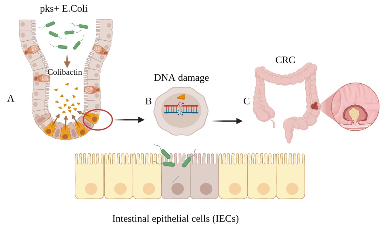
We briefly integrated the current evidence on the mechanism of CRC induced by the E. coli into Figure 2. There were three crucial phases of CRC development: [74,75,76].
A. E. coli produces a cyclomodulin called colibactin.
B. The CpG deamination signature is the most prevalent mutational process in human colon cancer and causes C>T mutations at methylated CG sites. Whereas the increasing number of in vivo studies has been determining the colibactin-attributable alterations in colorectal cancer. The WGS study reveals a distinct mutational signature caused by colibactin, as human intestinal organoids are exposed to genotoxic E. coli via repeated luminal injections over 5 months. As stated in the study, colibactin promotes DNA alkylation in the intestinal epithelial cells, leading to the formation of interstrand cross-links and DNA double-strand breaks. These crosslinks could be resolved in various ways, including induction of DSBs, Nucleotide Excision Repair, or translation synthesis, which in turn may lead to various mutational outcomes [77,75].
C. This may result in the development of genomic instability and mutations in the prominent oncogenes and tumor suppressor genes. Over the years, the accumulation of DNA damage may drive both the initiation and progression of CRC, transforming normal colonic epithelial cells into malignant tumor cells Figure 2. Figure 2 was independently created based on information reviewed in the literature. This figure was also created in BioRender Software, and the publication license was obtained.
It is well supported that E. coli cause double-strand DNA breaks and genomic instability in host cells by extensive in vitro and in vivo studies. However, the exact other underlying mechanism could not be fully established to determine the impact of E. coli on colon carcinogenesis, especially metastasis, and its interaction with a specific diet.
Discussion
In our study, the exact prevalence of E. coli and its significant relationship with CRC are inconsistent across the selected articles. However, the incidence of E. coli was higher in cancer tissues than in healthy tissues. This pattern is consistent with other studies. As reported in the literature, the incidence of E. coli was lower in healthy cohorts than in case groups: 22% in the USA [78]; 18.5% in Sweden [79,24]; and 4.35% in Malaysia [52]. Dietary differences could explain this prevalence of evidence of inconsistency. For instance, the western diet has been related to a higher incidence of colorectal cancer containing E. coli [29,80]. In addition, a prospective Japanese study found that energy-adjusted intakes of cruciferous vegetables and vitamin C were significantly associated with a higher prevalence of E. coli. Nevertheless, other factors, such as age, sex, and comorbidity, associated with the prevalence of E. coli are unknown [81]. To our knowledge, these dietary factors are not considered traditional risk factors for colorectal cancer [82,83]. But could have a role in the presence of E. coli; however, the reported findings are unstable [81]. Furthermore, more research is needed to establish evidence on the factors underlying the prevalence of E. coli.
We found that E. coli is more enriched in early-onset cancer than late-onset cancer. Marcos Díaz-Gay et al. found that late-onset cases showed enrichment for signatures similar to those observed in age-dependent changes in normal colorectal crypts. In contrast, colibactin-induced signatures were enriched in early-onset cancers. In addition, colibactin was associated with an earlier age of onset, an effect more evident in the distal colon and rectum [84]. Lee-six et al. found that the colibactin-related mutation signature in normal colonic crypts was most prevalent in younger children aged less than 10 years [70]. During the early life stage, our gut microbiome is still evolving, and it is considered a sensitive period to extrinsic influences. Therefore, it could interpret the association between APC: c.835-8 A>G and E. coli in EOCRCs [85]. Yen Lin Chu et al. reported that colibactin-producing bacteria are less prevalent in EOCRC than in LOCRC. Therefore, this finding should be further confirmed in larger cohorts and a longitudinal study to determine how early mutagenic exposure of E. coli influences later life and induces CRC [59].
Limitation
Our review included articles published since 2021, potentially excluding earlier relevant studies. In addition, the prevalence of E. coli strains was insufficient to support a worldwide prevalence estimate. It depended on geography, age groups in the population, and detection methods. We could not clearly establish causality regarding the overreach of E. coli in cancer due to a limited number of published articles. Therefore, to prove the relationship between E. coli and CRC, large prospective studies in more controlled clinical settings are necessary.
Conclusion
We evaluated the main characteristics of E. coli and its impact on CRC development, and reviewed key features of pathogenic E. coli strains. Since the evolution of virulence in E. coli strains has been studied, horizontal gene transfer, which is crucial for the spread of the pks genomic island, has been shown to play a key role in this evolution. Therefore, colibactin, encoded by a pks, has been regarded as a key player in carcinogenesis, particularly in CRC. Several studies have proposed a primary underlying mechanism: colibactin promotes DNA alkylation in intestinal epithelial cells, thereby inducing DNA double-strand breaks. However, in human studies, results were inconsistent, and a large cohort study should be conducted to further enrich the current evidence. Furthermore, clinical translation, such as the use of screening or therapeutic biomarkers, requires prospective validation and standardized detection methods.
Conflicts of Interest
The authors declare no conflicts of interest.
Funding Source
The authors did not receive any external funding for this study.
Acknowledgments
The authors have no acknowledgements to declare.
Institutional Review Board (IRB)
This review study did not require ethical approval.
Large Language Model
No artificial intelligence tools or large language models were used to generate this manuscript.
Authors Contribution
BU contributed to project administration, visualization, and conceptualization. CB contributed to methodology and writing-original draft. TS contributed to visualization and writing-original draft. GHD contributed to resources. YT contributed to data curation. TB contributed to writing-review and editing and supervision. GD contributed to visualization and resources. TL contributed to supervision. BB contributed to project administration and conceptualization.
Data Availability
This article is a review of previously published literature and did not generate or analyze any new primary dataset. Therefore, no new data are available. The sources supporting the findings of this review are included in the reference list.
References
- Bray F., Ferlay J., Soerjomataram I., Siegel R. L., Torre L. A., Jemal A.. Global cancer statistics 2018: GLOBOCAN estimates of incidence and mortality worldwide for 36 cancers in 185 countries. CA Cancer J Clin. 2018;68(6):394-424. doi:10.3322/caac.21492 PMID: 30207593
- Globocan. Global Cancer Observatory: Cancer Today.
- Fu M., Li Y., Wang J.. Incidence and Mortality of Colorectal Cancer in Asia in 2022 and Projections for 2050. J Gastroenterol Hepatol. 2025;40(5):1143-1156. doi:10.1111/jgh.16910 PMID: 40018878
- Tsedendorj Y., Zorigtbaatar S., Davaasuren N. E., Dondov G., Ganbat T., Lonjid T., Batsaikhan A., Tudev U., Batsaikhan B.. Incidence and Mortality due to Colorectal Cancer in Mongolia, 2018-2022. Asian Pac J Cancer Prev. 2024;25(7):2305-2310. doi:10.31557/APJCP.2024.25.7.2305 PMID: 39068562 PMCID: PMC11480595
- Lichtenstein P., Holm N. V., Verkasalo P. K., Iliadou A., Kaprio J., Koskenvuo M., Pukkala E., Skytthe A., Hemminki K.. Environmental and heritable factors in the causation of cancer--analyses of cohorts of twins from Sweden, Denmark, and Finland. N Engl J Med. 2000;343(2):78-85. doi:10.1056/NEJM200007133430201 PMID: 10891514
- Mucci L. A., Hjelmborg J. B., Harris J. R., Czene K., Havelick D. J., Scheike T., Graff R. E., Holst K., Moller S., Unger R. H., McIntosh C., Nuttall E., Brandt I., Penney K. L., Hartman M., Kraft P., Parmigiani G., Christensen K., Koskenvuo M., Holm N. V., Heikkila K., Pukkala E., Skytthe A., Adami H. O., Kaprio J., Nordic Twin Study of Cancer Collaboration. Familial Risk and Heritability of Cancer Among Twins in Nordic Countries. JAMA. 2016;315(1):68-76. doi:10.1001/jama.2015.17703 PMID: 26746459 PMCID: PMC5498110
- McNabb S., Harrison T. A., Albanes D., Berndt S. I., Brenner H., Caan B. J., Campbell P. T., Cao Y., Chang-Claude J., Chan A., Chen Z., English D. R., Giles G. G., Giovannucci E. L., Goodman P. J., Hayes R. B., Hoffmeister M., Jacobs E. J., Joshi A. D., Larsson S. C., Le Marchand L., Li L., Lin Y., Mannisto S., Milne R. L., Nan H., Newton C. C., Ogino S., Parfrey P. S., Petersen P. S., Potter J. D., Schoen R. E., Slattery M. L., Su Y. R., Tangen C. M., Tucker T. C., Weinstein S. J., White E., Wolk A., Woods M. O., Phipps A. I., Peters U.. Meta-analysis of 16 studies of the association of alcohol with colorectal cancer. Int J Cancer. 2020;146(3):861-873. doi:10.1002/ijc.32377 PMID: 31037736 PMCID: PMC6819207
- Botteri E., Borroni E., Sloan E. K., Bagnardi V., Bosetti C., Peveri G., Santucci C., Specchia C., van den Brandt P., Gallus S., Lugo A.. Smoking and Colorectal Cancer Risk, Overall and by Molecular Subtypes: A Meta-Analysis. Am J Gastroenterol. 2020;115(12):1940-1949. doi:10.14309/ajg.0000000000000803 PMID: 32773458
- Socol C. T., Chira A., Martinez-Sanchez M. A., Nunez-Sanchez M. A., Maerescu C. M., Mierlita D., Rusu A. V., Ruiz-Alcaraz A. J., Trif M., Ramos-Molina B.. Leptin Signaling in Obesity and Colorectal Cancer. Int J Mol Sci. 2022;23(9). doi:10.3390/ijms23094713 PMID: 35563103 PMCID: PMC9102849
- Veettil S. K., Wong T. Y., Loo Y. S., Playdon M. C., Lai N. M., Giovannucci E. L., Chaiyakunapruk N.. Role of Diet in Colorectal Cancer Incidence: Umbrella Review of Meta-analyses of Prospective Observational Studies. JAMA Netw Open. 2021;4(2):e2037341. doi:10.1001/jamanetworkopen.2020.37341 PMID: 33591366 PMCID: PMC7887658
- Morrison D. S., Parr C. L., Lam T. H., Ueshima H., Kim H. C., Jee S. H., Murakami Y., Giles G., Fang X., Barzi F., Batty G. D., Huxley R. R., Woodward M.. Behavioural and metabolic risk factors for mortality from colon and rectum cancer: analysis of data from the Asia-Pacific Cohort Studies Collaboration. Asian Pac J Cancer Prev. 2013;14(2):1083-7. doi:10.7314/apjcp.2013.14.2.1083 PMID: 23621191
- Kikuchi N., Nishiyama T., Sawada T., Wang C., Lin Y., Watanabe Y., Tamakoshi A., Kikuchi S.. Perceived Stress and Colorectal Cancer Incidence: The Japan Collaborative Cohort Study. Sci Rep. 2017;7:40363. doi:10.1038/srep40363 PMID: 28091607 PMCID: PMC5238416
- Sung H., Ferlay J., Siegel R. L., Laversanne M., Soerjomataram I., Jemal A., Bray F.. Global Cancer Statistics 2020: GLOBOCAN Estimates of Incidence and Mortality Worldwide for 36 Cancers in 185 Countries. CA Cancer J Clin. 2021;71(3):209-249. doi:10.3322/caac.21660 PMID: 33538338
- Pearlman R., Frankel W. L., Swanson B., Zhao W., Yilmaz A., Miller K., Bacher J., Bigley C., Nelsen L., Goodfellow P. J., Goldberg R. M., Paskett E., Shields P. G., Freudenheim J. L., Stanich P. P., Lattimer I., Arnold M., Liyanarachchi S., Kalady M., Heald B., Greenwood C., Paquette I., Prues M., Draper D. J., Lindeman C., Kuebler J. P., Reynolds K., Brell J. M., Shaper A. A., Mahesh S., Buie N., Weeman K., Shine K., Haut M., Edwards J., Bastola S., Wickham K., Khanduja K. S., Zacks R., Pritchard C. C., Shirts B. H., Jacobson A., Allen B., de la Chapelle A., Hampel H., Ohio Colorectal Cancer Prevention Initiative Study Group. Prevalence and Spectrum of Germline Cancer Susceptibility Gene Mutations Among Patients With Early-Onset Colorectal Cancer. JAMA Oncol. 2017;3(4):464-471. doi:10.1001/jamaoncol.2016.5194 PMID: 27978560 PMCID: PMC5564179
- Ho C. H., Cheng K. C., Chao C. M., Lai C. C., Chiang S. R., Chen C. M., Liao K. M., Wang J. J., Lee P. H., Hung C. M., Tai C. M., Chiu C. C.. Does radiotherapy increase the risk of colorectal cancer among prostate cancer patients? A large population-based study. J Cancer. 2020;11(21):6204-6212. doi:10.7150/jca.44726 PMID: 33033503 PMCID: PMC7532509
- Taylor D. P., Burt R. W., Williams M. S., Haug P. J., Cannon-Albright L. A.. Population-based family history-specific risks for colorectal cancer: a constellation approach. Gastroenterology. 2010;138(3):877-85. doi:10.1053/j.gastro.2009.11.044 PMID: 19932107 PMCID: PMC2831153
- Roshandel G., Ghasemi-Kebria F., Malekzadeh R.. Colorectal Cancer: Epidemiology, Risk Factors, and Prevention. Cancers (Basel). 2024;16(8). doi:10.3390/cancers16081530 PMID: 38672612 PMCID: PMC11049480
- Badero O. J., Meribole E. S., Omokore O., Quadri I. O., Kingdom P., Ifeanyichukwu O. C., Ogunnoiki S. O., Samuel-Ogunnoiki P. M., Adeyoola O., Osibowale B., Chima-Ogbuiyi N., Buari M. O., Umeh J., Adeyemi A.. Gut Microbiota and Colorectal Cancer: Is Microbial Dysbiosis in Carcinogenesis an Emerging Risk Factor?. Cureus. 2026;18(1):e102283. doi:10.7759/cureus.102283 PMID: 41602516 PMCID: PMC12834471
- Gacesa R., Kurilshikov A., Vich Vila A., Sinha T., Klaassen M. A. Y., Bolte L. A., Andreu-Sanchez S., Chen L., Collij V., Hu S., Dekens J. A. M., Lenters V. C., Bjork J. R., Swarte J. C., Swertz M. A., Jansen B. H., Gelderloos-Arends J., Jankipersadsing S., Hofker M., Vermeulen R. C. H., Sanna S., Harmsen H. J. M., Wijmenga C., Fu J., Zhernakova A., Weersma R. K.. Environmental factors shaping the gut microbiome in a Dutch population. Nature. 2022;604(7907):732-739. doi:10.1038/s41586-022-04567-7 PMID: 35418674
- Gomaa E. Z.. Human gut microbiota/microbiome in health and diseases: a review. Antonie Van Leeuwenhoek. 2020;113(12):2019-2040. doi:10.1007/s10482-020-01474-7 PMID: 33136284
- Sobhani I., Amiot A., Le Baleur Y., Levy M., Auriault M. L., Van Nhieu J. T., Delchier J. C.. Microbial dysbiosis and colon carcinogenesis: could colon cancer be considered a bacteria-related disease?. Therap Adv Gastroenterol. 2013;6(3):215-29. doi:10.1177/1756283X12473674 PMID: 23634186 PMCID: PMC3625019
- Tlaskalova-Hogenova H., Stepankova R., Hudcovic T., Tuckova L., Cukrowska B., Lodinova-Zadnikova R., Kozakova H., Rossmann P., Bartova J., Sokol D., Funda D. P., Borovska D., Rehakova Z., Sinkora J., Hofman J., Drastich P., Kokesova A.. Commensal bacteria (normal microflora), mucosal immunity and chronic inflammatory and autoimmune diseases. Immunol Lett. 2004;93(2-3):97-108. doi:10.1016/j.imlet.2004.02.005 PMID: 15158604
- Gaab M. E., Lozano P. O., Ibanez D., Manese K. D., Riego F. M., Tiongco R. E., Albano P. M.. A Meta-Analysis on the Association of Colibactin-Producing pks+ Escherichia coli with the Development of Colorectal Cancer. Lab Med. 2023;54(1):75-82. doi:10.1093/labmed/lmac072 PMID: 35960765
- Hall A. B., Tolonen A. C., Xavier R. J.. Human genetic variation and the gut microbiome in disease. Nat Rev Genet. 2017;18(11):690-699. doi:10.1038/nrg.2017.63 PMID: 28824167
- Gomes T. A., Elias W. P., Scaletsky I. C., Guth B. E., Rodrigues J. F., Piazza R. M., Ferreira L. C., Martinez M. B.. Diarrheagenic Escherichia coli. Braz J Microbiol. 2016;47 Suppl 1(Suppl 1):3-30. doi:10.1016/j.bjm.2016.10.015 PMID: 27866935 PMCID: PMC5156508
- Palaniappan R. U., Zhang Y., Chiu D., Torres A., Debroy C., Whittam T. S., Chang Y. F.. Differentiation of Escherichia coli pathotypes by oligonucleotide spotted array. J Clin Microbiol. 2006;44(4):1495-501. doi:10.1128/JCM.44.4.1495-1501.2006 PMID: 16597882 PMCID: PMC1448672
- Mitsuoka T., Hayakawa K.. [The fecal flora in man. I. Composition of the fecal flora of various age groups].
- Penders J., Thijs C., Vink C., Stelma F. F., Snijders B., Kummeling I., van den Brandt P. A., Stobberingh E. E.. Factors influencing the composition of the intestinal microbiota in early infancy. Pediatrics. 2006;118(2):511-21. doi:10.1542/peds.2005-2824 PMID: 16882802
- Gordon D. M., Cowling A.. The distribution and genetic structure of Escherichia coli in Australian vertebrates: host and geographic effects. Microbiology (Reading). 2003;149(Pt 12):3575-3586. doi:10.1099/mic.0.26486-0 PMID: 14663089
- Ochoa T. J., Barletta F., Contreras C., Mercado E.. New insights into the epidemiology of enteropathogenic Escherichia coli infection. Trans R Soc Trop Med Hyg. 2008;102(9):852-6. doi:10.1016/j.trstmh.2008.03.017 PMID: 18455741 PMCID: PMC2575077
- Kralicek S. E., Sitaraman L. M., Kuprys P. V., Harrington A. T., Ramakrishna B., Osman M., Hecht G. A.. Clinical Manifestations and Stool Load of Atypical Enteropathogenic Escherichia coli Infections in United States Children and Adults. Gastroenterology. 2022;163(5):1321-1333. doi:10.1053/j.gastro.2022.07.064 PMID: 35948108 PMCID: PMC9613550
- Majowicz S. E., Scallan E., Jones-Bitton A., Sargeant J. M., Stapleton J., Angulo F. J., Yeung D. H., Kirk M. D.. Global incidence of human Shiga toxin-producing Escherichia coli infections and deaths: a systematic review and knowledge synthesis. Foodborne Pathog Dis. 2014;11(6):447-55. doi:10.1089/fpd.2013.1704 PMID: 24750096 PMCID: PMC4607253
- Vieira Nadia, Bates Sarah J., Solberg Owen D., Ponce Karina, Howsmon Rebecca, Cevallos William, Trueba Gabriel, Riley L. E. E., Eisenberg Joseph N. S.. High Prevalence of Enteroinvasive Escherichia Coli Isolated in a Remote Region of Northern Coastal Ecuador. The American Journal of Tropical Medicine and Hygiene. 2007;76(3):528-533. doi:10.4269/ajtmh.2007.76.528
- van den Beld M. J., Reubsaet F. A.. Differentiation between Shigella, enteroinvasive Escherichia coli (EIEC) and noninvasive Escherichia coli. Eur J Clin Microbiol Infect Dis. 2012;31(6):899-904. doi:10.1007/s10096-011-1395-7 PMID: 21901636
- Khalil I. A., Troeger C., Blacker B. F., Rao P. C., Brown A., Atherly D. E., Brewer T. G., Engmann C. M., Houpt E. R., Kang G., Kotloff K. L., Levine M. M., Luby S. P., MacLennan C. A., Pan W. K., Pavlinac P. B., Platts-Mills J. A., Qadri F., Riddle M. S., Ryan E. T., Shoultz D. A., Steele A. D., Walson J. L., Sanders J. W., Mokdad A. H., Murray C. J. L., Hay S. I., Reiner R. C., Jr.. Morbidity and mortality due to shigella and enterotoxigenic Escherichia coli diarrhoea: the Global Burden of Disease Study 1990-2016. Lancet Infect Dis. 2018;18(11):1229-1240. doi:10.1016/S1473-3099(18)30475-4 PMID: 30266330 PMCID: PMC6202441
- Shah Nipam, DuPont Herbert L., Ramsey David J.. Global Etiology of Travelers’ Diarrhea: Systematic Review from 1973 to the Present. The American Journal of Tropical Medicine and Hygiene. 2009;80(4):609-614. doi:10.4269/ajtmh.2009.80.609
- Chowdhury F., Islam M. T., Ahmmed F., Akter A., Mwebia M. B., Im J., Rickett N. Y., Mbae C. K., Aziz A. B., Ongadi B., Khanam F., Khan A. I., Firoj M. G., Rahman S. I. A., Park S. E., Haile K., Mwangi M., Ngugi B., Behute M. G., Kering K., Agampodi S., Kanungo S., Zaman K., Kariuki S., Qadri F., Clemens J. D.. Epidemiological and Clinical Features of Enterotoxigenic Escherichia coli (ETEC) Diarrhea in an Urban Slum in Dhaka, Bangladesh. Open Forum Infect Dis. 2025;12(7):ofaf375. doi:10.1093/ofid/ofaf375 PMID: 40689249 PMCID: PMC12272338
- Khuntia H. K., Manna P., Barik D. K., Biswas S., Bramha P. K., Pati S., Ranjit M., Bal M., Kerketta A. S.. High prevalence of enterotoxigenic Escherichia coli strains in hospitalized diarrhea patients: a preliminary study from a cholera-endemic area in India (2022). Front Microbiol. 2025;16:1470783. doi:10.3389/fmicb.2025.1470783 PMID: 40535018 PMCID: PMC12174433
- Wanke C. A., Schorling J. B., Barrett L. J., Desouza M. A., Guerrant R. L.. Potential role of adherence traits of Escherichia coli in persistent diarrhea in an urban Brazilian slum. Pediatr Infect Dis J. 1991;10(10):746-51. doi:10.1097/00006454-199110000-00006 PMID: 1658721
- Mathewson J. J., Jiang Z. D., Zumla A., Chintu C., Luo N., Calamari S. R., Genta R. M., Steephen A., Schwartz P., DuPont H. L.. HEp-2 cell-adherent Escherichia coli in patients with human immunodeficiency virus-associated diarrhea. J Infect Dis. 1995;171(6):1636-9. doi:10.1093/infdis/171.6.1636 PMID: 7769307
- Peng Zhong, Wang Xiangru, Huang Jiangqing, Li Bin. Pathogenic Escherichia coli. Molecular Medical Microbiology. 2024:1065-1096. doi:10.1016/b978-0-12-818619-0.00069-1
- Abbasi Pejman, Kargar Mohammad, Doosti Abbas, Mardaneh Jalal, Ghorbani-Dalini Sadegh, Dehyadegari Mohammad Ali. Molecular Detection of Diffusely Adherent Escherichia coli Strains Associated with Diarrhea in Shiraz, Iran. Archives of Pediatric Infectious Diseases. 2016;5(2). doi:10.5812/pedinfect.37629
- Spano L. C., Sadovsky A. D. I., Segui P. N., Saick K. W., Kitagawa S. M. S., Pereira F. E. L., Fagundes-Neto U., Scaletsky I. C. A.. Age-specific prevalence of diffusely adherent Escherichia coli in Brazilian children with acute diarrhoea. J Med Microbiol. 2008;57(Pt 3):359-363. doi:10.1099/jmm.0.47660-0 PMID: 18287300
- Baqui A. H., Sack R. B., Black R. E., Haider K., Hossain A., Alim A. R., Yunus M., Chowdhury H. R., Siddique A. K.. Enteropathogens associated with acute and persistent diarrhea in Bangladeshi children less than 5 years of age. J Infect Dis. 1992;166(4):792-6. doi:10.1093/infdis/166.4.792 PMID: 1527413
- Medina M., Castillo-Pino E.. An introduction to the epidemiology and burden of urinary tract infections. Ther Adv Urol. 2019;11:1756287219832172. doi:10.1177/1756287219832172 PMID: 31105774 PMCID: PMC6502976
- Terlizzi M. E., Gribaudo G., Maffei M. E.. UroPathogenic Escherichia coli (UPEC) Infections: Virulence Factors, Bladder Responses, Antibiotic, and Non-antibiotic Antimicrobial Strategies. Front Microbiol. 2017;8:1566. doi:10.3389/fmicb.2017.01566 PMID: 28861072 PMCID: PMC5559502
- Glik J., Kawecki M., Gazdzik T., Nowak M.. The impact of the types of microorganisms isolated from blood and wounds on the results of treatment in burn patients with sepsis. Pol Przegl Chir. 2012;84(1):6-16. doi:10.2478/v10035-012-0002-7 PMID: 22472489
- Lee P., Sin E., Yip K. T., Ng K.. A 10-Year Study of Neonatal Sepsis from Tuen Mun Hospital, Hong Kong. Pathogens. 2025;14(3). doi:10.3390/pathogens14030276 PMID: 40137761 PMCID: PMC11945203
- Wijetunge D. S., Gongati S., DebRoy C., Kim K. S., Couraud P. O., Romero I. A., Weksler B., Kariyawasam S.. Characterizing the pathotype of neonatal meningitis causing Escherichia coli (NMEC). BMC Microbiol. 2015;15:211. doi:10.1186/s12866-015-0547-9 PMID: 26467858 PMCID: PMC4606507
- Kamali Dolatabadi R., Feizi A., Halaji M., Fazeli H., Adibi P.. The Prevalence of Adherent-Invasive Escherichia coli and Its Association With Inflammatory Bowel Diseases: A Systematic Review and Meta-Analysis. Front Med (Lausanne). 2021;8:730243. doi:10.3389/fmed.2021.730243 PMID: 34926490 PMCID: PMC8678049
- Kostic A. D., Xavier R. J., Gevers D.. The microbiome in inflammatory bowel disease: current status and the future ahead. Gastroenterology. 2014;146(6):1489-99. doi:10.1053/j.gastro.2014.02.009 PMID: 24560869 PMCID: PMC4034132
- Iyadorai T., Mariappan V., Vellasamy K. M., Wanyiri J. W., Roslani A. C., Lee G. K., Sears C., Vadivelu J.. Prevalence and association of pks+ Escherichia coli with colorectal cancer in patients at the University Malaya Medical Centre, Malaysia. PLoS One. 2020;15(1):e0228217. doi:10.1371/journal.pone.0228217 PMID: 31990962 PMCID: PMC6986756
- de Klaver W., de Wit M., Bolijn A., Tijssen M., Delis-van Diemen P., Lemmens M., Spaander M. C., Dekker E., van Leerdam M. E., Coupe V. M., van Boxtel R., Clevers H., Carvalho B., Meijer G. A.. Polyketide synthase positive Escherichia coli one-time measurement in stool is not informative of colorectal cancer risk in a screening setting. J Pathol. 2024;263(2):217-225. doi:10.1002/path.6276 PMID: 38551073
- Liu K., Yang X., Zeng M., Yuan Y., Sun J., He P., Sun J., Xie Q., Chang X., Zhang S., Chen X., Cai L., Xie Y., Jiao X.. The Role of Fecal Fusobacterium nucleatum and pks(+) Escherichia coli as Early Diagnostic Markers of Colorectal Cancer. Dis Markers. 2021;2021:1171239. doi:10.1155/2021/1171239 PMID: 34853619 PMCID: PMC8629656
- Oliero M., Hajjar R., Cuisiniere T., Fragoso G., Calve A., Dagbert F., Loungnarath R., Sebajang H., Schwenter F., Wassef R., Ratelle R., De Broux E., Richard C. S., Santos M. M.. Prevalence of pks + bacteria and enterotoxigenic Bacteroides fragilis in patients with colorectal cancer. Gut Pathog. 2022;14(1):51. doi:10.1186/s13099-022-00523-y PMID: 36578036 PMCID: PMC9798702
- Joo J. E., Chu Y. L., Georgeson P., Walker R., Mahmood K., Clendenning M., Meyers A. L., Como J., Joseland S., Preston S. G., Diepenhorst N., Toner J., Ingle D. J., Sherry N. L., Metz A., Lynch B. M., Milne R. L., Southey M. C., Hopper J. L., Win A. K., Macrae F. A., Winship I. M., Rosty C., Jenkins M. A., Buchanan D. D.. Intratumoral presence of the genotoxic gut bacteria pks(+) E. coli, Enterotoxigenic Bacteroides fragilis, and Fusobacterium nucleatum and their association with clinicopathological and molecular features of colorectal cancer. Br J Cancer. 2024;130(5):728-740. doi:10.1038/s41416-023-02554-x PMID: 38200234 PMCID: PMC10912205
- Miyasaka T., Yamada T., Uehara K., Sonoda H., Matsuda A., Shinji S., Ohta R., Kuriyama S., Yokoyama Y., Takahashi G., Iwai T., Takeda K., Ueda K., Kanaka S., Ohashi R., Yoshida H.. Pks-positive Escherichia coli in tumor tissue and surrounding normal mucosal tissue of colorectal cancer patients. Cancer Sci. 2024;115(4):1184-1195. doi:10.1111/cas.16088 PMID: 38297479 PMCID: PMC11007018
- Chen B., Ramazzotti D., Heide T., Spiteri I., Fernandez-Mateos J., James C., Magnani L., Graham T. A., Sottoriva A.. Contribution of pks(+) E. coli mutations to colorectal carcinogenesis. Nat Commun. 2023;14(1):7827. doi:10.1038/s41467-023-43329-5 PMID: 38030613 PMCID: PMC10687070
- Chu Y. L., Georgeson P., Clendenning M., Mahmood K., Walker R., Como J., Joseland S., Preston S. G., Rice T., Lynch B. M., Milne R. L., Southey M. C., Giles G. G., Phipps A. I., Hopper J. L., Win A. K., Rosty C., Macrae F. A., Winship I., Jenkins M. A., Buchanan D. D., Joo J. E.. Intratumoural pks(+)Escherichia coli is associated with risk of metachronous colorectal cancer and adenoma development in people with Lynch syndrome. EBioMedicine. 2025;114:105661. doi:10.1016/j.ebiom.2025.105661 PMID: 40158390 PMCID: PMC11995779
- Groisman E. A., Ochman H.. Pathogenicity islands: bacterial evolution in quantum leaps. Cell. 1996;87(5):791-4. doi:10.1016/s0092-8674(00)81985-6 PMID: 8945505
- Addington E., Sandalli S., Roe A. J.. Current understandings of colibactin regulation. Microbiology (Reading). 2024;170(2). doi:10.1099/mic.0.001427 PMID: 38314762 PMCID: PMC10924459
- Putze J., Hennequin C., Nougayrede J. P., Zhang W., Homburg S., Karch H., Bringer M. A., Fayolle C., Carniel E., Rabsch W., Oelschlaeger T. A., Oswald E., Forestier C., Hacker J., Dobrindt U.. Genetic structure and distribution of the colibactin genomic island among members of the family Enterobacteriaceae. Infect Immun. 2009;77(11):4696-703. doi:10.1128/IAI.00522-09 PMID: 19720753 PMCID: PMC2772509
- Sadeghi M., Mestivier D., Sobhani I.. Contribution of pks+ Escherichia coli (E. coli) to Colon Carcinogenesis. Microorganisms. 2024;12(6). doi:10.3390/microorganisms12061111 PMID: 38930493 PMCID: PMC11205849
- Bian X., Plaza A., Zhang Y., Muller R.. Two more pieces of the colibactin genotoxin puzzle from Escherichia coli show incorporation of an unusual 1-aminocyclopropanecarboxylic acid moiety. Chem Sci. 2015;6(5):3154-3160. doi:10.1039/c5sc00101c PMID: 28706687 PMCID: PMC5490422
- Mousa J. J., Yang Y., Tomkovich S., Shima A., Newsome R. C., Tripathi P., Oswald E., Bruner S. D., Jobin C.. MATE transport of the E. coli-derived genotoxin colibactin. Nat Microbiol. 2016;1:15009. doi:10.1038/nmicrobiol.2015.9 PMID: 27571755 PMCID: PMC5704960
- Tang-Fichaux M., Chagneau C. V., Bossuet-Greif N., Nougayrede J. P., Oswald E., Branchu P.. The Polyphosphate Kinase of Escherichia coli Is Required for Full Production of the Genotoxin Colibactin. mSphere. 2020;5(6). doi:10.1128/mSphere.01195-20 PMID: 33328353 PMCID: PMC7771237
- Pino M. S., Chung D. C.. The chromosomal instability pathway in colon cancer. Gastroenterology. 2010;138(6):2059-72. doi:10.1053/j.gastro.2009.12.065 PMID: 20420946 PMCID: PMC4243705
- Martincorena I., Raine K. M., Gerstung M., Dawson K. J., Haase K., Van Loo P., Davies H., Stratton M. R., Campbell P. J.. Universal Patterns of Selection in Cancer and Somatic Tissues. Cell. 2017;171(5):1029-1041 e21. doi:10.1016/j.cell.2017.09.042 PMID: 29056346 PMCID: PMC5720395
- Vogelstein B., Papadopoulos N., Velculescu V. E., Zhou S., Diaz L. A., Jr., Kinzler K. W.. Cancer genome landscapes. Science. 2013;339(6127):1546-58. doi:10.1126/science.1235122 PMID: 23539594 PMCID: PMC3749880
- Lee-Six H., Olafsson S., Ellis P., Osborne R. J., Sanders M. A., Moore L., Georgakopoulos N., Torrente F., Noorani A., Goddard M., Robinson P., Coorens T. H. H., O'Neill L., Alder C., Wang J., Fitzgerald R. C., Zilbauer M., Coleman N., Saeb-Parsy K., Martincorena I., Campbell P. J., Stratton M. R.. The landscape of somatic mutation in normal colorectal epithelial cells. Nature. 2019;574(7779):532-537. doi:10.1038/s41586-019-1672-7 PMID: 31645730
- Nougayrede J. P., Homburg S., Taieb F., Boury M., Brzuszkiewicz E., Gottschalk G., Buchrieser C., Hacker J., Dobrindt U., Oswald E.. Escherichia coli induces DNA double-strand breaks in eukaryotic cells. Science. 2006;313(5788):848-51. doi:10.1126/science.1127059 PMID: 16902142
- Wirbel J., Pyl P. T., Kartal E., Zych K., Kashani A., Milanese A., Fleck J. S., Voigt A. Y., Palleja A., Ponnudurai R., Sunagawa S., Coelho L. P., Schrotz-King P., Vogtmann E., Habermann N., Nimeus E., Thomas A. M., Manghi P., Gandini S., Serrano D., Mizutani S., Shiroma H., Shiba S., Shibata T., Yachida S., Yamada T., Waldron L., Naccarati A., Segata N., Sinha R., Ulrich C. M., Brenner H., Arumugam M., Bork P., Zeller G.. Meta-analysis of fecal metagenomes reveals global microbial signatures that are specific for colorectal cancer. Nat Med. 2019;25(4):679-689. doi:10.1038/s41591-019-0406-6 PMID: 30936547 PMCID: PMC7984229
- Xue M., Kim C. S., Healy A. R., Wernke K. M., Wang Z., Frischling M. C., Shine E. E., Wang W., Herzon S. B., Crawford J. M.. Structure elucidation of colibactin and its DNA cross-links. Science. 2019;365(6457). doi:10.1126/science.aax2685 PMID: 31395743 PMCID: PMC6820679
- Cuevas-Ramos G., Petit C. R., Marcq I., Boury M., Oswald E., Nougayrede J. P.. Escherichia coli induces DNA damage in vivo and triggers genomic instability in mammalian cells. Proc Natl Acad Sci U S A. 2010;107(25):11537-42. doi:10.1073/pnas.1001261107 PMID: 20534522 PMCID: PMC2895108
- Pleguezuelos-Manzano C., Puschhof J., Rosendahl Huber A., van Hoeck A., Wood H. M., Nomburg J., Gurjao C., Manders F., Dalmasso G., Stege P. B., Paganelli F. L., Geurts M. H., Beumer J., Mizutani T., Miao Y., van der Linden R., van der Elst S., Genomics England Research Consortium, Garcia K. C., Top J., Willems R. J. L., Giannakis M., Bonnet R., Quirke P., Meyerson M., Cuppen E., van Boxtel R., Clevers H.. Mutational signature in colorectal cancer caused by genotoxic pks(+) E. coli. Nature. 2020;580(7802):269-273. doi:10.1038/s41586-020-2080-8 PMID: 32106218 PMCID: PMC8142898
- Rosendahl Huber A., Pleguezuelos-Manzano C., Puschhof J., Ubels J., Boot C., Saftien A., Verheul M., Trabut L. T., Groenen N., van Roosmalen M., Ouyang K. S., Wood H., Quirke P., Meijer G., Cuppen E., Clevers H., van Boxtel R.. Improved detection of colibactin-induced mutations by genotoxic E. coli in organoids and colorectal cancer. Cancer Cell. 2024;42(3):487-496 e6. doi:10.1016/j.ccell.2024.02.009 PMID: 38471458
- Dougherty M. W., Jobin C.. Shining a Light on Colibactin Biology. Toxins (Basel). 2021;13(5). doi:10.3390/toxins13050346 PMID: 34065799 PMCID: PMC8151066
- Dejea C. M., Fathi P., Craig J. M., Boleij A., Taddese R., Geis A. L., Wu X., DeStefano Shields C. E., Hechenbleikner E. M., Huso D. L., Anders R. A., Giardiello F. M., Wick E. C., Wang H., Wu S., Pardoll D. M., Housseau F., Sears C. L.. Patients with familial adenomatous polyposis harbor colonic biofilms containing tumorigenic bacteria. Science. 2018;359(6375):592-597. doi:10.1126/science.aah3648 PMID: 29420293 PMCID: PMC5881113
- Eklof V., Lofgren-Burstrom A., Zingmark C., Edin S., Larsson P., Karling P., Alexeyev O., Rutegard J., Wikberg M. L., Palmqvist R.. Cancer-associated fecal microbial markers in colorectal cancer detection. Int J Cancer. 2017;141(12):2528-2536. doi:10.1002/ijc.31011 PMID: 28833079 PMCID: PMC5697688
- Arima K., Zhong R., Ugai T., Zhao M., Haruki K., Akimoto N., Lau M. C., Okadome K., Mehta R. S., Vayrynen J. P., Kishikawa J., Twombly T. S., Shi S., Fujiyoshi K., Kosumi K., Ogata Y., Baba H., Wang F., Wu K., Song M., Zhang X., Fuchs C. S., Sears C. L., Willett W. C., Giovannucci E. L., Meyerhardt J. A., Garrett W. S., Huttenhower C., Chan A. T., Nowak J. A., Giannakis M., Ogino S.. Western-Style Diet, pks Island-Carrying Escherichia coli, and Colorectal Cancer: Analyses From Two Large Prospective Cohort Studies. Gastroenterology. 2022;163(4):862-874. doi:10.1053/j.gastro.2022.06.054 PMID: 35760086 PMCID: PMC9509428
- Iwasaki M., Kanehara R., Yamaji T., Katagiri R., Mutoh M., Tsunematsu Y., Sato M., Watanabe K., Hosomi K., Kakugawa Y., Ikematsu H., Hotta K., Kunisawa J., Wakabayashi K., Matsuda T.. Association of Escherichia coli containing polyketide synthase in the gut microbiota with colorectal neoplasia in Japan. Cancer Sci. 2022;113(1):277-286. doi:10.1111/cas.15196 PMID: 34779109 PMCID: PMC8748232
- Keum N., Giovannucci E.. Global burden of colorectal cancer: emerging trends, risk factors and prevention strategies. Nat Rev Gastroenterol Hepatol. 2019;16(12):713-732. doi:10.1038/s41575-019-0189-8 PMID: 31455888
- Song M., Garrett W. S., Chan A. T.. Nutrients, foods, and colorectal cancer prevention. Gastroenterology. 2015;148(6):1244-60 e16. doi:10.1053/j.gastro.2014.12.035 PMID: 25575572 PMCID: PMC4409470
- Diaz-Gay M., Dos Santos W., Moody S., Kazachkova M., Abbasi A., Steele C. D., Vangara R., Senkin S., Wang J., Fitzgerald S., Bergstrom E. N., Khandekar A., Otlu B., Abedi-Ardekani B., de Carvalho A. C., Cattiaux T., Penha R. C. C., Gaborieau V., Chopard P., Carreira C., Cheema S., Latimer C., Teague J. W., Mukeriya A., Zaridze D., Cox R., Albert M., Phouthavongsy L., Gallinger S., Malekzadeh R., Niavarani A., Miladinov M., Eric K., Milosavljevic S., Sangrajrang S., Curado M. P., Aguiar S., Reis R. M., Reis M. T., Romagnolo L. G., Guimaraes D. P., Holcatova I., Kalvach J., Vaccaro C. A., Pinero T. A., Swiatkowska B., Lissowska J., Roszkowska-Purska K., Huertas-Salgado A., Shibata T., Shiba S., Sangkhathat S., Chitapanarux T., Roshandel G., Ashton-Prolla P., Damin D. C., de Oliveira F. H., Humphreys L., Lawley T. D., Perdomo S., Stratton M. R., Brennan P., Alexandrov L. B.. Geographic and age variations in mutational processes in colorectal cancer. Nature. 2025;643(8070):230-240. doi:10.1038/s41586-025-09025-8 PMID: 40267983 PMCID: PMC12221974
- Arrieta M. C., Stiemsma L. T., Amenyogbe N., Brown E. M., Finlay B.. The intestinal microbiome in early life: health and disease. Front Immunol. 2014;5:427. doi:10.3389/fimmu.2014.00427 PMID: 25250028 PMCID: PMC4155789