Incidental Bilateral Pulmonary Emboli and Reactive Lymphadenopathy Secondary to Pancreatic Duct Leak in a Patient with Pancreatic Pseudocyst: A Case Report
Ghadi MoubarakⒾ1*, Yasamin Yazdani1, Umair Rehman2, Robert Anderson3, Catherine DavisⒾ4, Yixiao Chen1, Camli Al-Sadek1, Hema Atluri1
- 1Department of Internal Medicine, Baylor University Medical Center, Dallas, TX, USA
- 2Department of Radiology, Baylor University Medical Center, Dallas, TX, USA
- 3Department of Gastroenterology, Baylor University Medical Center, Dallas, TX, USA
- 4Department of Surgical Oncology, Baylor University Medical Center, Dallas, TX, USA
Abstract
Pulmonary embolism (PE) is a rare complication of acute pancreatitis (AP). We report a 40-year-old male who presented with a left breast lump and was found to have mild hyperprolactinemia and elevated pancreatic enzymes. Initial workup with abdominopelvic CT scan revealed incidental lung-base emboli, deemed at low risk given no right ventricular (RV) strain and negative biomarkers. Other findings on CT included large abdominal ascites, pancreatic pseudocyst, and diffuse lymphadenopathy. Cytologies from ascites, pancreatic sampling, and lymph node biopsy were negative for malignancy. A pancreatic duct leak was identified and successfully stented, resulting in the resolution of the pancreatic pseudocyst and lymphadenopathy. Given the severity of these incidental findings, clinicians should consider PE when unexplained tachycardia or hypoxemia occurs and recognize that incidental PE can be found on cross-sectional imaging.
Keywords: Acute pancreatitis, Lymphadenopathy, Pseudocyst, Pulmonary embolism
Article information
Introduction
Acute pancreatitis (AP) is an inflammatory disorder of the pancreas characterized by a wide spectrum of clinical severity, ranging from mild self-limited episodes to severe disease that can trigger a systemic inflammatory response [1]. Extrapancreatic manifestations, notably diffuse lymphadenopathy, have been previously reported- particularly in the setting of autoimmune pancreatitis or malignancy [2]. Vascular complications occur in approximately one-quarter of patients with AP and include hemorrhage, arterial thrombosis, and splanchnic venous thrombosis [3]. Pulmonary embolism (PE) is a rarely reported complication of AP [4,5,6]. In fact, PE can be diagnostically challenging in patients with AP, given that its clinical presentation can be masked by the systemic inflammatory response syndrome (SIRS) and pain. Here we report a patient who presented with AP and was found to have diffuse lymphadenopathy and bilateral PE.
Case Presentation
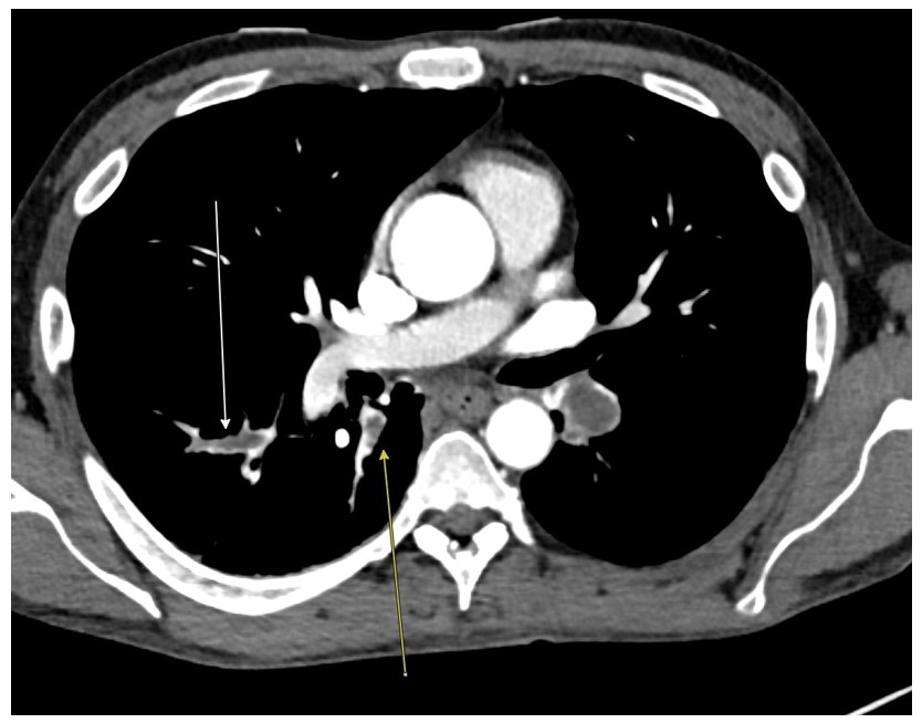
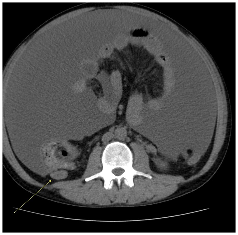
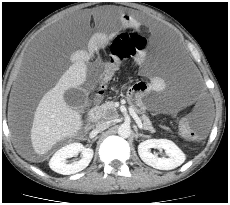
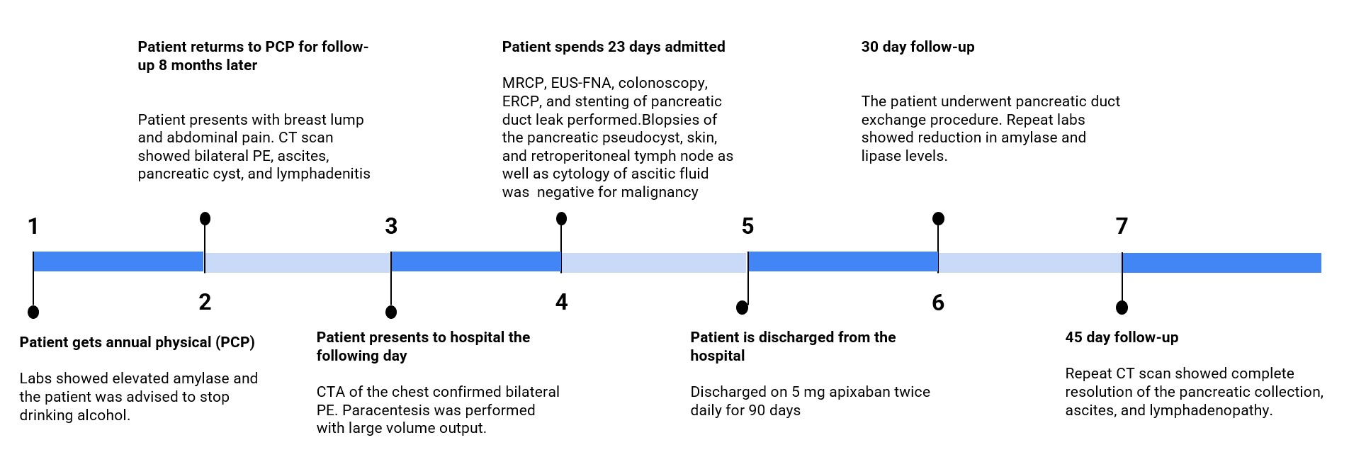
A 40-year-old man with no significant past medical or surgical history presented to his primary care physician (PCP) for his annual physical exam. Labs were remarkable for elevated amylase at 143 U/L. Patient admits to drinking beer daily since he was 21 years old and was advised to cut down on his alcohol use. Eight months later, the patient presented to his PCP with a 2-week history of a painless left breast lump. He also reported mild abdominal pain, abdominal distension, and an unintentional weight loss of 10 pounds over the same period. He denied fever, chest pain, shortness of breath, nausea, vomiting, or diarrhea. Initial laboratory studies were significant for elevated amylase and lipase to 839 U/L and 1,753 U/L, respectively. Prolactin was mildly elevated to 20.6 ng/mL. Both the mammogram and the brain MRI were unremarkable, with no evidence of malignancy. A CT scan of the abdomen and pelvis revealed bilateral lower lobe PE (Figure 1) without right ventricular strain, inferior vena cava (IVC) thrombus, large volume abdominal and pelvic ascites, paraoesophageal and retroperitoneal lymph nodes (Figure 2), and a 1.8 cm pseudocyst within the pancreatic uncinate (Figure 3). The PCP subsequently informed the patient of these findings and advised immediate hospital evaluation.
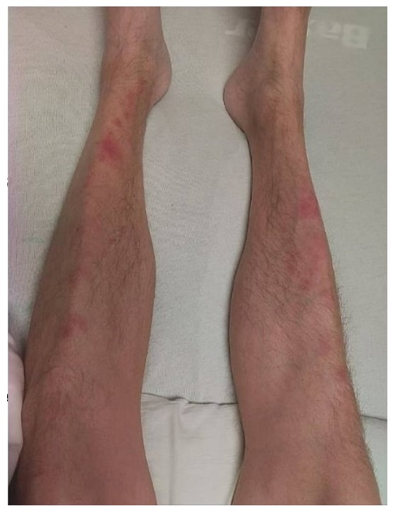
On admission, the patient was afebrile, tachycardic, tachypneic, and saturating well on room air. Physical examination revealed a palpable left breast lump, abdominal distension with diffuse tenderness on palpation, and multiple red, firm, and non-tender nodules on the lower extremities (Figure 4). Relevant laboratory findings included white blood cells (WBC) , platelets , ferritin 2,781 ng/mL, sodium 127 mEq/L, aspartate aminotransferase 79, alanine aminotransferase 96, and erythrocyte sedimentation rate 39. A CT Pulmonary Angiogram (CTPA) confirmed the findings of bilateral lower lobe PE. Biomarkers of PE, including high-sensitivity troponin and BNP, were within normal range. An echocardiogram was performed and demonstrated a left ventricular ejection fraction (LVEF) of 60% with no evidence of right ventricular (RV) strain. Therefore, the incidental PE was deemed low risk, and the patient was started on a heparin infusion. A right upper-quadrant ultrasound was negative for gallstones. The patient denies recent abdominal trauma or a family history of pancreatitis. Ascitic fluid was high in amylase (> 7,500) with a serum-to-ascites albumin gradient of 1.0, a lactic acid dehydrogenase level of 783 U/L, and a WBC count of 3,228 cells//mm with 27% neutrophils. No malignant cells were seen, and the patient’s Gram stain and acid-fast bacilli smear results were negative, with the culture remaining sterile. Cancer markers, including the cancer antigen 19-9, carcinoembryonic antigen (CEA), alpha fetoprotein, and beta HCG, were normal.
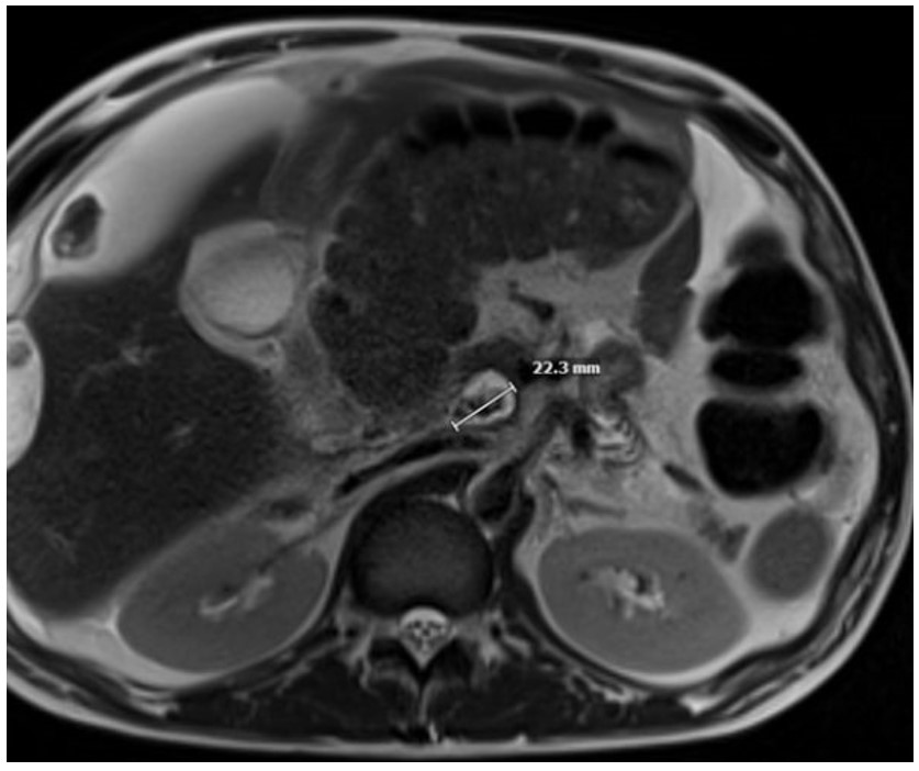
Magnetic Resonance Cholangiopancreatography (MRCP) showed a pancreatic pseudocyst (Figure 5) with nodularity adjacent to the ascending colon, along with the previously noted abnormalities seen on CT imaging. Subsequently, an endoscopic ultrasound-guided fine needle aspiration (EUS-FNA) of the pancreatic mass was performed, and cytopathology was negative for malignant cells. Colonoscopy revealed a normal colon, and no biopsies were obtained. A retroperitoneal lymph node biopsy was also negative for malignancy. A biopsy of the lower extremity lesions was consistent with pancreatic panniculitis, without evidence of malignancy. Due to the recurrent ascites, a MRCP with secretin stimulation was performed, confirming the presence of a pancreatic duct leak. An Endoscopic Retrograde Cholangiopancreatography (ERCP) was subsequently attempted; however, closure of the leak was not possible due to extensive peripancreatic inflammation. Infectious workup, including blood cultures and -D-glucan, was negative. A leukemia/lymphoma panel was also negative. Comprehensive rheumatologic workup – including antinuclear antibody (ANA), extractable nuclear antigen (ENA) panel, complement levels, total immunoglobulin (IgG) and subclasses (IgG1 – 4), lupus anticoagulant, -glycoprotein, and anticardiolipin antibodies – was unremarkable.
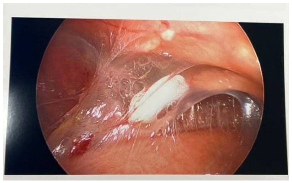
The patient subsequently underwent a diagnostic laparoscopy, which revealed extensive filmy adhesions tethering the bowel to the abdominal wall, without evidence of peritoneal carcinomatosis (Figure 6). No biopsies were taken, given extensive adhesions with risk to bowel and visceral injury. A decision was made to provide a pancreatic rest by placing a peritoneal drain and a Dobhoff tube for post-pyloric feeding, along with initiation of an octreotide drip. A week later, a repeat ERCP confirmed the presence of a pancreatic pseudocyst, and two pancreatic stents were placed into the ventral pancreatic duct, terminating into the pseudocyst. The patient was discharged on a direct oral anticoagulant, Apixaban 5 mg twice a day for 90 days. The hypercoagulable state that led to the formation of PE was deemed to be provoked by the AP, and therefore, a 3-month course of Apixaban was deemed appropriate. At his 30-day follow-up, pancreatic duct stent exchange was performed, and repeat labs demonstrated improvement in amylase and lipase levels with resolved thrombocytosis. A repeat CT scan at 45-day follow-up demonstrated complete resolution of the pancreatic pseudocyst, as well as resolution of the ascites and lymphadenopathy (Figure 7).
Discussion
PE is a very rare complication of AP. The underlying mechanism is likely multifactorial and may reflect extrinsic communication of the pseudocyst with the adjacent vasculature triggering the formation of a thrombus through the release of pancreatic enzymes along with intrinsic vascular changes due to hypercoagulability and proteolytic damage [7,8]. In our case, other possible mechanisms include reactive thrombosis or systemic inflammation. Our experience suggests that PE may be an underrecognized complication of AP, as most reported cases, including ours, were found incidentally after the diagnosis of AP had already been established [5]. In their review of reported cases of AP with PE, Yan et al. demonstrated that early identification and timely treatment of PE were associated with favorable outcomes, with no reported fatalities [5].
Lymphadenopathy may occur in autoimmune pancreatitis or when AP is the initial manifestation of an underlying malignancy, particularly lymphoma [2,9]. In fact, peripancreatic and hilar lymphadenopathy occur in approximately 80% of patients with type 1 autoimmune pancreatitis [2]. In our case, severe systemic inflammation likely led to reactive lymphadenopathy, as autoimmune and malignant etiologies were effectively ruled out and given full resolution on repeat CT after pancreatic leak control. However, complete tissue exclusion was not performed to confirm the latter hypothesis.
Prolactin is primarily synthesized and secreted by the pituitary gland but can also be produced by immune cells, including T cells, B cells, and macrophages- in response to cytokines [10]. In men, hyperprolactinemia can present with gynecomastia, erectile dysfunction, or decreased libido. In our case, the patient presented with a breast lump and mild hyperprolactinemia in the setting of AP, likely representing a nonspecific finding and reflecting an exaggerated immune response to systemic inflammation .
Pancreatic panniculitis is a rare but important systemic manifestation of pancreatic disease and supports our finding of pancreatic enzyme leakage. It results from circulating enzymes, particularly lipase, causing subcutaneous fat necrosis [11]. In our case, the evidence of pancreatic panniculitis helped unify our diagnosis of pancreatic duct leak with ascites and systemic inflammation, serving as an important clinical clue to ongoing pancreatic injury.
Conclusion
In conclusion, this case illustrates that incidental pulmonary embolism and reactive lymphadenopathy can accompany pancreatic duct leak and pancreatic ascites. Early recognition of these associations may help avoid diagnostic uncertainty, and treatment of the underlying pancreatic leak may lead to resolution of systemic findings.
Conflicts of Interest
The authors declare that they have no financial or non-financial competing interests, personal relationships, institutional affiliations, or other relationships or activities that could have influenced, or be perceived to have influenced, the conduct, interpretation, or reporting of this case report. All authors are responsible for the accuracy and completeness of this declaration.
Funding Source
The authors declare that no specific grant or funding was received for this research from any public, commercial, or not-for-profit funding agency.
Acknowledgments
None.
Informed Consent
Informed consent was obtained from the patient for publication of this case report and any accompanying clinical details and images. All efforts have been made to protect the patient’s identity, and no directly identifying personal information is included in the manuscript.
Large Language Model
None.
Authors Contribution
All authors were involved in the collection of data and drafting of the manuscript. All authors gave final approval of the version to be published and agreed to be accountable for aspects of the work.
Data Availability
Data sharing is not applicable to this article, as it is based on a single-patient case report, and no datasets were generated or analyzed.
References
- Valverde-Lopez F., Martinez-Cara J. G., Redondo-Cerezo E.. Acute pancreatitis. Med Clin (Barc). 2022;158(11):556-563. doi:10.1016/j.medcli.2021.12.012 PMID: 35277268
- Hamano H., Arakura N., Muraki T., Ozaki Y., Kiyosawa K., Kawa S.. Prevalence and distribution of extrapancreatic lesions complicating autoimmune pancreatitis. J Gastroenterol. 2006;41(12):1197-205. doi:10.1007/s00535-006-1908-9 PMID: 17287899
- Kalas M. A., Leon M., Chavez L. O., Canalizo E., Surani S.. Vascular complications of pancreatitis. World J Clin Cases. 2022;10(22):7665-7673. doi:10.12998/wjcc.v10.i22.7665 PMID: 36158481 PMCID: PMC9372863
- Ohta H., Hachiya T.. A case of inferior vena cava thrombosis and pulmonary embolism secondary to acute exacerbation of chronic pancreatitis: a rare finding in radionuclide venography. Ann Nucl Med. 2002;16(2):147-9. doi:10.1007/BF02993720 PMID: 12043910
- Yan L. L., Li S. W., Fang Y. X., Yan X. D., He B. L.. A Good Prognosis of Patients with Acute Pancreatitis Combined with Pulmonary Embolism: Early Identification and Intervention. Curr Med Imaging. 2024;20:e15734056275789. doi:10.2174/0115734056275789231129102057 PMID: 38389370
- Zhang Q., Zhang Q. X., Tan X. P., Wang W. Z., He C. H., Xu L., Huang X. X.. Pulmonary embolism with acute pancreatitis: a case report and literature review. World J Gastroenterol. 2012;18(6):583-6. doi:10.3748/wjg.v18.i6.583 PMID: 22363127 PMCID: PMC3280406
- Lungarella G., Gardi C., de Santi M. M., Luzi P.. Pulmonary vascular injury in pancreatitis: evidence for a major role played by pancreatic elastase. Exp Mol Pathol. 1985;42(1):44-59. doi:10.1016/0014-4800(85)90017-6 PMID: 3843961
- Piercy K. T., Ayerdi J., Geary R. L., Hansen K. J., Edwards M. S.. Acute pancreatitis: A complication associated with rheolytic mechanical thrombectomy of deep venous thrombosis. J Vasc Surg. 2006;44(5):1110-3. doi:10.1016/j.jvs.2006.06.030 PMID: 17098552
- Saif M. W., Khubchandani S., Walczak M.. Secondary pancreatic involvement by a diffuse large B-cell lymphoma presenting as acute pancreatitis. World J Gastroenterol. 2007;13(36):4909-11. doi:10.3748/wjg.v13.i36.4909 PMID: 17828824 PMCID: PMC4611771
- Barrett R., Narasimhulu C. A., Parthasarathy S.. Adrenergic hormones induce extrapituitary prolactin gene expression in leukocytes-potential implications in obesity. Sci Rep. 2018;8(1):1936. doi:10.1038/s41598-018-20378-1 PMID: 29386559 PMCID: PMC5792634
- Rongioletti F., Caputo V.. Pancreatic panniculitis. G Ital Dermatol Venereol. 2013;148(4):419-25. PMID: 23900163