Life-Threatening Pulmonary Embolism in a Patient with Suspected Antiphospholipid Syndrome and Severe Thrombocytopenia: A Case Report
Ahmed HassanⒾ1*, Mohamed Hamouda Elkasaby2, Mostafa Sabry Elshahat1, Omar Nasr Elshafey1, Islam Ebeid3, Ahmed AliⒾ1
- 1Department of Cardiology, Suez Medical Complex, Egypt Healthcare Authority, Suez, Egypt
- 2Faculty of Medicine, Al-Azhar University, Cairo, Egypt
- 3Department of Cardiology, National Institute of Diabetes and Endocrinology, Giza, Egypt.
Abstract
Antiphospholipid syndrome (APS) is an autoimmune disorder characterized by vascular thrombosis and obstetric complications in the presence of persistent antiphospholipid antibodies (aPL). Pulmonary embolism (PE), a frequent thrombotic manifestation of APS, can be life-threatening when associated with right ventricular dysfunction. We present the case of a 33-year-old woman with tachypnea, tachycardia, hypoxemia, and syncope. An ECG revealed characteristics of PE (S1Q3T3), and imaging confirmed intermediate-high-risk PE with right ventricular dysfunction, including a total occlusion of the left pulmonary artery and partial obstruction of the right pulmonary artery. Laboratory studies demonstrated aPL positivity alongside severe thrombocytopenia, significantly elevating her bleeding risk with standard anticoagulation therapy. This case highlights the diagnostic and therapeutic challenges in managing suspect APS-related PE with concomitant thrombocytopenia.
Keywords: Pulmonary embolism, Antiphospholipid syndrome, Thrombocytopenia
Article information
Introduction
Antiphospholipid syndrome (APS) is an acquired autoimmune disorder causing hypercoagulability, presenting with pregnancy complications, venous/arterial thromboembolism, thrombocytopenia, and microvascular disease due to circulating antiphospholipid antibodies (aPL) [1,2]. Available data indicate an annual incidence of 1-2 per 100,000 population and a prevalence of 40-50 per 100,000 adults based on population studies [3,4].
Deep venous thrombosis (DVT) represents the predominant venous manifestation in APS, occurring in 30-40% of patients, with lower extremity involvement being most common [5,6,7]. Pulmonary embolism affects 11-20% of individuals and serves as the initial presentation in 9-12% of cases, typically secondary to lower limb thrombosis rather than occurring as an isolated event [8]. These complications can progress to portal hypertension and cirrhosis, presenting with variable severity from asymptomatic disease to fulminant hepatic failure [9].
We report a challenging clinical case involving a patient with intermediate-high-risk pulmonary embolism and associated right heart strain requiring anticoagulation therapy, complicated by concurrent thrombocytopenia that significantly increased the risk of bleeding. Written informed consent was obtained from the patient to report this case.
Case Presentation
A 33-year-old woman presented to the emergency department (June 11, 2025) with 2 days of dyspnea, palpitations, and tinnitus, followed by syncopal episodes.
Clinical Findings: Physical examination revealed blood pressure (95/60), tachycardia (113 bpm), tachypnea (26 breaths/minute), and hypoxemia (SpO 84% on room air), with a significant medical history including autoimmune hepatitis, liver cirrhosis, gastric varices treated by endoscopy, and pre-eclampsia complicated by preterm delivery and fetal demise at 28 weeks; her family history was notable for SLE (sister) and breast cancer (mother).
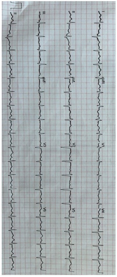
Laboratory Results: Laboratory evaluation revealed anemia, severe thrombocytopenia, prolonged PTT, and elevated troponin levels (48 ng/L), with detailed results summarized in Table 1. ECG demonstrated sinus tachycardia with an S1Q3T3 pattern (prominent S wave in I, Q wave in III, and negative T wave in III) (Figure 1).
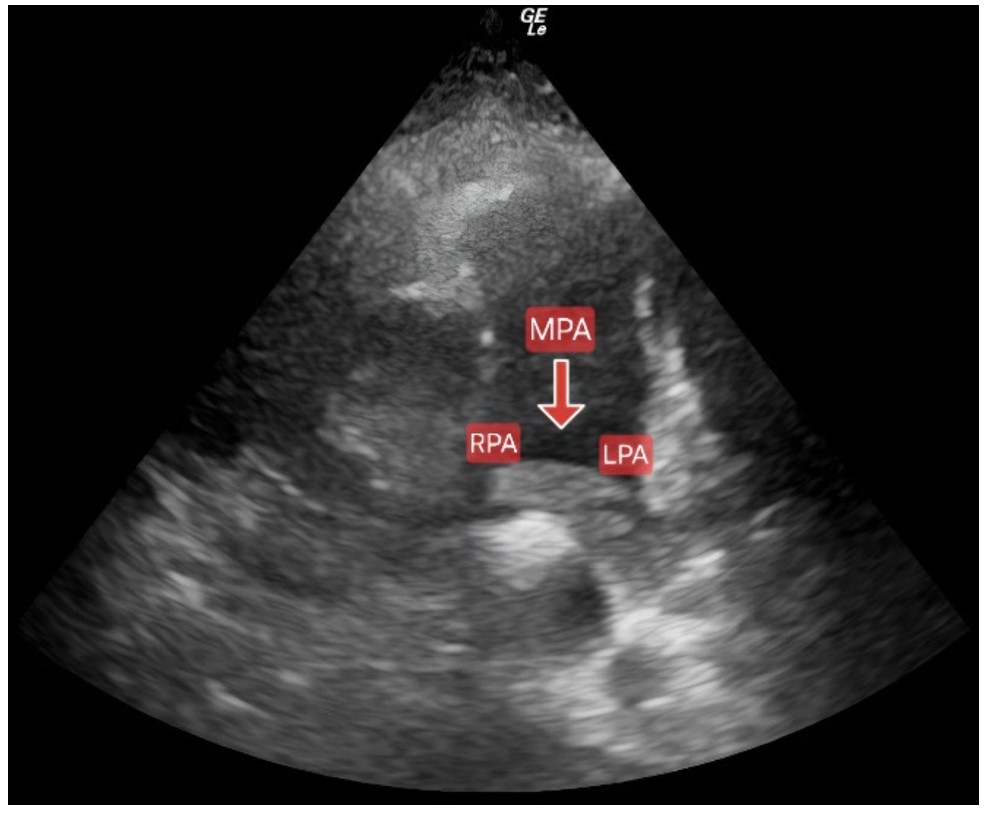
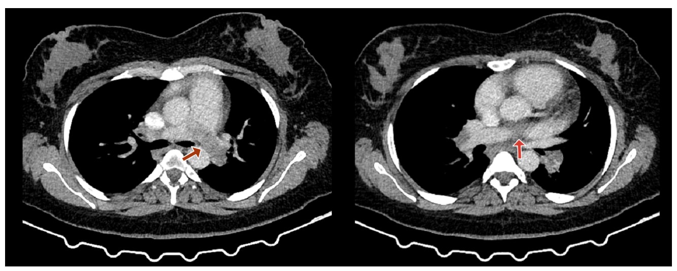
Imaging Studies: Echocardiography demonstrated preserved left ventricular function (ejection fraction 64%) with mild right heart dilation. Right ventricular systolic pressure (RVSP) measured 55 mmHg on June 12, and the RV/LV ratio was above 1. The D-shaped septum confirmed right heart strain. Imaging identified dilatation of the main pulmonary artery and thrombus occluding the left pulmonary artery with partial right pulmonary artery obstruction Figure 2. CT pulmonary angiography confirmed massive hypodense thrombi at the main pulmonary artery bifurcation, extending into left pulmonary arteries and proximal branches, with lesser right-sided involvement Figure 3. Duplex ultrasound showed a short segment of the right superficial femoral vein with partially compressible lumen and hyper- to isoechoic thrombus with partial recanalization, consistent with chronic DVT. No acute thrombotic component was identified. Pelvic abdominal ultrasound revealed cirrhotic changes and splenomegaly (15 cm). CT brain and carotid/vertebral Doppler studies showed normal findings.
Coagulation studies demonstrated a weakly positive direct Coombs test. Serologic evaluation revealed elevated antinuclear antibodies (ANA; index 27.70) and increased anticardiolipin IgG levels (19.60 GPL U/mL). The dRVVT screen ratio was 2.0, with a confirm ratio of 1.55. Comprehensive coagulation and immunological laboratory results are summarized in Table 2.
Treatment: The patient received a continuous unfractionated heparin infusion for two weeks, beginning with an initial bolus of 5,600 IU, followed by weight-based dosing at 18 IU/kg/hour (1,260 IU/hour for a 70 kg patient). Anticoagulation was closely monitored using aPTT, with a target range of 60 – 80 seconds, and dose adjustments as needed. Warfarin 7 mg daily was initiated prior to discharge with a 5-day overlap with heparin, targeting a therapeutic INR range of 2 – 3. The patient was also prescribed bisoprolol 5 mg daily and pantoprazole 40 mg daily. Corticosteroid therapy was initiated on hospital day 1 (day of admission) with methylprednisolone 1 g daily for 5 days, followed by oral prednisolone 60 mg daily. Respiratory therapy comprised ipratropium 500 mcg and budesonide 0.5 mg twice daily. No platelet transfusions were administered during hospitalization, as there was no active bleeding and platelet counts remained above the transfusion threshold .
Follow-up: Following initiation of UFH and corticosteroid therapy on Day 1, the patient showed gradual clinical improvement, with resolution of dyspnea, tachycardia, and hypoxemia over the first week. Platelet counts recovered gradually from 24 ×10³/µL at presentation to 50 ×10³/µL by Day 11, alongside a reduction in RVSP from 55 mmHg to 33 mmHg on echocardiography. Warfarin was introduced on Day 14 with a 5-day heparin overlap; therapeutic INR was achieved by Day 19, at which point heparin was discontinued. By Day 23, the patient was ambulatory and symptom-free, with a platelet count of 140,000/µL and an INR of 2.2. At outpatient review on September 12, 2025, lupus anticoagulant positivity was confirmed on repeat testing (dRVVT screen ratio 2.0, confirm ratio 1.55). Bone marrow biopsy on December 1, 2025, showed mildly hypercellular marrow with adequate megakaryocytes and no infiltration.
Discussion
Pulmonary embolism (PE) remains a major cause of cardiovascular morbidity and mortality, and early risk stratification is crucial to guide management [10]. The presence of APS, a systemic autoimmune disorder associated with a hypercoagulable state, further increases the risk of VTE, including PE, often in young individuals without classic cardiovascular risk factors. Although PE management is generally well established, treatment becomes considerably more challenging when thrombotic and bleeding risks coexist, as in this patient with suspected APS. Intermediate-high-risk PE is characterized by hemodynamic stability, RV dysfunction, and elevated troponin, which typically warrants prompt anticoagulation as the first-line intervention [11]. Here, we report a case of intermediate- high-risk PE in a young woman with suspected APS and severe thrombocytopenia.
The patient’s presentation with acute dyspnea, syncope, tachycardia, and hypoxemia is highly suggestive of an acute cardiopulmonary event, with PE being a leading differential diagnosis in this clinical context. Notably, syncope is recognized as a marker of hemodynamic compromise in PE, often indicating significant RV dysfunction or obstruction of major pulmonary arteries. The presence of an S1Q3T3 pattern on ECG, while non-specific, is a classic indicator of acute RV strain and, when coupled with the clinical presentation, further increases the pre-test probability for PE. Echocardiographic findings of RV dilation, septal flattening, and elevated RVSP provided additional objective evidence of RV dysfunction, satisfying ESC criteria for intermediate-high-risk PE and necessitating urgent confirmatory imaging. CTPA, the current gold standard for PE diagnosis, confirmed massive thrombotic obstruction of the left pulmonary artery [10,12]. Importantly, beyond the acute presentation, the patient’s history of autoimmune hepatitis, liver cirrhosis, pre-eclampsia, and fetal demise raised early suspicion for an underlying prothrombotic condition, particularly APS [12]. In this case, the patient exhibited both significant anatomical obstruction on CTPA and functional impairment of the RV, fulfilling the criteria for intermediate-high-risk PE [12]. The subsequent identification of lupus anticoagulant and anticardiolipin antibodies strongly supported a suspected diagnosis of APS, offering a unifying explanation for both the thrombotic event and the associated thrombocytopenia, and further underscoring the need for long-term anticoagulation and close follow-up.
Thrombocytopenia is a recognized, though often underappreciated, hematologic manifestation of APS. Its pathogenesis is multifactorial, primarily involving immune-mediated platelet destruction, impaired platelet production, and increased consumption in the context of active thrombosis. In this patient, thrombocytopenia is likely multifactorial, with contributions not only from APS but also from underlying autoimmune hepatitis and portal hypertension. Autoimmune hepatitis, as part of a broader autoimmune diathesis, may contribute to immune-mediated platelet destruction, while portal hypertension and splenomegaly associated with liver cirrhosis promote splenic sequestration and reduced thrombopoietin production, further aggravating thrombocytopenia [13].
| Parameter | D1 | D2 | D3 | D4 | D5 | D6 | D7 | D8 | D9 | D10 | D11 | D12 | D14 | D15 | D17 | D19 | D21 | D22 | D23 | Reference Range |
|---|---|---|---|---|---|---|---|---|---|---|---|---|---|---|---|---|---|---|---|---|
| HB | 10.7 | 9.8 | 10 | 10.2 | 10 | 10.3 | 10.3 | 10.5 | 10.5 | 10.6 | 10.6 | 10.7 | 10.9 | 10.7 | 10.8 | 10.8 | 10.8 | 10.8 | 10.8 | 12-15 g/dL |
| TLC | 5 | - | - | 4.2 | - | 5.9 | - | - | 7.4 | - | - | 7.3 | 9.2 | - | 7.6 | - | - | - | 6.1 | 4000-11000 cells/uL |
| PLT | 24 | 25 | 24 | 25 | 28 | 30 | 30 | 35 | 40 | 44 | 50 | 60 | 90 | 110 | 110 | 114 | 127 | 130 | 140 | 150000-450000/uL |
| INR | 1.2 | 1.2 | 1.2 | 1.4 | 1.3 | 1.3 | 1.4 | 1.5 | 1.7 | 1.9 | 2 | 2 | 2.1 | 2.1 | 2.5 | 2.3 | 2.2 | 2.4 | 2.2 | 0.9-1.1 |
| PTT | 58.49 | 58 | 60 | 60 | 62 | 63 | 65 | 65 | 66 | 67 | 67 | 68 | 68 | 70 | 75 | 73 | 73 | 76 | 70 | 25-37 |
| CRP | 5 | - | - | - | - | - | - | 7 | - | - | - | - | - | - | - | - | - | - | - | 1 mg/dL |
| Urea | 26 | - | 29 | 32 | - | 55 | 55 | 55 | 48 | 27 | 46 | 36 | 52 | - | 38 | - | - | - | - | 7-30 mg/dL |
| Creat | 1.2 | 0.6 | 0.7 | 1.01 | - | 1.26 | 1.2 | 0.7 | 0.7 | 0.8 | 1.02 | 0.8 | 0.9 | - | 0.8 | - | - | - | - | 0.6-1.1 mg/dL |
| Na | 135 | - | - | 138 | - | 137 | - | - | 138 | - | 135 | - | 135 | - | 140 | - | 134 | - | 138 | 135-145 mEq/L |
| K | 3.9 | - | - | 3.9 | - | - | 4.3 | - | 3.5 | - | 4.2 | - | - | 4.2 | - | - | - | - | 4 | 3.5-5 mEq/L |
| ALT | 27 | - | - | - | - | 32 | - | - | 32 | 31 | - | - | - | - | 36 | - | - | - | - | 7-55 U/L |
| AST | 23 | - | - | - | - | 29 | - | - | - | - | 16 | - | - | 29 | - | 18 | - | - | 22 | 10-40 U/L |
HB, hemoglobin; TLC, total leukocyte count; PLT, platelet count; INR, international normalized ratio; PTT, partial thromboplastin time; CRP, C-reactive protein; Na, sodium; K, potassium; ALT, alanine aminotransferase; AST, aspartate aminotransferase.
| Section A: Antiphospholipid Antibody (aPL) Tests | ||||
|---|---|---|---|---|
| Test / Parameter | June 13, 2025 | Sep 12, 2025 | Reference Range | Units |
| Anti-Cardiolipin IgG | 19.60 | — | < 10.0 | GPL U/mL |
| Anti-Cardiolipin IgM | 3.4 | — | < 7.0 | MPL U/mL |
| Antinuclear Antibody (ANA) | 27.70 | — | < 10.0 | Index |
| Section B: Lupus Anticoagulant (LA) Screening & Confirmatory Tests | ||||
| Screening | ||||
| Lupus Anticoagulant (LAC) — clotting time | 36.00 | — | 34–44 | seconds |
| dRVVT Screen Ratio | — | 2.00 | < 1.2 | ratio |
| Confirmatory | ||||
| dRVVT Confirm Ratio | — | 1.55 | 1.20 | ratio |
| LAC Normalized Ratio | — | 1.30 | < 1.2 | ratio |
| Section C: Complement Levels | ||||
| C3 | 91 | — | 90–180 | mg/dL |
| C4 | 13 | — | 15–40 | mg/dL |
| Section D: Coagulation & Thrombophilia Screen | ||||
| Protein C Activity | — | 64.3 | 72–160 | % |
| Free Protein S Antigen | — | 55.5 | 60–150 | % |
| Antithrombin III Activity | — | 99 | 80–120 | % |
| Factor V Leiden Mutation | — | Not detected | Not detected | — |
| Section E: Other Investigations | ||||
| Direct Coombs Test | Weakly positive | — | Negative | — |
Bone Marrow Biopsy (December 1, 2025): Mildly hypercellular marrow with trilineage hematopoiesis, mild erythrocyte hyperplasia, and adequate megakaryocytes with a mild increase in reticular fibers. No infiltration by non-hematopoietic cells. Abbreviations: aPL, antiphospholipid antibodies; ANA, antinuclear antibody; dRVVT, dilute Russell's viper venom time; LAC, lupus anticoagulant; GPL, IgG phospholipid units; MPL, IgM phospholipid units; ‘—’, test not performed at that time point.
While mild to moderate thrombocytopenia is frequently observed in APS, severe thrombocytopenia is uncommon, occurring in approximately 10 – 15% of cases, and presents a major therapeutic dilemma, particularly in the setting of acute VTE or PE [14]. Management of anticoagulation in APS patients with severe thrombocytopenia remains a significant challenge, as current APS guidelines do not provide clear recommendations for this scenario [14]. However, the coexistence of life-threatening thrombosis requiring anticoagulation or thrombolysis and a markedly elevated bleeding risk due to profound thrombocytopenia and varices represents an uncommon but significant therapeutic dilemma [10,12].
Given absolute contraindications to thrombolysis, an individualized conservative strategy was employed. Despite the complexity of intermediate-high-risk PE in the context of severe thrombocytopenia and variceal bleeding risk, cautious parenteral heparin anticoagulation, followed by transition to a vitamin K antagonist according to recent AHA/ACC PE guideline recommendations [15], combined with corticosteroid therapy for immune-mediated thrombocytopenia, was both safe and effective. Reperfusion therapy was not indicated due to hemodynamic stability and elevated bleeding risk. Systemic and catheter-directed thrombolysis were contraindicated by severe thrombocytopenia and cirrhosis; the presence of previously treated gastric varices further elevated the bleeding risk, though detailed endoscopic assessment was not available at the time of presentation. Surgical embolectomy and ECMO were unnecessary, and IVC filter placement was deferred given the feasibility of anticoagulation. Notably, this approach resulted in gradual improvement of platelet counts, enabling the continuation of full anticoagulation without major bleeding complications. The patient’s subsequent clinical and radiological improvement, including resolution of hypoxia, reduction in RVSP, and partial thrombus resolution, highlights the success of this carefully tailored treatment plan. This case demonstrates that, even in the most complex and intermediate-high-risk PE presentations, favorable outcomes can be achieved through judicious modification of standard treatment protocols based on individual patient factors and multidisciplinary input.
This case has several limitations. Anti- glycoprotein I antibodies were not assessed due to laboratory constraints. However, persistent lupus anticoagulant positivity (elevated dRVVT screen/confirm ratios and anticardiolipin IgG) supports the diagnosis of antiphospholipid syndrome in the appropriate clinical context. A complete hemolysis workup was not available; therefore, Evans syndrome could not be definitively excluded. Advanced anticoagulation monitoring (e.g., chromogenic factor X levels) was also unavailable, and clinical findings and current recommendations guided management. Despite these limitations, the favorable outcome supports the individualized therapeutic approach in this complex suspected APS-associated PE case.
Conclusion
We report a complex case of intermediate-high-risk pulmonary embolism in a young woman with suspected APS and severe thrombocytopenia. The coexistence of life-threatening thrombosis and contraindications to standard reperfusion posed major diagnostic and therapeutic challenges. A tailored, multidisciplinary strategy with cautious anticoagulation and immunomodulatory therapy resulted in a favorable outcome.
Conflicts of Interest
The authors declare no competing interests that could have influenced the objectivity or outcome of this research.
Funding Source
The authors declare that no specific grant or funding was received for this research from any public, commercial, or not-for-profit funding agency.
Acknowledgments
The authors sincerely acknowledge Mohamed Helmy Soliman for his valuable assistance in contacting the patient, providing relevant investigation results, and printing the CT angiography images for clinical documentation.
Informed Consent
Written informed consent was obtained from the patient for publication of this case report and any accompanying images.
Large Language Model
None.
Authors Contribution
AH contributed to case identification, data curation, validation, and writing of the original draft. MHE was involved in writing the original draft. MSE and ONE contributed to data curation, validation, and references. IE participated in writing, review, and editing. AA provided supervision and contributed to writing, review, and editing.
Data Availability
No new datasets were generated or analyzed during the current study. All relevant clinical information supporting the findings of this case report is included in the article. Additional details are not publicly available to protect patient privacy and confidentiality.
References
- Gaspar P., Sciascia S., Tektonidou M. G.. Epidemiology of antiphospholipid syndrome: macro- and microvascular manifestations. Rheumatology (Oxford). 2024;63(SI):SI24-SI36. doi:10.1093/rheumatology/kead571 PMID: 38320589 PMCID: PMC10846913
- Miyakis S., Lockshin M. D., Atsumi T., Branch D. W., Brey R. L., Cervera R., Derksen R. H., PG D. E. Groot, Koike T., Meroni P. L., Reber G., Shoenfeld Y., Tincani A., Vlachoyiannopoulos P. G., Krilis S. A.. International consensus statement on an update of the classification criteria for definite antiphospholipid syndrome (APS). J Thromb Haemost. 2006;4(2):295-306. doi:10.1111/j.1538-7836.2006.01753.x PMID: 16420554
- Dabit J. Y., Valenzuela-Almada M. O., Vallejo-Ramos S., Duarte-Garcia A.. Epidemiology of Antiphospholipid Syndrome in the General Population. Curr Rheumatol Rep. 2022;23(12):85. doi:10.1007/s11926-021-01038-2 PMID: 34985614 PMCID: PMC8727975
- Sciascia S., Radin M., Unlu O., Erkan D., Roccatello D.. Infodemiology of antiphospholipid syndrome: Merging informatics and epidemiology. Eur J Rheumatol. 2018;5(2):92-95. doi:10.5152/eurjrheum.2018.17105 PMID: 30185355 PMCID: PMC6072687
- Bertero M. T., Bazzan M., Carignola R., Montaruli B., Silvestro E., Sciascia S., Vaccarino A., Baldovino S., Roccatello D., Antiphospholipid Piedmont Consortium. Antiphospholipid syndrome in northwest Italy (APS Piedmont Cohort): demographic features, risk factors, clinical and laboratory profile. Lupus. 2012;21(7):806-9. doi:10.1177/0961203312446974 PMID: 22635240
- Cervera R., Piette J. C., Font J., Khamashta M. A., Shoenfeld Y., Camps M. T., Jacobsen S., Lakos G., Tincani A., Kontopoulou-Griva I., Galeazzi M., Meroni P. L., Derksen R. H., de Groot P. G., Gromnica-Ihle E., Baleva M., Mosca M., Bombardieri S., Houssiau F., Gris J. C., Quere I., Hachulla E., Vasconcelos C., Roch B., Fernandez-Nebro A., Boffa M. C., Hughes G. R., Ingelmo M., Euro-Phospholipid Project Group. Antiphospholipid syndrome: clinical and immunologic manifestations and patterns of disease expression in a cohort of 1,000 patients. Arthritis Rheum. 2002;46(4):1019-27. doi:10.1002/art.10187 PMID: 11953980
- Sevim E., Zisa D., Andrade D., Sciascia S., Pengo V., Tektonidou M. G., Ugarte A., Gerosa M., Belmont H. M., Zamorano M. A. A., Fortin P. R., Ji L., Efthymiou M., Cohen H., Branch D. W., de Jesus G. R., Andreoli L., Petri M., Rodriguez E., Cervera R., Knight J. S., Atsumi T., Willis R., Roubey R., Bertolaccini M. L., Erkan D., Barbhaiya M., APS ACTION Investigators. Characteristics of Patients With Antiphospholipid Antibody Positivity in the APS ACTION International Clinical Database and Repository. Arthritis Care Res (Hoboken). 2022;74(2):324-335. doi:10.1002/acr.24468 PMID: 32986935 PMCID: PMC10725727
- Shi M., Gao W., Jin Y., Zhu J., Liu Y., Wang T., Li C.. Antiphospholipid Syndrome-Related Pulmonary Embolism: Clinical Characteristics and Early Recognition. Front Cardiovasc Med. 2022;9:872523. doi:10.3389/fcvm.2022.872523 PMID: 35898271 PMCID: PMC9309373
- Espinosa G., Font J., Garcia-Pagan J. C., Tassies D., Reverter J. C., Gaig C., Cervantes F., Cervera R., Bosch J., Ingelmo M.. Budd-Chiari syndrome secondary to antiphospholipid syndrome: clinical and immunologic characteristics of 43 patients. Medicine (Baltimore). 2001;80(6):345-54. doi:10.1097/00005792-200111000-00001 PMID: 11704712
- Konstantinides S. V., Meyer G., Becattini C., Bueno H., Geersing G. J., Harjola V. P., Huisman M. V., Humbert M., Jennings C. S., Jimenez D., Kucher N., Lang I. M., Lankeit M., Lorusso R., Mazzolai L., Meneveau N., Ni Ainle F., Prandoni P., Pruszczyk P., Righini M., Torbicki A., Van Belle E., Zamorano J. L., ESC Scientific Document Group. 2019 ESC Guidelines for the diagnosis and management of acute pulmonary embolism developed in collaboration with the European Respiratory Society (ERS). Eur Heart J. 2020;41(4):543-603. doi:10.1093/eurheartj/ehz405 PMID: 31504429
- Ambati A., Knight J. S., Zuo Y.. Antiphospholipid syndrome management: a 2023 update and practical algorithm-based approach. Curr Opin Rheumatol. 2023;35(3):149-160. doi:10.1097/BOR.0000000000000932 PMID: 36866678 PMCID: PMC10364614
- Tektonidou M. G., Andreoli L., Limper M., Amoura Z., Cervera R., Costedoat-Chalumeau N., Cuadrado M. J., Dorner T., Ferrer-Oliveras R., Hambly K., Khamashta M. A., King J., Marchiori F., Meroni P. L., Mosca M., Pengo V., Raio L., Ruiz-Irastorza G., Shoenfeld Y., Stojanovich L., Svenungsson E., Wahl D., Tincani A., Ward M. M.. EULAR recommendations for the management of antiphospholipid syndrome in adults. Ann Rheum Dis. 2019;78(10):1296-1304. doi:10.1136/annrheumdis-2019-215213 PMID: 31092409 PMCID: PMC11034817
- Peck-Radosavljevic M.. Thrombocytopenia in chronic liver disease. Liver Int. 2017;37(6):778-793. doi:10.1111/liv.13317 PMID: 27860293
- Zuily S., Cervera R., Foret T., Bertocchi S., Tincani A.. Thrombocytopenia in antiphospholipid syndrome: Is anticoagulation and/or antiaggregation always required?. Autoimmun Rev. 2024;23(1):103417. doi:10.1016/j.autrev.2023.103417 PMID: 37619905
- Writing Committee Members, Creager M. A., Barnes G. D., Giri J., Mukherjee D., Jones W. S., Burnett A. E., Carman T., Casanegra A. I., Castellucci L. A., Clark S. M., Cushman M., de Wit K., Eaves J. M., Fang M. C., Goldberg J. B., Henkin S., Johnston-Cox H., Kadavath S., Kadian-Dodov D., Keeling W. B., Klein A. J. P., Li J., McDaniel M. C., Moores L. K., Piazza G., Prenger K. S., Pugliese S. C., Ranade M., Rosovsky R. P., Russo F., Secemsky E. A., Sista A. K., Tefera L., Weinberg I., Westafer L. M., Young M. N.. 2026 AHA/ACC/ACCP/ACEP/CHEST/SCAI/SHM/SIR/SVM/SVN Guideline for the Evaluation and Management of Acute Pulmonary Embolism in Adults: A Report of the American College of Cardiology/American Heart Association Joint Committee on Clinical Practice Guidelines. Circulation. 2026;153(12):e977-e1051. doi:10.1161/CIR.0000000000001415 PMID: 41712677