Cellular Neurothekeoma with Atypical Histologic Features in the Thigh: A Case Report
Abdulkarim HasanⒾ1,2*, Khalid Nafie1, Okba Merabet1, Stephen Osasan1, Samira Attar1,3, Mohammed S. Abdelwahed2,4, Ali Nagi5
- 1Department of Pathology and Laboratory, Prince Mishari bin Saud Hospital, Al-Baha Health Cluster, Ministry of Health, Baljurashi, Saudi Arabia
- 2Department of Pathology, Faculty of Medicine, Al-Azhar University, Cairo, Egypt
- 3Department of Pathology, Lakhdari Mohamed ElAkhdar Hospital, Touggourt, Algeria
- 4Department of Basic Medical Sciences, Pathology Division, College of Medicine, University of Jeddah, Jeddah, Saudi Arabia
- 5Department of Surgery, Prince Mishari bin Saud Hospital, Al-Baha Health Cluster, Ministry of Health, Baljurashi, Saudi Arabia
Abstract
Neurothekeoma is a rare, benign neoplastic process that most commonly presents on the head and neck of young adults. We report a case of cellular neurothekeoma in a middle-aged woman with a thigh lesion showing histologic features that closely mimicked malignant tumors, particularly undifferentiated pleomorphic sarcoma, sarcomatoid carcinoma, and amelanotic melanoma.
A dermal/subcutaneous thigh lesion demonstrated marked cytologic atypia and an infiltrative growth pattern on histologic evaluation. Immunohistochemistry supported the diagnosis of cellular neurothekeoma: tumor cells were positive for CD10 and showed very focal MITF staining, and negative for S100, SOX10, desmin, EMA, AE1/AE3, SMA, NSE, and factor XIIIa. The lesion was completely excised with clear margins. At 12 months of clinical follow-up, no recurrence was observed.
Cellular neurothekeoma should be considered in the differential diagnosis of atypical dermal soft-tissue masses with worrisome histologic features. Ancillary studies, particularly immunohistochemistry, are essential to exclude aggressive mimics and avoid overtreatment, although conclusions remain limited by the single-case nature of this report and the available literature.
Keywords: Cellular neurothekeoma, Immunohistochemistry, Neurothekeoma, Soft tissue tumor, Case report
Article information
Introduction
Cellular neurothekeoma is a benign cutaneous tumor that was first introduced in 1969 by Harkin and Reed, who described “nerve sheath myxoma” as a myxoid tumor of presumed neural origin [1]. Gallager and Helwig later reclassified these lesions as neurothekeoma [2,3]. Historically, neurothekeoma and nerve sheath myxoma were considered synonymous and thought to be of nerve sheath origin; however, several subsequent studies have suggested that cellular neurothekeoma is more likely a fibrohistiocytic lesion and histopathologically distinct from nerve sheath myxoma [3,4]. The histogenesis of cellular neurothekeoma remains uncertain and is a topic of debate [4].
Clinically, neurothekeoma is a slow-growing, often asymptomatic dermal swelling. It has been divided into three main histologic subtypes: myxoid, cellular, and mixed [3]. The myxoid subtype (corresponding to classic nerve sheath myxoma) is now considered a separate entity rather than a subtype of neurothekeoma [3,4]. In contrast, cellular neurothekeomas tend to pose a greater diagnostic challenge, particularly because of their histologic resemblance to other dermal and subcutaneous tumors and their lack of a completely specific immunophenotype [5].
| Marker | Neurothekeoma | Melanoma | Undifferentiated pleomorphic sarcoma | Sarcomatoid carcinoma |
|---|---|---|---|---|
| MITF | + | + | +/- | - |
| CD10 | + | - | + | - |
| NSE | + | - | - | - |
| S100 | - | + | - | - |
| MelanA | - | + | - | - |
| HMB45 | - | + | - | - |
| CD68 | - | - | + | - |
| SMA | - | - | - | -/+ |
| Desmin | - | - | - | - |
| EMA | - | - | - | + |
| AE1/AE3 | - | - | - | + |
| Factor XIIIa | - | - | + | - |
MITF, microphthalmia-associated transcription factor; NSE, neuron-specific enolase; SMA, smooth muscle actin; EMA, epithelial membrane antigen.
Classically, cellular neurothekeoma shows a dermal lobular growth pattern composed of nests and fascicles of epithelioid and/or spindled cells arranged in a swirling or fascicular pattern, with oval nuclei, pinpoint nucleoli, and abundant eosinophilic cytoplasm [6]. Nevertheless, several atypical features have been described, including cytologic atypia, prominent myxoid stroma, infiltrative growth, vascular or perineural invasion, plexiform architecture, and dense hyalinized stroma [7]. When such atypical features are present, the diagnosis of cellular neurothekeoma is particularly challenging, and worrisome lesions such as melanoma, undifferentiated pleomorphic sarcoma, and sarcomatoid carcinoma must be carefully excluded. Atypical cellular neurothekeomas, characterized by larger size, infiltrative borders, deep extension, or an increased mitotic rate, have been reported only in small series and case reports, and their biological behavior remains incompletely defined. Most lesions occur on the head and neck, shoulders, and upper extremities, whereas thigh involvement appears to be uncommon. In this context, we present a case of cellular neurothekeoma with markedly atypical histologic features and an infiltrative pattern arising at an uncommon anatomical site (thigh) in a middle-aged woman. In large series, most lesions have been reported on the head and neck, shoulder and upper extremities, whereas thigh involvement appears distinctly less frequent. This case underscores the diagnostic pitfalls posed by this tumor and highlights the value of a targeted immunohistochemical panel in establishing the correct diagnosis.
Case Presentation
A 40-year-old woman with no history of local trauma, prior surgery at the site, or exogenous hormonal therapy presented with a painless, well-defined mass on the left thigh that had slowly enlarged over 6 months. Examination revealed a firm dermal/subcutaneous soft-tissue nodule measuring 2.5 × 2.0 cm, with no overlying epidermal changes and no palpable regional lymphadenopathy. The initial clinical differential diagnosis included dermatofibroma and dermatofibrosarcoma protuberans. Preoperative ultrasound demonstrated a well-circumscribed superficial lesion without deep fascial or muscular involvement; therefore, MRI was not pursued. The lesion was excised in its entirety under local anesthesia and submitted for histopathologic evaluation. Histology showed an infiltrative atypical spindle-cell dermal neoplasm, prompting an immunohistochemical work-up. The combined morphologic and immunophenotypic findings, supported by external tertiary-center expert review, were consistent with cellular neurothekeoma. At 12 months of follow-up, there was no evidence of recurrence.
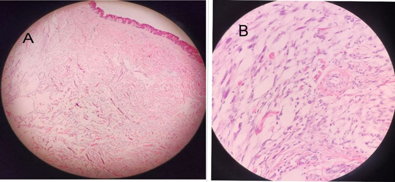
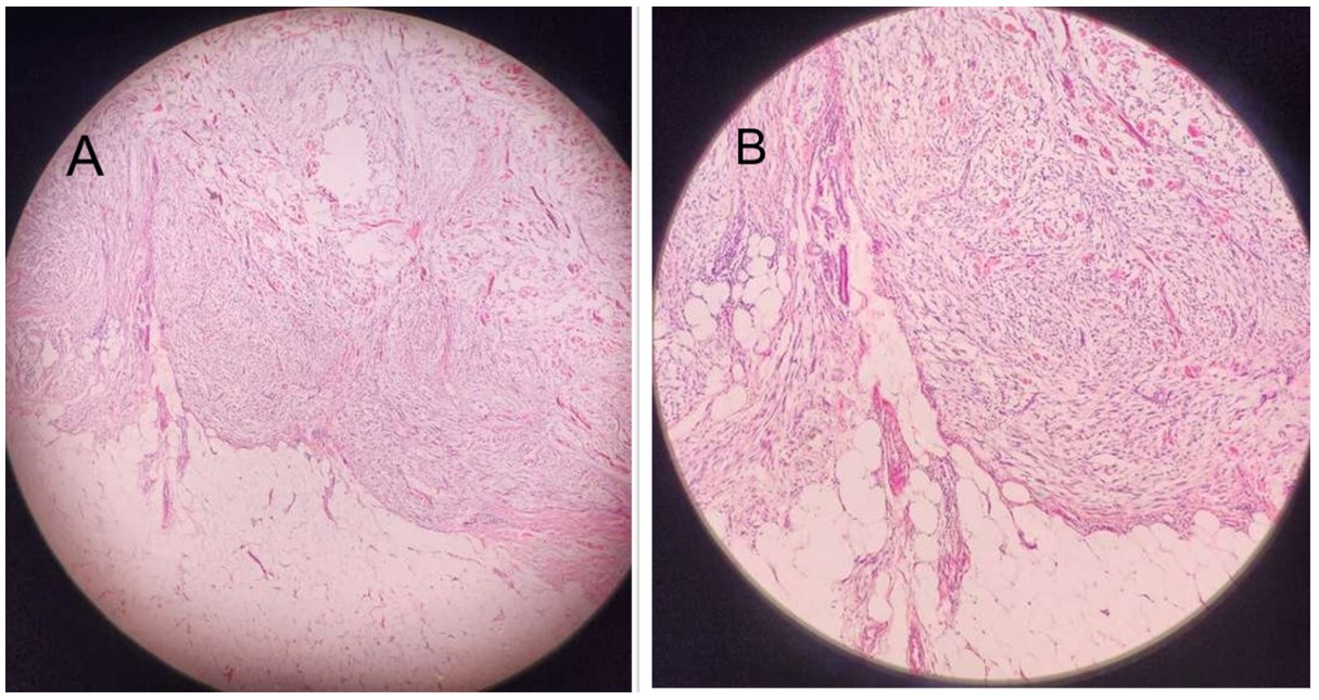
Microscopic examination of haematoxylin and eosin–stained sections showed a poorly circumscribed dermal spindle-cell neoplasm with focal superficial extension into the subcutis Figure 1. The tumor was composed of elongated cells arranged in nests, interconnecting fascicles, and scattered single cells, displaying an infiltrative pattern with extension between adnexal structures and splitting of reticular collagen bundles. The neoplastic cells demonstrated cytologic atypia, including enlarged hyperchromatic nuclei with occasional nucleoli, and focally vacuolated cytoplasm Figure 2. Mitotic activity was 3–5 mitoses per 10 high-power fields, without atypical mitotic figures. No geographic necrosis or conspicuous multinucleated giant cells were identified. Lymphovascular invasion and perineural invasion were absent. The lesion was completely excised, with the closest histologic margin measuring 1 mm. In view of the cytologic atypia and infiltrative pattern, the main histologic considerations were undifferentiated pleomorphic sarcoma, sarcomatoid carcinoma, and amelanotic melanoma.
Immunohistochemical studies were performed, and the paraffin blocks and representative slides were reviewed at a tertiary centre by a soft-tissue pathology expert. The tumour cells showed diffuse positivity for CD10, with very focal nuclear staining for MITF. They were negative for S100, desmin, EMA, SOX10, AE1/AE3, SMA, NSE, and factor XIIIa. Based on the morphologic and immunohistochemical findings, undifferentiated pleomorphic sarcoma was considered unlikely because of the lack of marked pleomorphism and multinucleation and the absence of factor XIIIa expression, while sarcomatoid carcinoma was excluded by the negative cytokeratin AE1/AE3 and EMA staining. Amelanotic melanoma was also considered an unlikely diagnosis given the histologic appearance and the absence of S100 and SOX10 expression. Taken together, these findings supported the diagnosis of cellular neurothekeoma.
The benign nature of the diagnosis and the low but not entirely negligible risk of local recurrence were explained, and the patient expressed relief and understanding of the surveillance plan. No recurrence was detected during the 12-month follow-up.
Discussion
Cellular neurothekeoma is a benign neoplastic soft-tissue lesion that represents a diagnostic challenge for both clinicians and pathologists. It predominantly affects women in the second to third decades of life. It typically presents as an erythematous, well-defined, solitary, slow-growing papule or nodule less than 2 cm in diameter [8,7]. In the large series by Fetsch et al., which included 178 tumors, most lesions were located on the head and neck, arms, and shoulders [9].
In contrast, our patient was a 40-year-old woman with a painless, relatively larger (2.5 cm) lesion situated at an unusual anatomical location (thigh). The pathogenesis of cellular neurothekeoma remains unclear. Some authors have proposed a possible triggering role for trauma or high estrogen levels in at least a subset of cases [10,11,8]. The broad clinical and histologic differential diagnosis includes not only benign and malignant neoplasms such as dermatofibroma, dermatofibrosarcoma protuberans, atypical fibroxanthoma, undifferentiated pleomorphic sarcoma, sarcomatoid carcinoma, and melanoma, but also reactive processes such as nodular fasciitis [9,8,7]. In the series by Fetsch et al., neurothekeoma was not suspected clinically in any of the 176 evaluable patients, underscoring that this entity is primarily a histologic and immunohistochemical diagnosis [9].
Typical neurothekeomas are well circumscribed on histological examination and consist of a dermal proliferation of spindle and stellate cells arranged in concentric or nested patterns, with generally bland cytologic features and limited mitotic activity. Focal nuclear enlargement, hyperchromasia, or multinucleation may be seen in some cases, but these changes are usually modest [12,13].
Typical (not absolute) immunohistochemical patterns of cellular neurothekeoma, compared with other lesions with atypical features, are shown in Table 1 [12].
A subset of cellular neurothekeomas, however, exhibits atypical or “worrisome” features, including larger tumor size, deeper extension into subcutaneous fat or skeletal muscle, vascular or perineural invasion, diffuse infiltrative borders, a high mitotic index ( mitoses per 10 high-power fields), and marked pleomorphism [14,15,7,13]. These atypical lesions are collectively referred to as atypical cellular neurothekeoma, and their biological behavior remains incompletely characterized due to the limited number of reported cases and relatively short follow-up [14,15,13]. Cases with one or more of these features are commonly referred to as atypical cellular neurothekeoma. Our case meets criteria for the atypical subset because it was >2 cm, had infiltrative borders, and showed increased mitotic activity, thereby fitting within this atypical subset.
The lesion in our patient showed several features that raised concern for malignancy, including poor circumscription, an infiltrative growth pattern, and cytologic atypia with mitotic figures. Conversely, there was no marked pleomorphism, necrosis, or prominent multinucleated giant-cell component, findings that would be more typical of undifferentiated pleomorphic sarcoma or other high-grade sarcomas [9,3,7]. In this setting, immunohistochemistry played a crucial role in refining the diagnosis.
Cellular neurothekeoma classically shows positivity for CD10, NKI/C3, and NSE, with variable expression of MITF and CD68, and is typically negative for S100 protein, SOX10, desmin, and cytokeratins [9,7]. In our case, NKI/C3, which is frequently reported as positive in cellular neurothekeoma, was not performed in our laboratory. Still, the tumor cells were diffusely positive for CD10, with very focal MITF staining, and were negative for S100, SOX10, AE1/AE3, EMA, desmin, SMA, NSE, and factor XIIIa. The absence of S100 and SOX10 expression effectively excluded conventional melanocytic lesions, including amelanotic melanoma, and the lack of cytokeratin (AE1/AE3) and EMA staining argued strongly against sarcomatoid carcinoma, as shown in the literature [9,8,7]. Undifferentiated pleomorphic sarcoma usually expresses vimentin and CD68 and may show factor XIIIa positivity, reflecting histiocytic differentiation [9,3,7]The negative factor XIIIa in our case, combined with the absence of diffuse pleomorphism and multinucleation, made this diagnosis unlikely.
Although NSE positivity is commonly reported in cellular neurothekeoma, negative or only focal expression has also been documented, and immunophenotypic variability is increasingly recognized, particularly in atypical examples. In practice, NSE is often included within initial panels for dermal spindle/epithelioid lesions because it can provide supportive evidence of neuroectodermal/perineural differentiation in the appropriate morphologic context; however, NSE lacks specificity, and its diagnostic utility in cellular neurothekeoma is adjunctive rather than decisive. Accordingly, NSE negativity does not exclude cellular neurothekeoma, and interpretation should prioritize the overall immunoprofile and the exclusion of key malignant mimics [12,9,7]. In this context, the overall immunoprofile in our patient remained compatible with cellular neurothekeoma.
Table 1 summarizes the typical immunohistochemical profile of cellular neurothekeoma and its main malignant mimics, including melanoma, undifferentiated pleomorphic sarcoma, and sarcomatoid carcinoma [12]. The pattern seen in our lesion, CD10 positivity with focal MITF and absence of S100, cytokeratins, EMA, and factor XIIIa, fits best with cellular neurothekeoma and was discordant with the profiles expected for melanoma, undifferentiated pleomorphic sarcoma, or sarcomatoid carcinoma.
In the present case, several clinicopathological features are noteworthy. First, the tumor occurred in a middle-aged woman, whereas most cellular neurothekeomas are reported in younger patients in the second and third decades of life [9,8,7]. Second, the lesion involved the thigh, an uncommon site compared with the more typical head and neck, shoulder, and upper-extremity locations [9,8,7]. Finally, the tumor exhibited a distinctly infiltrative growth pattern and significant cytological atypia that closely mimicked high-grade sarcoma and carcinoma; however, the overall morphologic and immunohistochemical profile remained most consistent with cellular neurothekeoma.
Most reported cellular neurothekeomas, including those with atypical features, have followed a benign course, with rare local recurrences and no convincing evidence of distant metastasis [14,12,9,15,7,13]. Nevertheless, the limited number of atypical cases and relatively short follow-up warrant cautious clinical surveillance. Our case adds to the growing body of evidence that histologic aggressiveness (cytologic atypia and infiltrative borders) in cellular neurothekeoma does not invariably imply clinically aggressive behavior, provided that other malignant entities are carefully excluded. However, this conclusion is based on a single case with 12 months of follow-up and on small published series, and longer-term data from larger cohorts are needed.
Molecular and cytogenetic studies were not performed in this case. To date, no recurrent or pathognomonic genetic alteration has been established for cellular neurothekeoma, and molecular data remain limited. In this context, the recently reported heterozygous NF1 c.4333-2dup variant in a neurothekeoma is an isolated finding of uncertain significance, but may contribute to clarifying the genetic background of these tumors [16].
Conclusion
Clinicians and pathologists should include cellular neurothekeoma in the differential diagnosis of dermal and subcutaneous soft-tissue masses, even when they arise at uncommon sites and demonstrate marked cytological atypia with an infiltrative growth pattern. In such cases, histologic features alone may strongly suggest high-grade malignancy. A carefully selected immunohistochemical panel, interpreted in the light of morphology and clinical context, may help avoid misdiagnosis and unnecessary aggressive treatment. This case supports the view that atypical histologic features do not necessarily indicate malignancy and should not, in isolation, be interpreted as evidence of aggressive biological potential. Accurate diagnosis requires careful clinicopathologic correlation and a targeted immunohistochemical panel to exclude malignant mimics. Given that outcome data in cellular neurothekeoma remain limited and follow-up is variable across the literature, additional well-characterized cases with longer surveillance and, where available, molecular studies are needed to better define the full biological spectrum of this tumor.
Conflicts of Interest
The authors declare no competing interests that could have influenced the objectivity or outcome of this research.
Funding Source
The authors declare that no specific grant or funding was received for this research from any public, commercial, or not-for-profit funding agency.
Acknowledgments
The authors are grateful to the Department of Histopathology at King Faisal Specialist Hospital, Riyadh, for their professional consultation and guidance.
Informed Consent
Informed consent was obtained from the patient for participation in this report.
Large Language Model
The authors declared that generative AI tools (ChatGPT, OpenAI) were used to assist in language refinement and grammar checking; the authors reviewed and verified all content and took full responsibility for integrity and accuracy.
Authors Contribution
All authors contributed to conception/design, data acquisition, drafting, and critical revision; all approved the final version.
Data Availability
The data supporting the findings of this case report are not publicly available in order to protect patient privacy and confidentiality. All relevant clinical, histopathological, and immunohistochemical information supporting the conclusions of this report is included within the article. Additional de-identified details may be made available from the corresponding author upon reasonable request and subject to institutional and ethical considerations.
References
- Harkin James C, Reed Richard Jay. Tumors of the peripheral nervous system. 1969.
- Vered M., Fridman E., Carpenter W. M., Buchner A.. Classic neurothekeoma (nerve sheath myxoma) and cellular neurothekeoma of the oral mucosa: immunohistochemical profiles. J Oral Pathol Med. 2011;40(2):174-80. doi:10.1111/j.1600-0714.2010.00952.x PMID: 20923450
- Gallager R. L., Helwig E. B.. Neurothekeoma--a benign cutaneous tumor of neural origin. Am J Clin Pathol. 1980;74(6):759-64. doi:10.1093/ajcp/74.6.759 PMID: 7446487
- Murphrey M., Huy Nguyen A., White K. P., Krol A., Bernert R., Yarbrough K.. Pediatric cellular neurothekeoma: Seven cases and systematic review of the literature. Pediatr Dermatol. 2020;37(2):320-325. doi:10.1111/pde.14043 PMID: 31930561
- Wilson A. D., Rigby H., Orlando A.. Atypical cellular neurothekeoma--a diagnosis to be aware of. J Plast Reconstr Aesthet Surg. 2008;61(2):186-8. doi:10.1016/j.bjps.2007.01.078 PMID: 17709307
- Chen K. H., Fang C. L., Chen C. C.. Cellular-type neurothekeoma over the chest wall of a woman. Tzu Chi Med J. 2016;28(1):33-34. doi:10.1016/j.tcmj.2015.11.002 PMID: 28757716 PMCID: PMC5509170
- Stratton J., Billings S. D.. Cellular neurothekeoma: analysis of 37 cases emphasizing atypical histologic features. Mod Pathol. 2014;27(5):701-10. doi:10.1038/modpathol.2013.190 PMID: 24186141
- Ojha Hare Ram, Verma Neha, Srivastava Himanshu, Raguram Shubha. Cellular neurothekeoma of the eyelid: A rare tumor with a common clinical presentation. Indian Journal of Ophthalmology - Case Reports. 2024;4(2):395-397. doi:10.4103/ijo.Ijo_1075_23
- Fetsch J. F., Laskin W. B., Hallman J. R., Lupton G. P., Miettinen M.. Neurothekeoma: an analysis of 178 tumors with detailed immunohistochemical data and long-term patient follow-up information. Am J Surg Pathol. 2007;31(7):1103-14. doi:10.1097/PAS.0b013e31802d96af PMID: 17592278
- de Giorgi V., Alfaioli B., Franchi A., Gori A., Sestini S., Papi F., Lotti T.. Cellular neurothekeoma in a girl: could oestrogens favour the development and growth of this rare tumour?. J Eur Acad Dermatol Venereol. 2008;22(9):1149-50. doi:10.1111/j.1468-3083.2008.02595.x PMID: 18462302
- Navarrete-Dechent C., Curi-Tuma M., Marin C., Gonzalez S., Sandoval-Osses M.. Cellular neurothekeoma: case report and its (un) relation with nerve sheath myxoma. An Bras Dermatol. 2015;90(3 Suppl 1):156-9. doi:10.1590/abd1806-4841.20153402 PMID: 26312702 PMCID: PMC4540536
- D'Alessandris N., Picchetto A., Pignataro M. G., Cerbelli B., Manzo D., Rocca C. D., d'Amati G., Pernazza A.. Atypical cellular neurothekeoma (ACN) of the elderly: case report and brief review of the literature. Pathologica. 2020;112(4):210-213. doi:10.32074/1591-951X-104 PMID: 33393524 PMCID: PMC8183354
- Wick Mark Robert. Cutaneous Tumors and Pseudotumors of the Head and Neck. Gnepp's Diagnostic Surgical Pathology of the Head and Neck. 2021:1012-1097. doi:10.1016/b978-0-323-53114-6.00014-6
- Bhatia S., Chu P., Weinberg J. M.. Atypical cellular neurothekeoma. Dermatol Surg. 2003;29(11):1154-7. doi:10.1046/j.1524-4725.2003.29356.x PMID: 14641347
- Hübner-Garretón María Luz, Ñanco Meléndez Claudio, Muñoz Montes Carlos, Riveros Marivil Francisca. Diagnóstico diferencial de un tumor de partes blandas en el brazo. Piel. 2025;40(1):26-31. doi:10.1016/j.piel.2024.04.004
- de la Guardia V., Castro-Perez E., Porcell A. I., de Tena-Davila S. G., Pacheco M.. Atypical cellular neurothekeoma: a case report with a novel NF1 mutation. Diagn Pathol. 2024;19(1):151. doi:10.1186/s13000-024-01578-y PMID: 39578817 PMCID: PMC11583657