Late-Onset Mesh Migration Following Hernia Repair: Two Cases of Enteric Fistulization
Mateo Londoño BarrientosⒾ1*, Laura Alvarez HerreraⒾ1, Daniel MontoyaⒾ1, Carlos LópezⒾ2, Carlos DelgadoⒾ2
- 1Medicine Student, Universidad EIA, Medellín, Colombia.
- 2General Surgeon, Hospital Pablo Tobón Uribe, Medellín, Colombia
Abstract
Mesh migration is a rare but serious late complication of hernia repair, often presenting years or even decades after implantation. Its clinical manifestations are nonspecific, making diagnosis challenging and frequently delayed until advanced complications occur. We report two female patients with a history of multiple abdominal surgeries and prior mesh implantation who developed severe complications related to mesh migration. The first case involved an 86-year-old woman presenting with acute right lower quadrant pain; intraoperative findings revealed mesh erosion into the transverse colon and distal ileum, forming an entero-colonic fistula and abscess. Despite staged surgical management, her course was complicated by necrotizing esophagitis and fatal hemodynamic collapse. The second case involved a 61-year-old woman with Crohn’s disease, in whom mesh erosion into the small bowel produced recurrent fistulization and adhesive syndrome, requiring multiple laparotomies and partial mesh removal. She survived but was discharged with a high-output external enterocutaneous fistula under multidisciplinary follow-up. These cases illustrate the potential for late-onset mesh migration to cause life-threatening complications, including multiorgan involvement and persistent fistulas, long after hernia repair. They highlight the importance of maintaining clinical suspicion in patients with prior mesh implantation presenting with nonspecific abdominal symptoms, and raise questions about the need for long-term surveillance strategies and the exploration of safer prosthetic materials to reduce the risk of catastrophic outcomes.
Keywords: Entero-Colonic Fistula, Hernia, Ventral, Mesh Migration, Postoperative Complications, Case Report
Article information
Introduction
The repair of ventral and incisional hernias with prosthetic mesh has proven effective in reducing recurrence rates and is now considered the standard of care in most cases [1]. Nevertheless, the use of synthetic materials has also introduced a spectrum of complications which, although uncommon, may lead to severe consequences, particularly when they appear in a delayed fashion [2].
Among these, mesh migration is a rare but significant complication that poses diagnostic and therapeutic challenges. It often presents insidiously and may occur years after the initial hernioplasty. Reporting such cases helps improve early recognition of nonspecific symptoms in patients with a history of mesh repair, adds to the evidence on the long-term risks of mesh implants, and emphasizes the importance of extended follow-up [3,4,5].
In this context, we present two adult female patients with an unusually late presentation of mesh migration, one with fatal multiorgan involvement, and the second one with a persistent high-output enterocutaneous fistula because of an incomplete mesh removal. These cases underscore the importance of considering this diagnosis in acute abdominal presentations among patients with remote surgical histories and highlight the need to improve long-term safety strategies for mesh-based hernia repair.
Case 1
An 86-year-old female with a history of rectal carcinoma in remission, previously treated surgically, and multiple abdominal procedures including hysterectomy, proctosigmoidectomy, and several herniorrhaphies with mesh placement, the most recent performed twenty years earlier, presented to the emergency department with one week of severe right iliac fossa pain and the sensation of an abdominal mass.
On admission, she was hemodynamically stable. Physical examination revealed a painful, erythematous, warm, incarcerated abdominal wall hernia in the right lower quadrant, adjacent to the inguinal region, with associated abdominal guarding. Laboratory tests showed elevated acute-phase reactants, anemia, and leukocytosis Table 1.
| Laboratory findings on admission | Patient 1 | Patient 2 | Reference value |
|---|---|---|---|
| Hemoglobin (g/dL) | 9.3 | 10 | 12-15 |
| Hematocrit (%) | 27.7 | 30.1 | 36-46 |
| Leucocytes (μL) | 14,100 | 17,000 | 4,000-10,000 |
| Neutrophils (%) | 91.3% | 90% | 40-70 |
| Lymphocytes (%) | 4.5% | 5% | 20-45 |
| Platelets (μL) | 363,000 | 350,000 | 150,000-400,000 |
| Creatinine (mg/dL) | 1.06 | 0.6 | 0.5-1.1 |
| BUN (mg/dL) | 29.8 | 25 | 7-20 |
| C-reactive protein (CRP) | 37.69 | 34 | <5 |
L, microliters; BUN, blood urea nitrogen; CRP, C-reactive protein.
A clinical diagnosis of incisional hernia in the right lower quadrant, related to prior abdominal surgery, was established. Given the acute presentation and signs of peritonitis, no preoperative imaging was performed, and the patient was taken emergently to exploratory laparotomy.
Intraoperative findings included a large mass in the right iliac fossa with overlying inflammatory skin and soft tissue changes; multiple meshes of different materials were placed in several planes of the abdominal wall, one of which had eroded into the transverse colon and distal ileum, creating a fistula between the two structures and perforating into the lateral hernial sac. This accounted for the inflammatory changes and abscess formation. Fascial involvement across multiple layers of the abdominal wall and a large lateral hernial defect were also identified Figure 1.
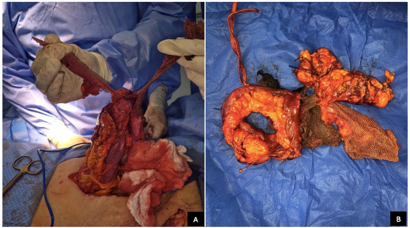
Broad-spectrum intravenous antibiotic therapy was initiated upon admission due to clinical suspicion of intra-abdominal infection and elevated inflammatory markers. Surgical management included segmental resection of the colon and small bowel, removal of multiple mesh fragments, and debridement of the abdominal wall Figure 1. Given the patient’s critical condition, bowel continuity was deferred, and intraoperative tissue samples from the distal ileum and adjacent adipose tissue were obtained for both aerobic and anaerobic cultures. Histopathological analysis revealed severe acute inflammation consistent with secondary peritonitis; however, no pathogenic microorganisms were isolated.
The patient remained in the intensive care unit (ICU) for 11 days, during which she required four additional surgical procedures, including restoration of bowel continuity, peritoneal washouts, and repair of anastomotic leakage and dehiscence. Continuous abdominal drainage with negative-pressure wound therapy (VAC system) was instituted, and broad-spectrum antibiotic therapy with piperacillin–tazobactam was continued.
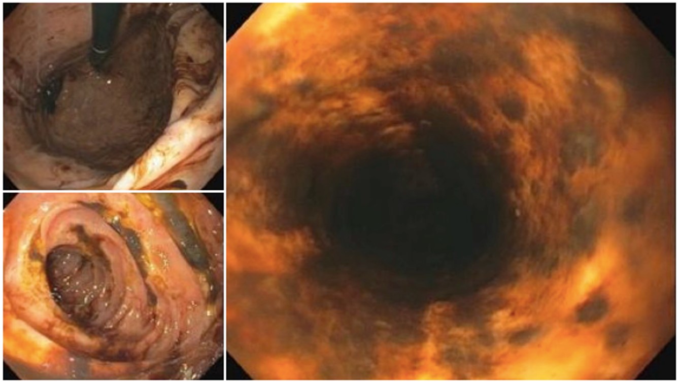
Two days after her last surgery, she developed multiple episodes of hematemesis with acute anemia. Upper gastrointestinal endoscopy revealed a cricopharyngeal tear of probable emetic origin and severe necrotizing ulcerative esophagitis (“black esophagus”) Figure 2. Despite intensive management, she experienced rapid hemodynamic deterioration and died one day later. No autopsy was performed. Based on the clinical course, her death was presumed to be secondary to complications of prolonged critical illness, likely associated with peritonitis, hemodynamic instability, and ischemic injury, rather than a direct consequence of mesh migration itself.
Case 2
A 61-year-old female with Crohn’s disease under infliximab therapy, and a history of hysterectomy, oophorectomy, surgery for adhesive small bowel obstruction, and two previous mesh herniorrhaphies, the last one performed 16 years ago, presented to the emergency department with acute abdominal pain and peritoneal signs. At presentation, there was no clinical or biochemical evidence of active Crohn’s disease; she had no gastrointestinal or extraintestinal manifestations suggestive of a flare; recent fecal calprotectin levels were within the normal range; and infliximab levels were therapeutic, with no significant anti-drug antibodies. Laboratory tests revealed anemia and leukocytosis Table 1. Abdominal computed tomography (CT) demonstrated adhesive syndrome without evidence of fistulization.
Given her clinical deterioration, an emergency exploratory laparotomy was performed. Intraoperatively, a polypropylene mesh was identified, originally placed in the retromuscular plane and secured with sutures, which had eroded into a loop of small bowel, resulting in an enterocutaneous fistula involving the abdominal wall. Extensive adhesiolysis was performed, followed by resection of approximately 30 cm of small intestine and a termino-terminal anastomosis. Complete mesh removal was not feasible due to dense integration of the prosthetic material into the abdominal wall.
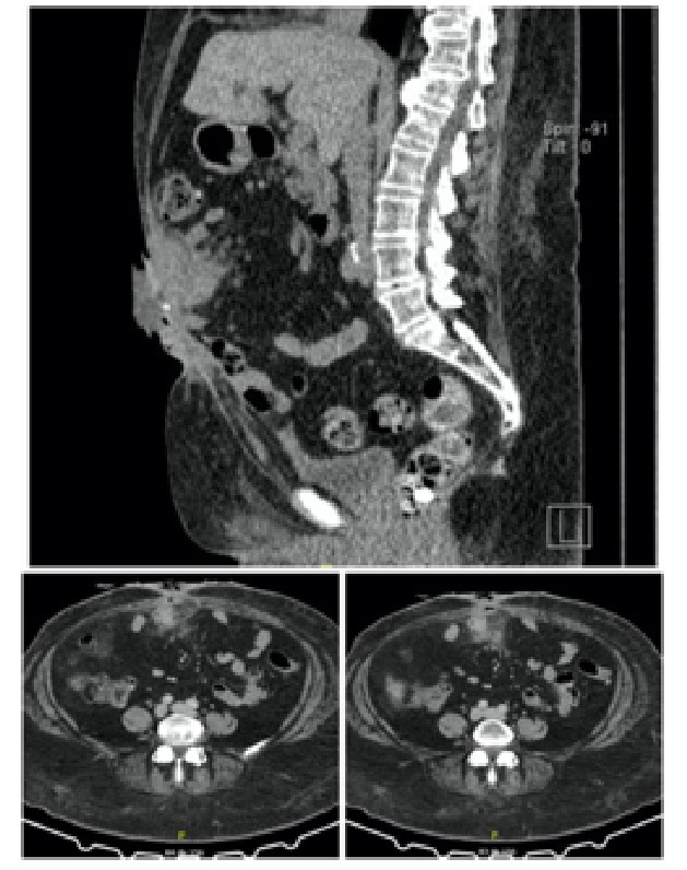
During her hospital stay, the patient developed postoperative ileus and a surgical site infection caused by Escherichia coli. Owing to clinical suspicion of an anastomotic leak, a re-laparotomy was undertaken, which revealed severe adhesive disease. The previously constructed anastomosis was found to be intact; however, residual mesh fragments had perforated the small intestine, resulting in a 5-mm fistula, which was also documented in a new contrast-enhanced computed tomography Figure 3. Given the extensive adhesions and firm incorporation of the mesh into the abdominal wall, complete removal of the prosthetic material and definitive fistula closure were not possible. Consequently, partial mesh excision was performed, the fistula was exteriorized to the skin, and the abdominal wall was closed.
The patient was discharged after one month of in-hospital management with a high-output external enterocutaneous fistula. She was followed on an outpatient basis by a multidisciplinary team, including general surgeons, with laboratory monitoring every two weeks initially and subsequently every month, as well as nutritionists and a wound care clinic providing dressing changes three times per week. One month after discharge, she required readmission due to dehydration secondary to persistent high fistula output, without electrolyte imbalance, renal dysfunction, or nutritional deterioration; temporary total parenteral nutrition was reinitiated. Definitive surgical reconstruction, including fistula closure and removal of residual mesh material, was performed 5 months later. At the time of manuscript submission, she remains under outpatient follow-up without recurrent septic complications.
Discussion
Mesh migration remains an uncommon complication. In the limited literature on adverse events following mesh repair, cases of migration are rarely reported, and complete migration into the intestinal lumen is even more rare [3,4,5].
Although no standardized mechanism has been established, proposed theories suggest that mesh, as a foreign body, induces an inflammatory response that promotes displacement into adjacent tissues, potentially leading to fistula formation. During this process, the omentum encapsulates the mesh, while chronic inflammation and intestinal peristalsis facilitate erosion into hollow viscera. A primary mechanism of migration has also been described, where the mesh traverses tissues of lower resistance due to inadequate fixation or external mechanical forces [6,7,5]. In both cases, chronic inflammatory changes and dense adhesions were prominent intraoperative findings, supporting the proposed mechanisms of gradual erosion rather than acute displacement.
The role of implantation and fixation techniques in the risk of migration remains uncertain. Some studies suggest a higher risk when mesh is placed intraperitoneally or when sutureless, tension-free techniques are used. In such cases, the mesh may gradually shift due to gravity, intra-abdominal pressure, or biomechanical forces during ambulation; however, these associations lack statistical significance [8,7]. In Case 1, multiple mesh fragments of different materials were identified intraoperatively, located in distinct anatomical planes, suggesting repeated repairs and complex prior surgical interventions that may have favored chronic inflammation and progressive migration. In Case 2, intraperitoneal placement of a polypropylene mesh fixed with sutures was confirmed, which may have contributed to gradual erosion into the small bowel.
Additional factors such as mesh size, material, and shape may influence migration behavior. Manzini et al. reported that polyglactin meshes induce less inflammatory infiltration than polypropylene, potentially altering complication rates [5]. In our series, polypropylene mesh was clearly identified in Case 2, whereas Case 1 involved the removal of multiple mesh types, limiting precise material-specific correlations.
Patient-related factors may also modulate the risk and severity of mesh-related complications. Chronic inflammatory conditions such as Crohn’s disease have been associated with impaired tissue healing, increased susceptibility to fistula formation, and poorer surgical outcomes, particularly in patients receiving biologic therapies [9]. These factors may act synergistically with local mechanical or foreign-body–related processes to facilitate fistulization. In Case 2, the presence of Crohn’s disease and ongoing infliximab therapy therefore represents an important differential consideration. However, several findings support mesh migration as the primary etiologic mechanism in this patient. The fistula originated at the site of direct contact between residual polypropylene mesh fragments and the small bowel, was directly visualized intraoperatively, and correlated with imaging findings demonstrating prosthetic material adjacent to the fistulous tract. Furthermore, there was no evidence of active Crohn’s disease at the time of presentation. Accordingly, Crohn’s disease is interpreted as a contributing risk factor rather than the primary cause of fistula formation in this case.
Because migration is unpredictable, clinical manifestations vary depending on the affected organ. Reported presentations include enteric, enterovesical, and enterocutaneous fistulas, as well as chronic abdominal pain, anemia, abdominal masses, recurrent urinary tract infections, hematuria, bowel obstruction, perforation, and sepsis [10,2,11]. Our cases illustrate this spectrum, presenting with peritonitis and septic deterioration, and a persistent high-output enterocutaneous fistula.
Diagnosis depends on clinical suspicion and imaging. While incidental findings may arise during endoscopic procedures such as colonoscopy, computed tomography is generally the modality of choice. Its sensitivity, however, is limited by mesh material, and in some cases, the diagnosis can only be confirmed intraoperatively [3,6,2].
Management requires prompt surgical intervention to prevent deterioration. The standard approach consists of complete mesh removal combined with partial or total resection of the involved organ [3]. However, in our cases, complete mesh excision was not achievable due to extensive adhesions and firm integration of the prosthetic material into the abdominal wall, underscoring the clinical impact of this limitation and the complexity of surgical management in advanced presentations.
Patient-specific factors may also influence the risk of mesh-related complications and clinical outcomes. Case 2 had Crohn’s disease and was receiving infliximab therapy, both of which are associated with impaired tissue healing and increased susceptibility to fistula formation. In Case 1, advanced age and a history of multiple prior abdominal surgeries may have contributed to severe adhesive disease, delayed diagnosis, and poor clinical evolution. These comorbidities likely acted synergistically with mesh migration to worsen outcomes [12,13,14].
This report has limitations. It describes only two cases and is retrospective. Detailed information regarding original mesh implantation techniques and materials was incomplete, and long-term post-discharge follow-up was limited. These factors restrict the generalizability of our findings but reflect real-world challenges in evaluating late mesh-related complications.
Conclusion
In both cases presented, the definitive diagnosis was established only at exploratory laparotomy, highlighting the diagnostic challenge posed by mesh migration. As described in the literature, a staged surgical approach was attempted; however, the clinical outcomes differed significantly, with one patient developing extensive multiorgan involvement and succumbing to the disease, while the other evolved with a persistent enterocutaneous fistula.
These cases highlight the importance of maintaining a high index of suspicion in patients with a history of ventral or incisional hernia repair with mesh, regardless of the time elapsed since the original surgery. Early recognition and timely surgical intervention are essential to prevent severe, potentially life-threatening complications. Finally, these cases underscore the need to continue evaluating the long-term safety of mesh materials and implantation techniques, particularly in complex or high-risk patients, to reduce the incidence of serious late complications, such as mesh migration.
Conflicts of Interest
The authors declare no conflicts of interest.
Funding Source
The authors declare no funding sources.
Acknowledgments
The authors would like to thank Hospital Pablo Tobón Uribe and Universidad EIA for their institutional support and for facilitating this research.
Informed Consent
Written informed consent for publication of anonymized clinical data and accompanying images was obtained directly from the second patient. In the first case, informed consent was obtained from the patient’s next of kin due to the patient’s death.
Large Language Model
The authors declare that generative artificial intelligence (AI) tools (ChatGPT) were used to assist in language refinement and grammar checking during the preparation of this manuscript. The authors reviewed and verified all content, and they take full responsibility for the integrity and accuracy of the manuscript.
Authors Contribution
CAL and CAD contributed to the conception and design of the study. ML and DM were responsible for data acquisition and also participated in data analysis and interpretation. LA drafted the manuscript. CAD critically revised the manuscript for important intellectual content. All authors approved the final version of the manuscript.
Data Availability
Data supporting the findings of this study are available from the corresponding author upon reasonable request.
References
- Kokotovic D., Bisgaard T., Helgstrand F.. Long-term Recurrence and Complications Associated With Elective Incisional Hernia Repair. JAMA. 2016;316(15):1575-1582. doi:10.1001/jama.2016.15217 PMID: 27750295
- Gandhi D., Marcin S., Xin Z., Asha B., Kaswala D., Zamir B.. Chronic abdominal pain secondary to mesh erosion into cecum following incisional hernia repair: a case report and literature review. Ann Gastroenterol. 2011;24(4):321-324. PMID: 24713759 PMCID: PMC3959323
- Agrawal A., Avill R.. Mesh migration following repair of inguinal hernia: a case report and review of literature. Hernia. 2006;10(1):79-82. doi:10.1007/s10029-005-0024-8 PMID: 16258705
- LeBlanc K. A., Whitaker J. M., Bellanger D. E., Rhynes V. K.. Laparoscopic incisional and ventral hernioplasty: lessons learned from 200 patients. Hernia. 2003;7(3):118-24. doi:10.1007/s10029-003-0117-1 PMID: 12942345
- Manzini G., Henne-Bruns D., Kremer M.. Severe complications after mesh migration following abdominal hernial repair: report of two cases and review of literature. GMS Interdiscip Plast Reconstr Surg DGPW. 2019;8:Doc09. doi:10.3205/iprs000135 PMID: 31275799 PMCID: PMC6545489
- Chowbey P. K., Pithawala M., Khullar R., Sharma A., Soni V., Baijal M.. Totally extraperitoneal repair of inguinal hernia: A case for bilateral repair. J Minim Access Surg. 2006;2(3):171-3. doi:10.4103/0972-9941.27733 PMID: 21187991 PMCID: PMC2999780
- Jallali M., Chaouch M. A., Zenati H., Hassine H. B., Gafsi B., Noomen F.. Complications unveiled: A detailed case report on mesh migration post-incisional hernia repair. Int J Surg Case Rep. 2024;121:109976. doi:10.1016/j.ijscr.2024.109976 PMID: 38954968 PMCID: PMC11263626
- Carrozza V., Ivaldi L., Ferro A., Gennaro M., Bronzino P., Gambino E., Guffanti P., Aimo I., Morino M., Revetria P.. [Inguinal hernia repair: an experimental study on mesh migration with the tension-free suturless technique. 10 year follow-up]. Minerva Chir. 2011;66(1):21-40. PMID: 21389922
- El Boghdady M., Ewalds-Kvist B. M., Laliotis A.. Abdominal hernia mesh repair in patients with inflammatory bowel disease: A systematic review. Langenbecks Arch Surg. 2022;407(7):2637-2649. doi:10.1007/s00423-022-02638-x PMID: 35947216 PMCID: PMC9640397
- Cardoso D., Rebanda J., Gois C.. Mesh Migration and Bowel Perforation as a Late Complication of Transabdominal Preperitoneal Laparoscopic Hernia Repair. Cureus. 2022;14(12):e32683. doi:10.7759/cureus.32683 PMID: 36561326 PMCID: PMC9767664
- Kalles V., Mekras A., Mekras D., Papapanagiotou I., Al-Harethee W., Sotiropoulos G., Liakou P., Kastania A., Piperos T., Mariolis-Sapsakos T.. De Garengeot's hernia: a comprehensive review. Hernia. 2013;17(2):177-82. doi:10.1007/s10029-012-0993-3 PMID: 22983696
- Billioud V., Ford A. C., Tedesco E. D., Colombel J. F., Roblin X., Peyrin-Biroulet L.. Preoperative use of anti-TNF therapy and postoperative complications in inflammatory bowel diseases: a meta-analysis. J Crohns Colitis. 2013;7(11):853-67. doi:10.1016/j.crohns.2013.01.014 PMID: 23523418
- ten Broek R. P., Issa Y., van Santbrink E. J., Bouvy N. D., Kruitwagen R. F., Jeekel J., Bakkum E. A., Rovers M. M., van Goor H.. Burden of adhesions in abdominal and pelvic surgery: systematic review and met-analysis. BMJ. 2013;347:f5588. doi:10.1136/bmj.f5588 PMID: 24092941 PMCID: PMC3789584
- Tsai L., McCurdy J. D., Ma C., Jairath V., Singh S.. Epidemiology and Natural History of Perianal Crohn's Disease: A Systematic Review and Meta-Analysis of Population-Based Cohorts. Inflamm Bowel Dis. 2022;28(10):1477-1484. doi:10.1093/ibd/izab287 PMID: 34792604 PMCID: PMC9527611