Oral Sodium Bicarbonate as Adjunctive Therapy for Suspected Propafenone Cardiotoxicity in a Hemodynamically Stable Patient: A Case Report with Literature Review
Alok Arora1, Ahmed ElsayedⒾ2*, Yousif Elsayed2, Wadah Jason Ayoub1, Alice ChingⒾ1
- 1Medical College of Wisconsin, Milwaukee, WI, USA
- 2Faculty of Medicine, Alexandria University, Alexandria, Egypt
Abstract
We present the case of a 74-year-old male with a history of paroxysmal atrial fibrillation (PAF) and chronic kidney disease (CKD) who presented with fatigue. During hospitalization, telemetry captured an episode of hemodynamically stable wide-complex tachycardia consistent with monomorphic ventricular tachycardia. A 12-lead ECG revealed atrioventricular dissociation and a widened QRS complex. After discontinuing propafenone and initiating oral sodium bicarbonate due to limited intravenous availability, ECG abnormalities gradually improved over several hours, and the rhythm reverted to baseline PAF. A subsequent laboratory test confirmed supratherapeutic propafenone levels. This case highlights a temporally associated improvement in suspected propafenone cardiotoxicity with oral sodium bicarbonate in a hemodynamically stable patient. Written informed consent for publication was obtained.
Keywords: CYP450 interaction, Monomorphic Ventricular Tachycardia, Propafenone, Sodium bicarbonate, Case Report
Article information
Introduction
Propafenone is a Class IC antiarrhythmic drug used to treat supraventricular arrhythmias such as atrial fibrillation and atrial flutter. It blocks fast inward sodium channels, slowing phase 0 depolarization and resulting in prolongation of the PR interval and QRS duration [1]. While Class IC agents primarily cause QRS widening, apparent QT prolongation may be observed when QRS duration is markedly prolonged; therefore, repolarization should be interpreted using QRS-adjusted metrics such as the JT or JTc interval when feasible [2]. Propafenone exhibits use-dependent sodium channel blockade and is proarrhythmic, with risks including ventricular tachycardia and sudden cardiac death, particularly in patients with structural heart disease or impaired drug metabolism [3]. In cases of sodium channel blocker-induced cardiotoxicity, intravenous hypertonic sodium bicarbonate is generally recommended, based primarily on experience with tricyclic antidepressants and Class I antiarrhythmics. If initial medical therapy fails, early venoarterial ECMO should be considered for life-saving hemodynamic support and to facilitate drug clearance [4,5]. The role of oral sodium bicarbonate in this setting remains poorly characterized.
Here, we describe a case of suspected propafenone-associated wide-complex tachycardia managed with oral sodium bicarbonate due to resource limitations and discuss the clinical rationale, limitations, and implications of this approach.
Case Presentation
A 74-year-old African American male with a history of paroxysmal atrial fibrillation (PAF), chronic kidney disease stage 3b (CKD 3b), a remote history of amiodarone-induced thyroiditis (AIT), and diastolic heart failure presented to the ED with fatigue. His home medications for PAF included metoprolol (50 mg twice daily) and immediate-release propafenone (150 mg twice daily). Baseline laboratory and electrolyte results at admission and post-bicarbonate are presented in Table 1.
Initial evaluation suggested community-acquired pneumonia, and the patient was started on ceftriaxone 2 g IV daily and doxycycline 100 mg orally twice daily. These antibiotics are not significant inhibitors of CYP2D6 or CYP3A4 and are unlikely to substantially affect propafenone metabolism.
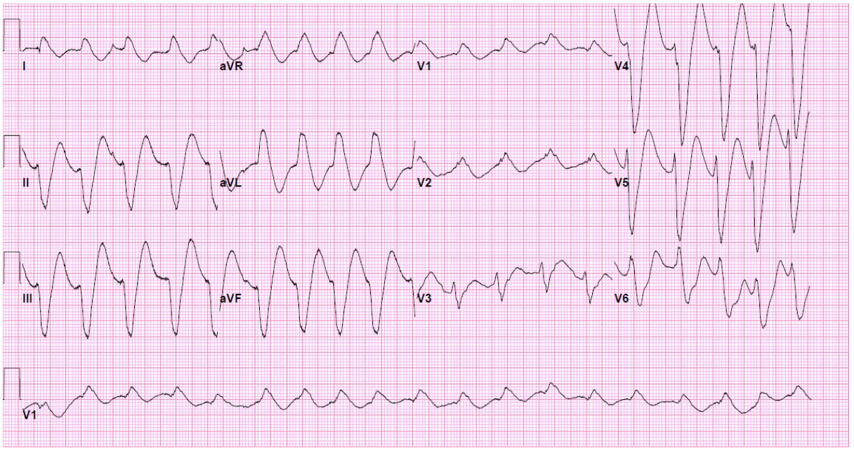
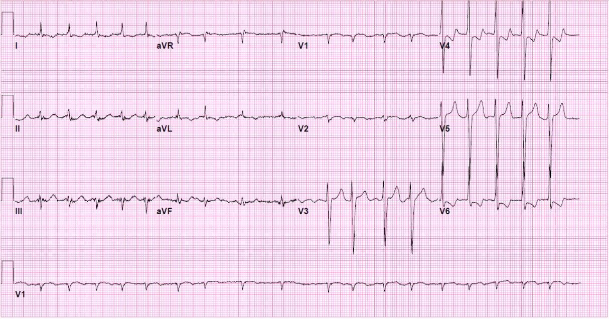
On hospital day two (hour 0), telemetry captured a regular wide-complex tachycardia at 130 bpm. The patient remained alert, normotensive, and asymptomatic. A 12-lead ECG demonstrated AV dissociation, left axis deviation, and QRS widening to 140 ms. Apparent QTc prolongation was attributed to QRS widening, as assessed by the JT formula (QT - QRS). Representative telemetry strips and 12-lead ECGs are included in Table 2, (Figure 1, Figure 2).
| Parameter | Admission | Hospital Day 2 (pre-bicarbonate) | 4 hrs. post-bicarbonate | 12 hrs. post-bicarbonate | Reference Range |
|---|---|---|---|---|---|
| ABG pH | 7.28 | 7.29 | 7.35 | 7.38 | 7.35-7.45 |
| HCO (mmol/L) | 11 | 12 | 18 | 22 | 22-29 |
| K (mmol/L) | 4.9 | 5.0 | 4.7 | 4.4 | 3.5-5.0 |
| Mg (mg/dL) | 2.0 | 1.9 | 2.0 | 2.1 | 1.7-2.3 |
| Na (mmol/L) | 138 | 137 | 140 | 139 | 135-145 |
| Cl (mmol/L) | 99 | 100 | 103 | 105 | 98-107 |
| BUN (mg/dL) | 30 | 32 | 31 | 30 | 6-20 |
| Creatinine (mg/dL) | 1.65 | 1.7 | 1.68 | 1.65 | 0.6-1.2 |
| Anion gap (mmol/L) | 27 | 28 | 20 | 12 | 8-12 |
ABG, arterial blood gas; pH, potential of hydrogen; HCO₃⁻, bicarbonate; K⁺, potassium; Mg²⁺, magnesium; Na⁺, sodium; Cl⁻, chloride; BUN, blood urea nitrogen; AG, anion gap; hrs, hours; pre-, before; mmol/L, millimoles per liter; mg/dL, milligrams per deciliter.
| Time (hrs.) | Event/Intervention | Rhythm | HR (bpm) | PR (ms) | QRS (ms) | QT (ms) | JT (ms) | Notes |
|---|---|---|---|---|---|---|---|---|
| -2 | Last propafenone dose | PAF | 80 | 180 | 110 | 420 | 310 | - |
| 0 | Index rhythm change detected | WCT | 130 | 200 | 140 | 520 | 380 | Patient asymptomatic; telemetry captured AV dissociation |
| +1 | Pre-bicarbonate labs | WCT | 130 | 200 | 140 | 520 | 380 | - |
| +3 | Oral sodium bicarbonate initiated (950 mg TID) | WCT | 130 | 190 | 130 | 500 | 370 | IV bicarbonate unavailable |
| +4 | Follow-up ECG | PAF | 100 | 180 | 115 | 430 | 315 | QRS narrowing; rhythm reverted toward baseline PAF |
| +6 | Serum propafenone level | PAF | 90 | 180 | 115 | 430 | 315 | 2.54 mcg/mL (Therapeutic range 0.5-2.0 mcg/mL) |
| +8 | Electrical cardioversion | PAF | 85 | 180 | 110 | 420 | 310 | Performed for symptomatic AF, not hemodynamic instability |
| +24 | Discharge | PAF | 80 | 180 | 110 | 420 | 310 | Discharged on dronedarone, metoprolol, reduced-dose oral bicarbonate |
WCT, wide-complex tachycardia; PAF, paroxysmal atrial fibrillation; AF, atrial fibrillation; AV, atrioventricular; ECG, electrocardiogram; HR, heart rate; PR, PR interval; QRS, QRS duration; QT, QT interval; JT, JT interval; IV, intravenous; TID, three times daily; hrs, hours; bpm, beats per minute; ms, milliseconds; mcg/mL, micrograms per milliliter.
The electrophysiology attending physician was consulted for clinical input and concurred in a diagnosis of monomorphic ventricular tachycardia based on persistent AV dissociation. Propafenone was discontinued. Five hours after the last dose (hour +3), oral sodium bicarbonate was initiated at 950 mg TID, the maximum allowed dose. Several factors guided the decision to use oral therapy: IV bicarbonate was expected to be unavailable for 6-8 hours; the patient had ongoing QRS widening (140 ms) and telemetry evidence of WCT; and, although hemodynamically stable, the arrhythmia could deteriorate. Supportive care alone was deemed insufficient. Oral sodium bicarbonate is well absorbed, with peak plasma levels occurring within 1-2 hours, thereby providing gradual correction of acidosis and potentially improving sodium channel kinetics. Continuous monitoring of ECG, blood pressure, and electrolytes was implemented to detect adverse effects. One hour after initiation (hour +4), a follow-up ECG showed QRS narrowing to 115 ms and partial restoration of baseline PAF Figure 2.
Given CKD and the risk of alkalosis/volume overload, electrolytes and ABGs were checked every 4-6 hours initially; blood pressure was measured hourly for the first 6 hours, and telemetry was continuous. The patient remained hemodynamically stable; no alkalosis or fluid overload occurred, and serial ECGs demonstrated progressive QRS narrowing.
At hour +6 (8 hours after the last 150 mg dose), serum propafenone was 2.54 mcg/mL (therapeutic range 0.5-2.0 mcg/mL). In the context of potential CYP2D6 poor-metabolizer status and residual enzyme inhibition from prior amiodarone therapy, clearance is slowed, with a half-life up to 32 hours. This means that levels may remain supratherapeutic for ~32 hours. Importantly, the clinical significance must be interpreted in the context of ECG and hemodynamic data, as QRS widening and arrhythmogenic manifestations are more reliable indicators of toxicity than absolute serum concentrations. Therefore, while supportive of toxicity, the primary indicators guiding management were QRS prolongation and telemetry findings.
Transesophageal echocardiography (TEE) showed no thrombus, EF 55%, and mild diastolic dysfunction. Electrical cardioversion at hour +8 was performed for symptomatic AF, not for hemodynamic instability or ventricular arrhythmia. The patient was discharged on dronedarone, metoprolol, and reduced-dose oral bicarbonate. Alternative rhythm-control options, such as sotalol, were avoided due to renal impairment and QTc risk. Discharge planning emphasized careful follow-up and serum drug monitoring if future antiarrhythmic therapy is initiated.
Dronedarone was chosen for rhythm control in combination with metoprolol to avoid amiodarone, given the patient’s prior amiodarone-induced thyroiditis. Its use was selected with awareness of its moderate inhibition of CYP3A4 and P-glycoprotein, warranting careful monitoring for potential interactions should propafenone be reintroduced, as well as consideration of additive AV nodal effects with metoprolol. Alternative agents such as sotalol were considered less favorable due to underlying renal impairment and QTc risk, and the discharge plan emphasized close follow-up and ECG monitoring if future antiarrhythmic therapy is initiated.
Discussion
Propafenone toxicity is an uncommon but well-documented clinical phenomenon, most often reported in the setting of intentional overdose, impaired drug metabolism, or drug-drug interactions [6]. Manifestations include hypotension, bradyarrhythmias, atrioventricular block, wide-complex tachycardia, and ventricular arrhythmias, primarily mediated through fast sodium channel blockade [7,8,9]. Apparent QT prolongation on standard ECG measurements can result from QRS widening rather than from true repolarization delay. Repolarization should therefore be assessed using JT or JTc intervals, which subtract QRS duration from the QT interval to isolate ventricular repolarization [10,11]. In our patient, at the peak of wide-complex tachycardia, the QT interval was 520 ms, QRS 140 ms, yielding a JT of 380 ms, which remained within expected limits, indicating that repolarization was not markedly prolonged. This reframing clarifies that the observed QT prolongation was secondary to QRS widening, reducing the risk of misinterpretation.
Several reports and trials describe sustained monomorphic ventricular tachycardia and congestive heart failure associated with propafenone toxicity, particularly in patients with structural heart disease [12,13]. These findings align with the use-dependent nature of Class IC antiarrhythmic agents, in which increasing heart rates amplify sodium channel blockade and slow intraventricular conduction [14]. Here, QRS increased from 110 ms at baseline to 140 ms at 130 bpm, reverting to 110-115 ms with lower rates (~80-90 bpm) after oral bicarbonate, illustrating rate-dependent conduction slowing. Linking these observations to the patient’s actual heart rate and ECG intervals reinforces the clinical relevance of use-dependence in propafenone toxicity.
Management of Class I toxicity is largely extrapolated from TCA and flecainide experiences. Intravenous hypertonic sodium bicarbonate is standard; mechanisms include sodium loading, alkalinization, reducing drug binding, and channel dissociation facilitation [15,11].
Although intravenous therapy is standard, oral sodium bicarbonate was administered temporally coincident with QRS narrowing in this hemodynamically stable patient. Serial ECGs showed improvement within 4-5 hours of oral therapy, paralleling normalization of serum bicarbonate and pH. It is important to note that this improvement cannot be attributed with certainty to oral therapy.
Propafenone is largely cleared hepatically and has a half-life of 2-10 hours in extensive metabolizers and up to 32 hours in poor metabolizers [16,17]. Case reports suggest ECG abnormalities can resolve spontaneously within several hours to a day after discontinuation, depending on metabolism and comorbidities [18]. Thus, the temporal association may reflect natural resolution rather than a direct pharmacologic effect of oral bicarbonate. While improvement coincided with therapy, causality cannot be established; spontaneous recovery is plausible. This uncontrolled observation cannot distinguish between drug discontinuation, ongoing metabolism, and alkalinizing therapy.
CKD-associated metabolic acidosis may enhance sodium channel blockade by altering the ionized fraction of drugs and myocardial Electrophysiology. Acidosis reduces inotropy and alters Na/Ca handling, promoting delayed after-depolarizations and arrhythmias [19]. In this patient, baseline metabolic acidosis (pH 7.28-7.29, HCO3- 11-12 mmol/L) may have enhanced sodium channel blockade and contributed to arrhythmia susceptibility. Correction of acidosis with oral bicarbonate likely improved cardiac conduction via multiple mechanisms, including normalization of extracellular pH and ionized drug fractions.
This case contributes to the literature by describing a temporally associated improvement in ECG after oral sodium bicarbonate in a hemodynamically stable patient. While causality cannot be established, it highlights a pragmatic option when IV therapy is unavailable. Prospective studies are warranted to clarify the safety, efficacy, and pharmacokinetics of oral alkalinization in Class IC cardiotoxicity. To our knowledge, after a targeted search (updated on February 1, 2025) of the published literature (PubMed, Medline, Scopus, and Cochrane Central), this is the first case report demonstrating that oral sodium bicarbonate is a successful rescue therapy for propafenone toxicity.
Conclusion
Oral sodium bicarbonate may be a pragmatic option for suspected propafenone cardiotoxicity in hemodynamically stable patients when intravenous therapy is unavailable. Although a temporal association with QRS narrowing was observed, causality cannot be established. Elevated propafenone levels were likely attributable to residual CYP450 inhibition and potential poor-metabolizer status, rather than to CKD alone. Clinicians should exercise caution and closely monitor electrolytes, acid-base status, hemodynamics, and potential drug interactions when managing similar cases.
Conflicts of Interest
All authors declare they have no competing interests.
Funding Source
The authors did not receive support from any organization for the submitted work.
Acknowledgments
None.
Informed Consent
The authors confirm that patient written consent forms have been obtained for this article, including all accompanying clinical data and images.
Large Language Model
None.
Authors Contribution
All authors contributed equally. All authors reviewed and approved the final manuscript.
Data Availability
Data supporting the findings of this case report are not publicly available because they contain information that could compromise patient privacy. De-identified clinical data may be made available from the corresponding author upon reasonable request, subject to applicable ethical and privacy considerations and with any necessary institutional approvals.
References
- Paolini E., Stronati G., Guerra F., Capucci A.. Flecainide: Electrophysiological properties, clinical indications, and practical aspects. Pharmacol Res. 2019;148:104443. doi:10.1016/j.phrs.2019.104443 PMID: 31493514
- Mahendru D., Joshi H. D., Kowey P.. Dofetilide in atrial fibrillation: A comprehensive review. Indian Pacing Electrophysiol J. 2025;25(6):451-457. doi:10.1016/j.ipej.2025.11.013 PMID: 41319865 PMCID: PMC12790294
- Colangelo T., Johnson D., Ho R.. Flecainide-Induced Atrial Flutter With 1:1 Conduction Complicated by Ventricular Fibrillation After Electrical Cardioversion. Tex Heart Inst J. 2021;48(2). doi:10.14503/THIJ-19-7099 PMID: 34086956 PMCID: PMC8262829
- Di Grande A., Giuffrida C., Narbone G., Le Moli C., Nigro F., Di Mauro A., Pirrone G., Tabita V., Alongi B.. Management of sodium-channel blocker poisoning: the role of hypertonic sodium salts. Eur Rev Med Pharmacol Sci. 2010;14(1):25-30. PMID: 20184086
- Vu N. M., Hill T. E., Summers M. R., Vranian M. N., Faulx M. D.. Management of life-threatening flecainide overdose: A case report and review of the literature. HeartRhythm Case Rep. 2016;2(3):228-231. doi:10.1016/j.hrcr.2015.12.013 PMID: 28491675 PMCID: PMC5419747
- Denaro C. P., Benowitz N. L.. Poisoning due to class 1B antiarrhythmic drugs. Lignocaine, mexiletine and tocainide. Med Toxicol Adverse Drug Exp. 1989;4(6):412-28. doi:10.1007/BF03259923 PMID: 2513464
- Enginyurt Özgür, Karabulut Özer Melike, Çoruh Akyol Bestegül, Süleyman Halis. Detection of Mushroom Extract Use in Patients Coming to the Family Medicine Outpatient Clinic for Routine Check-up and the Relationship Between Changes in Their Clinics. Middle Black Sea Journal of Health Science. 2025;11(2):136-142. doi:10.19127/mbsjohs.1665455
- Haddad E., Collins M.. A case of propafenone toxicity in the setting of dehydration and acute kidney injury. Glob Cardiol Sci Pract. 2024;2024(4):e202438. doi:10.21542/gcsp.2024.38 PMID: 39351481 PMCID: PMC11439426
- Kim H. K., Kim S. S., Ki Y. J., Park K. H., Choi D. H.. Ventricular Tachycardia in Association with Propafenone Overdose. Acta Cardiol Sin. 2021;37(1):100-103. doi:10.6515/ACS.202101_37(1).20200914A PMID: 33488033 PMCID: PMC7814319
- Bogossian H., Linz D., Heijman J., Bimpong-Buta N. Y., Bandorski D., Frommeyer G., Erkapic D., Seyfarth M., Zarse M., Crijns H. J.. QTc evaluation in patients with bundle branch block. Int J Cardiol Heart Vasc. 2020;30:100636. doi:10.1016/j.ijcha.2020.100636 PMID: 32995475 PMCID: PMC7509006
- Merino J. L., Tamargo J., Blomstrom-Lundqvist C., Boriani G., Crijns Hjgm, Dobrev D., Goette A., Hohnloser S. H., Naccarelli G. V., Reiffel J. A., Tfelt-Hansen J., Martinez-Cossiani M., Camm A. J., Almendral Garrote J. M., Sredniawa B., Kulakowski P., Savelieva I., Potpara T., Gorenek B., Zamorano J. L.. Practical compendium of antiarrhythmic drugs: a clinical consensus statement of the European Heart Rhythm Association of the European Society of Cardiology. Europace. 2025;27(8). doi:10.1093/europace/euaf076 PMID: 40159403 PMCID: PMC12367031
- Darbar Dawood. Standard antiarrhythmic drugs. In: Cardiac electrophysiology: from cell to bedside. 2014.
- Podrid P. J., Anderson J. L.. Safety and tolerability of long-term propafenone therapy for supraventricular tachyarrhythmias. The Propafenone Multicenter Study Group. Am J Cardiol. 1996;78(4):430-4. doi:10.1016/s0002-9149(96)00332-3 PMID: 8752188
- Lei M., Wu L., Terrar D. A., Huang C. L.. Modernized Classification of Cardiac Antiarrhythmic Drugs. Circulation. 2018;138(17):1879-1896. doi:10.1161/CIRCULATIONAHA.118.035455 PMID: 30354657
- Lavonas E. J., Akpunonu P. D., Arens A. M., Babu K. M., Cao D., Hoffman R. S., Hoyte C. O., Mazer-Amirshahi M. E., Stolbach A., St-Onge M., Thompson T. M., Wang G. S., Hoover A. V., Drennan I. R., American Heart Association. 2023 American Heart Association Focused Update on the Management of Patients With Cardiac Arrest or Life-Threatening Toxicity Due to Poisoning: An Update to the American Heart Association Guidelines for Cardiopulmonary Resuscitation and Emergency Cardiovascular Care. Circulation. 2023;148(16):e149-e184. doi:10.1161/CIR.0000000000001161 PMID: 37721023
- Ferreira A. I., Macedo Silva V., Arieira C., Xavier S., Magalhaes J., Cotter J.. Propafenone-Induced Cholestatic Liver Injury: When Diagnosis Does Not Skip a Beat. GE Port J Gastroenterol. 2025;32(3):212-220. doi:10.1159/000541557 PMID: 40469208 PMCID: PMC12133138
- Han Yong Y, George Cindy, Garg Uttam, Oroszi Gabor. Use of therapeutic plasma exchange to enhance propafenone elimination following intentional drug overdose. Toxicology Cases for the Clinical and Forensic Laboratory. 2020:203-207.
- Yilmaz Tuğçe, Osoydan Satici Merve, İSlam Mehmet Muzaffer, ÖZdemİR Serdar, EroĞLu Serkan Emre, Aksel Gökhan. Spontaneous Resolution Of Arrhythmia in Propafenone Intoxication: A Rare Case Report. Eurasian Journal of Toxicology. 2022;4(1):29-31. doi:10.51262/ejtox.1080167
- Sinha F., Schweda F., Maier L. S., Wagner S.. Impact of Impaired Kidney Function on Arrhythmia-Promoting Cardiac Ion Channel Regulation. Int J Mol Sci. 2023;24(18). doi:10.3390/ijms241814198 PMID: 37762501 PMCID: PMC10532292