Calcinosis Universalis in Juvenile Dermatomyositis: An X-Ray Radiological Case Report
Maxwell AduⒾ1*, Mahama Bukari1, Felicia Philipa Aggrey1, Jacob Leonard Ago2, Abdul Rashid KarimⒾ3
- 1Komfo Anokye Teaching Hospital, Kumasi, Ghana
- 2Discipline of Medical Radiations, School of Health and Biomedical Sciences, RMIT University, Bundoora, Australia
- 3Spectra Health Imaging and Interventional Radiology, Kumasi, Ghana
Abstract
Calcinosis universalis is a rare and debilitating complication of juvenile dermatomyositis (JDM), characterized by extensive deposition of calcium salts in soft tissues. This case study presents a 12-year-old female patient with a known history of JDM who developed widespread calcinosis, documented through plain radiographic imaging. X-ray evaluation revealed diffuse sheet-like and nodular calcifications involving multiple soft tissue regions, correlating with the patient’s progressive musculoskeletal pain, reduced mobility, and recurrent skin ulcerations. Clinical correlations highlighted the significant impact of calcinosis on functional outcomes, disease morbidity, and quality of life. The discussion emphasizes the role of conventional radiography in identifying and monitoring the extent of calcinosis, while also considering therapeutic challenges and the limited effectiveness of available interventions. This case underscores the importance of early recognition, multidisciplinary management, and further research into targeted therapies to reduce disease burden in affected pediatric populations.
Keywords: Calcinosis universalis, Juvenile dermatomyositis, Plain radiography, Pediatric rheumatology, Soft tissue calcification
Article information
Introduction
Juvenile dermatomyositis (JDM) is a rare, autoimmune inflammatory myopathy of childhood characterized by proximal muscle weakness and distinctive cutaneous manifestations such as heliotrope rash and Gottron’s papules. The disease has a worldwide incidence of approximately 2-4 cases per million children annually, with variations in severity and outcomes across ethnic groups [1]. Although advances in immunosuppressive therapy have improved survival, chronic complications remain common, particularly in low-resource settings where delayed diagnosis and limited access to advanced care may affect outcomes [2].
One of the most disabling complications of JDM is calcinosis, the dystrophic deposition of calcium salts in soft tissues despite normal serum calcium and phosphate levels. The condition affects approximately 20-40% of patients, often manifesting after prolonged disease activity or suboptimal immunosuppression [3,4]. The most extensive and severe form, calcinosis universalis, is characterized by widespread, sheet-like or nodular calcium deposits involving the skin, subcutaneous tissue, fascia, and muscles [5]. These deposits can cause chronic pain, joint contractures, recurrent infections, and functional impairment, significantly impacting the patient’s quality of life [6].
The pathophysiology of calcinosis universalis in JDM remains incompletely understood. Still, it is thought to involve chronic inflammation, tissue hypoxia, mitochondrial damage, and local disturbances in calcium-phosphate homeostasis [7]. Case reports have suggested potential benefits from agents such as intravenous immunoglobulin (IVIg), bisphosphonates, colchicine, and low-dose warfarin, though evidence remains anecdotal and responses variable [8,5]. Surgical intervention is reserved for localized, symptomatic lesions that impair function or ulcerate [9].
Radiographic evaluation remains the cornerstone of diagnosis in low and middle-income countries, where access to advanced imaging modalities such as CT or MRI may be limited. Plain radiographs provide a simple, cost-effective, and reliable means of identifying the extent and distribution of calcific deposits [10]. Given the scarcity of documented cases from sub-Saharan Africa, reporting such presentations contributes valuable insight into the radiological spectrum, clinical course, and multidisciplinary management of calcinosis universalis in resource-limited settings.
This case report describes a 12-year-old Ghanaian girl diagnosed with JDM who subsequently developed calcinosis universalis, highlighting the clinical presentation, radiographic findings, management challenges, and implications for patient care in a low-resource healthcare environment.
Case Presentation
A 12-year-old Ghanaian girl from the Ashanti region of Ghana presented to the Komfo Anokye Teaching Hospital, Kumasi, with progressive limb pain, joint stiffness, and multiple firm subcutaneous nodules over the lower limbs. Her mother reported that the swellings had gradually increased in size over the preceding 18 months and were associated with pain, difficulty in ambulation, and reduced range of motion at the knees.
Past Medical and Disease Timeline
The patient was initially diagnosed with juvenile dermatomyositis (JDM) at age 10 years after a 6-month history of proximal muscle weakness, characteristic heliotrope rash, and Gottron’s papules. Diagnosis was established according to the 2017 EULAR/ACR classification criteria, which included elevated serum muscle enzymes, compatible electromyographic findings, and typical cutaneous features. Initial treatment consisted of oral prednisolone (2 mg/kg/day) and weekly methotrexate (15 mg/m²).
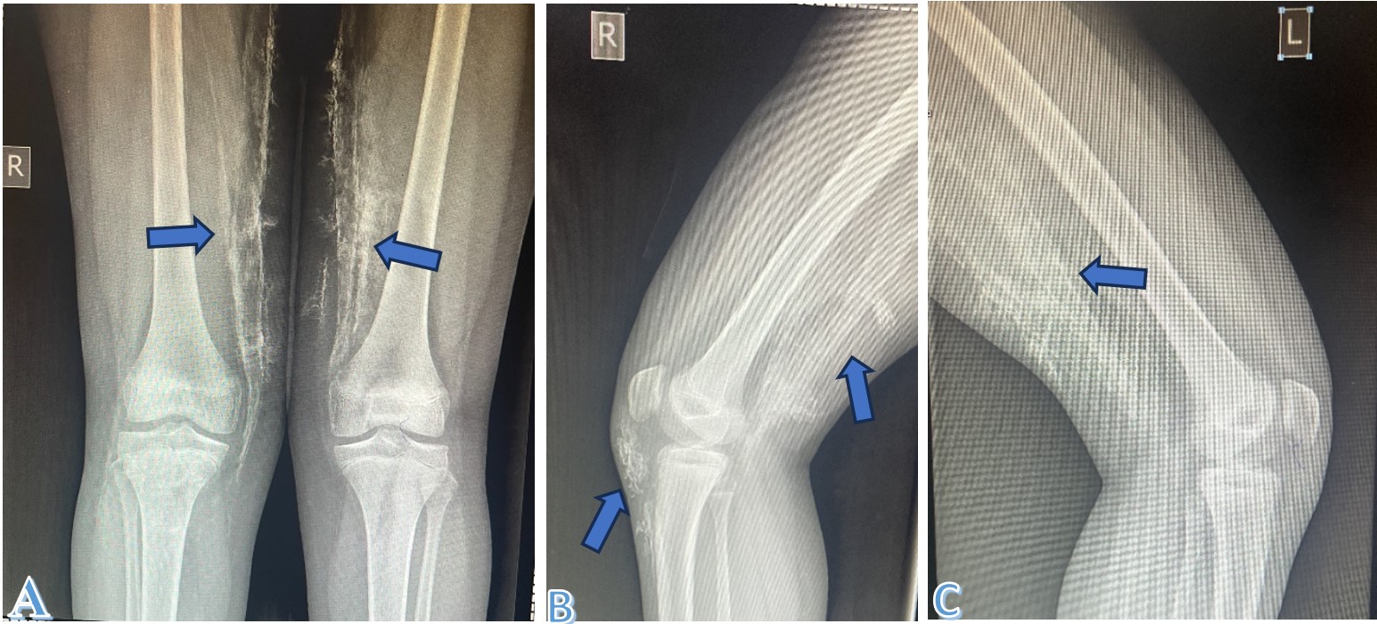
Despite partial improvement in muscle strength, she developed painful nodular lesions approximately 12 months after disease onset, consistent with calcinosis cutis. Over the next six months, the lesions coalesced and extended along fascial planes, resulting in diffuse, sheet-like calcification typical of calcinosis universalis.
Socioeconomic and Growth Context
The patient lives with her parents in a low-income suburban community. Household income is irregular, and medication supply interruptions occurred during follow-up. At presentation, her height and weight were below the 10th percentile for age (height = 132 cm; weight = 28 kg; BMI = 16.0 kg/m²), reflecting chronic disease and corticosteroid exposure. School attendance had declined due to mobility limitations and psychosocial distress related to the visible skin lesions.
Physical Examination
The patient appeared thin and chronically ill but alert and cooperative. Multiple hard, immobile subcutaneous plaques were palpable over the thigh regions. Several smaller nodules were noted along the extensor surfaces of the knees, with overlying skin induration and mild ulceration in some areas. No muscle tenderness was elicited. The joints of the lower limbs exhibited reduced range of motion secondary to pain and soft-tissue stiffness.
Muscle strength was graded using Manual Muscle Testing-8 (MMT-8) with a composite score of 64/80, and her Childhood Myositis Assessment Scale (CMAS) score was 42/52, indicating mild-to-moderate residual weakness. Functional disability was moderate (Childhood Health Assessment Questionnaire [CHAQ]= 1.1).
Laboratory Findings
Laboratory investigations revealed elevated serum creatine kinase (CK) = 486 U/L (reference 30–200 U/L) and aldolase = 9.5 U/L (reference 1.5–8.0 U/L). Erythrocyte sedimentation rate was 38 mm/hr. Serum calcium, phosphate, and parathyroid hormone levels were within normal limits, excluding metabolic causes of calcification. Renal and liver function tests were normal.
Testing for myositis-specific antibodies (anti-Mi-2, anti-TIF1-, anti-MDA5, anti-NXP2) was unavailable locally, and this limitation is acknowledged. Antinuclear antibody (ANA) was positive (1:160, speckled pattern).
Radiographic and Imaging Findings
Plain radiographs of the lower limbs demonstrated extensive, symmetrical, sheet-like calcifications along subcutaneous and intramuscular planes, extending from the distal femur to the proximal tibia and fibula region Figure 1. No periosteal reaction or bony involvement was observed.
Differential Diagnosis
Differentials considered included:
Treatment and Follow-up
The patient was managed by a multidisciplinary team comprising rheumatology, dermatology, radiology, physiotherapy, and pediatric specialists. Oral methotrexate was continued, and oral prednisolone was tapered. Because of extensive calcinosis and persistent pain, intravenous immunoglobulin (IVIg) (2 g/kg monthly × 3 cycles) was initiated, followed by a calcium and phosphate-restricted diet, colchicine 0.5 mg daily, and intensive physiotherapy for joint mobilization. Over 6 months, the patient reported reduced joint pain and improved mobility, with no new calcific lesions noted.
Pain VAS decreased from 7/10 to 3/10, MMT-8 improved to 72/80, and CMAS increased to 47/52. No major adverse drug reactions were observed. The family was counseled about the chronic nature of the condition and the importance of adherence to therapy and physiotherapy.
Patient and Family Perspective
The patient’s mother shared that before the diagnosis, she was deeply worried by her daughter’s persistent weakness and the appearance of hard swellings under the skin, which caused pain and made walking difficult. She expressed feelings of helplessness due to the family’s limited financial means and the long travel distance from their village to the hospital.
After several months of treatment and physiotherapy, she noticed significant improvement in her daughter’s ability to walk and participate in school activities. The family felt relieved that the pain and stiffness had lessened and that the child’s confidence was gradually returning. The mother emphasized that the hospital team’s continuous support and counseling helped them understand the importance of medication adherence and follow-up visits.
She also mentioned that seeing visible improvement despite their challenges gave her hope that other children in rural communities could recover if given timely and consistent medical care.
Follow-up Plan
The patient remains under quarterly follow-up with periodic laboratory monitoring and annual radiographic evaluation. Multidisciplinary support, including psychological counseling and nutritional monitoring, continues as part of long-term management.
Discussion
Calcinosis universalis is a rare and severe sequela of juvenile dermatomyositis (JDM), manifesting as diffuse calcium deposition in soft tissues and muscles. Although localized forms of calcinosis cutis are relatively common in JDM, the universalis variant remains exceptional and indicates chronic, uncontrolled inflammation or delayed therapeutic intervention [1]. Several reports in the literature, including early descriptions by Verger, Dabadie, and Serville (1961) [11] and Godeau et al. (1980) [12], documented similar diffuse calcifications as late-stage manifestations of dermatomyositis, often associated with prolonged disease duration and inadequate immunosuppression.
Comparison with Published Cases
In the current case, calcinosis developed approximately one year after JDM diagnosis, consistent with timelines reported in recent case studies. Ferjani et al (2024) [1] described a comparable pediatric case with extensive subcutaneous and fascial calcifications developing two years post-diagnosis, while Ostring et al. (2022) [13] reported a post–stem cell transplant patient who developed calcinosis universalis alongside hypercalcemia, underscoring the complexity of calcium metabolism in chronic inflammatory myopathies. Similarly, Jazayeri, Mehdizadeh, and Shahlaee (2012) [14] documented extensive calcinosis on skeletal radiographs, emphasizing that plain X-rays remain the most accessible diagnostic tool in resource-limited settings like Ghana.
Older reports have consistently highlighted that chronic disease activity, corticosteroid overuse, and vascular compromise promote calcium deposition [9,15]. The current case supports these findings, as the patient’s disease was only partially controlled during the initial treatment period due to irregular medication adherence - a reflection of socioeconomic barriers rather than therapeutic resistance.
Pathophysiology
The exact mechanism of calcinosis universalis remains unclear, but evidence points to a multifactorial process. Persistent inflammation and ischemia lead to tissue necrosis, releasing phosphate-rich vesicles that serve as nucleation sites for calcium salt precipitation [12]. Over time, these deposits coalesce into extensive plaques within muscles and subcutaneous tissue. Mitochondrial injury, local pH imbalance, and macrophage dysfunction also contribute to crystal formation [13]. This pathophysiology explains why aggressive early immunosuppression is essential to minimize chronic damage and calcinosis formation.
Radiological and Diagnostic Considerations
Plain radiography remains the cornerstone of diagnosis, revealing characteristic, sheet-like, or nodular calcifications along fascial planes and muscle groups [14]. Although CT and MRI provide superior spatial resolution, their use is often constrained in low- and middle-income settings due to cost and accessibility. In this case, radiographs were sufficient to delineate the full extent of calcification and monitor therapeutic response. These findings mirror those reported by [7], in which radiographs provided a reliable assessment of disease extent and response to therapy in an adult with calcinosis universalis.
Treatment and Management
The treatment of calcinosis universalis remains challenging, with no universally effective regimen. Reported interventions include immunosuppressive therapy, bisphosphonates, intravenous immunoglobulin (IVIg), diltiazem, colchicine, and surgical removal of localized deposits [16,17]. In our patient, the use of IVIg and colchicine was guided by reports from Touimy et al. (2013) [5] and Matsuoka et al. (1998) [16], who noted improvement in lesion progression and pain control. Terroso et al. (2013) [17] also observed partial resolution in adult dermatomyositis after combination therapy with corticosteroids and methotrexate. Conversely, Berger et al. (1987) [8] demonstrated benefit from low-dose warfarin, suggesting that modulation of calcium-binding proteins may reduce crystal formation; however, warfarin use in children remains controversial and requires careful monitoring.
Physiotherapy played a central role in this case, focusing on maintaining joint mobility, preventing contractures, and improving muscle strength. Similar functional recovery was reported by Boelch et al. (2015) [7], who found that rehabilitation significantly improved limb function after calcinosis reduction.
Prognosis and Psychosocial Impact
The prognosis of calcinosis universalis depends on the extent of soft-tissue involvement, therapeutic response, and access to multidisciplinary care. Persistent disease activity may lead to recurrent ulceration, infection, or joint contractures, as observed by Yun et al. (2006) and Cowley et al. (2022) [2,4]. Psychosocial effects - including stigma, anxiety, and school absenteeism - are frequently underreported but significantly impact quality of life. In our patient, parental support and counseling improved adherence and reduced distress, highlighting the importance of family-centered care.
Implications for Practice
This case underscores the need for early diagnosis, consistent immunosuppression, and multidisciplinary management in preventing severe calcinosis. In low-resource environments, where advanced imaging and antibody testing are often unavailable, careful clinical documentation, radiographic monitoring, and structured functional assessments remain crucial. Moreover, international collaborative registries could help establish evidence-based treatment protocols tailored to African pediatric populations.
Conclusion
Calcinosis universalis is an uncommon but devastating complication of juvenile dermatomyositis (JDM), often signifying chronic inflammation, delayed diagnosis, or suboptimal disease control. This case highlights how, even in resource-limited environments, careful clinical assessment, serial plain radiographs, and multidisciplinary collaboration can achieve meaningful improvement in pain, mobility, and function.
The presentation of a 12-year-old Akan girl from rural Ghana underscores the importance of documenting ethnicity, socioeconomic context, and healthcare access in JDM case reports, as these factors profoundly influence disease progression and outcomes. Plain radiography proved indispensable for both diagnosis and follow-up, demonstrating its enduring value where advanced imaging is unavailable.
Early and sustained immunosuppressive therapy, supported by physiotherapy and adjuncts such as intravenous immunoglobulin and colchicine, remains the mainstay of treatment. Psychological support for the patient and family is equally essential, given the disease’s cosmetic and functional burden. Continued reporting of such cases from Africa will strengthen global understanding of JDM phenotypes and inform context-appropriate management strategies.
Conflicts of Interest
The authors declare that they have no competing interests related to this case report.
Funding Source
This study received no specific grant from any funding agency in the public, commercial, or not-for-profit sectors. The authors personally supported all research and publication costs.
Acknowledgments
None.
Informed Consent
Informed consent was obtained from the patient’s guardian for the use of clinical and radiological information for academic and publication purposes. All identifying details have been anonymized to ensure patient confidentiality in accordance with ethical standards. The authors affirm that the patient’s guardian has reviewed this case report and has given permission for its publication.
Large Language Model
None.
Authors Contribution
AM conceptualized and drafted the case report, reviewed the literature, and prepared the radiological and clinical data. All authors contributed to the patient’s clinical management, data interpretation, and critical revision of the manuscript for intellectual content. All authors read and approved the final version of the manuscript.
Data Availability
The data supporting the findings of this study are derived from the patient’s clinical records and radiological investigations obtained during routine medical care. These data are not publicly available due to ethical and privacy considerations, as they contain information that could compromise patient confidentiality. De-identified data may be made available from the corresponding author upon reasonable request and subject to institutional and ethical approval.
References
- Ferjani M., Euch M. E.. Calcinosis universalis in juvenile dermatomyositis. Pan Afr Med J. 2024;48:132. doi:10.11604/pamj.2024.48.132.38591 PMID: 39554258 PMCID: PMC11567902
- Cowley S., Murphy A., Ea H. K., McCarthy G.. Dermatomyositis with calcinosis universalis. QJM. 2022;114(12):877-878. doi:10.1093/qjmed/hcab160 PMID: 34185088
- Chung C. H.. Calcinosis Universalis in Juvenile Dermatomyositis. Chonnam Med J. 2020;56(3):212-213. doi:10.4068/cmj.2020.56.3.212 PMID: 33014765 PMCID: PMC7520368
- Yun S. J., Lee J. B., Kim S. J., Lee S. C., Won Y. H., Kang H. C.. Calcinosis cutis universalis with joint contractures complicating juvenile dermatomyositis. Dermatology. 2006;212(4):401-3. doi:10.1159/000092301 PMID: 16707900
- Touimy M., Janani S., Rachidi W., Etaouil N., Mkinsi O.. Calcinosis universalis complicating juvenile dermatomyositis: improvement after intravenous immunoglobulin therapy. Joint Bone Spine. 2013;80(1):108-9. doi:10.1016/j.jbspin.2012.07.001 PMID: 22999904
- Posso-Osorio I., Echeverri A., Canas C. A., Bonilla-Abadia F.. Calcinosis universalis in multiple autoimmune syndrome. Joint Bone Spine. 2020;87(5):487. doi:10.1016/j.jbspin.2019.10.002 PMID: 31669809
- Boelch S. P., Barthel T., Goebel S., Rudert M., Plumhoff P.. Calcinosis Universalis of the Elbow: A Rare Case with Classical Presentation. Case Rep Orthop. 2015;2015:505420. doi:10.1155/2015/505420 PMID: 26579322 PMCID: PMC4633560
- Berger R. G., Featherstone G. L., Raasch R. H., McCartney W. H., Hadler N. M.. Treatment of calcinosis universalis with low-dose warfarin. Am J Med. 1987;83(1):72-6. doi:10.1016/0002-9343(87)90499-2 PMID: 3605184
- Gwinn J. L., Barnes G. R., Jr., Halvorson D., Burke E. C.. Radiological case of the month. Calcinosis universalis as a manifestation of dermatomyositis. Am J Dis Child. 1967;114(1):93-4. PMID: 4951532
- Lee C. C., Yang Y. H., Chiang B. L.. Clinical images: Calcinosis universalis complicating juvenile dermatomyositis. Arthritis Rheum. 2005;52(5):1626. doi:10.1002/art.21028 PMID: 15880809
- Verger P., Dabadie J., Serville F.. [Calcinosis universalis and chronic polymyositis in children; therapeutic considerations]. Arch Fr Pediatr. 1961;18:223-37. PMID: 13780858
- Godeau P., Herson S., Ghozlan R., Bletry O., Richard C., Herreman G.. [Calcinosis universalis associated with dermatomyositis. A report on five cases (author's transl)]. Sem Hop. 1980;56(45-46):1871-5. PMID: 6256893
- Ostring G., Prestidge T., Olding R., Mato H., Drake R., Cutfield W.. Calcium conundrums in juvenile dermatomyositis: Calcinosis universalis and hypercalcaemia post stem cell transplant. J Paediatr Child Health. 2022;58(7):1256-1258. doi:10.1111/jpc.15806 PMID: 34674313
- Jazayeri S. B., Mehdizadeh M., Shahlaee A.. Sketched x-rays: Calcinosis universalis. Eur J Pediatr. 2012;171(10):1577-8. doi:10.1007/s00431-012-1801-x PMID: 22847171
- Pohl W.. [Universal interstitial calcinosis in dermatomyositis]. Rofo. 1977;126(3):273-4. doi:10.1055/s-0029-1230580 PMID: 139350
- Matsuoka Y., Miyajima S., Okada N.. A case of calcinosis universalis successfully treated with low-dose warfarin. J Dermatol. 1998;25(11):716-20. doi:10.1111/j.1346-8138.1998.tb02490.x PMID: 9863284
- Terroso G., Bernardes M., Aleixo A., Madureira P., Vieira R., Bernardo A., Costa L.. Therapy of calcinosis universalis complicating adult dermatomyositis. Acta Reumatol Port. 2013;38(1):44-8. PMID: 24131911