Portal Venous Gas Following Laparoscopic Rectal Surgery: A Case Report of Internal Herniation without Bowel Necrosis
Ahmed TAHAⒾ1*, Enes KARADEMİRⒾ1, Uğur DUMANⒾ1, Ulaş KARABAYⒾ1, Deniz TİHANⒾ1
- 1Department of General Surgery, SBU, Bursa Yüksek İhtisas Training and Research Hospital, Bursa, Turkey
Abstract
The presence of free air (pneumatosis) in the main and intrahepatic portal veins generally indicates acute mesenteric ischaemia, often accompanied by bowel necrosis. Internal herniation (IH) is a rare but serious complication of laparoscopic rectal surgery, where the mesenteric window is deliberately left open. IH, followed by mechanical intestinal obstruction, can cause intestinal damage, ischaemia, and necrosis, which may manifest as hepatic portal venous gas (HPVG) on imaging. In cases linked to IH, mesenteric ischaemia often results in a severe and potentially fatal clinical course. Here, despite computerised tomography findings suggestive of advanced ischaemia, a young patient underwent successful surgical intervention for IH. Rapid diagnosis and prompt emergency surgery enabled the reduction of the herniation without bowel resection, preventing progression to irreversible complications. Although leaving mesenteric windows open maintains anastomotic perfusion, this practice may carry a risk of IH in selected patients. Importantly, no bowel resection was needed, illustrating the importance of early detection. Further comprehensive studies are recommended to explore this issue.
Keywords: Acute abdomen, Internal herniation, Low anterior resection, Mesenteric ischemia, Portal venous gas
Article information
Introduction
The presence of gas in the portal vein and/or its intrahepatic branches is most commonly associated with enteric bacterial translocation due to mesenteric ischemia or intestinal necrosis. However, other causes of HPVG include peritonitis, postoperative changes following abdominal surgery, intestinal necrosis, and gastrointestinal bleeding [1,2]. This condition is generally expected more in elderly patients presenting with an acute abdomen and as a result of thromboembolic events [3]. Fatal clinical course is more frequently observed in cases involving severe intestinal damage, delayed intervention, advanced age, complete obstruction due to intestinal pathologies, and comorbid cardiovascular diseases [1,3,2,4]. Similar results are expected in complete obstructions caused by IH and associated intestinal necrosis [5].
IH is a clinical condition characterised by the abnormal displacement of the small bowel through a mesenteric defect of varying aetiology, potentially leading to complete or near-complete intestinal obstruction. [6]. IH may arise congenitally, develop secondary to prior surgery, or result from intentionally preserved mesenteric openings during laparoscopic rectal surgery [5,7,6]. In cases of partial obstruction, patients may present with mild abdominal pain and vomiting, whereas complete obstruction is associated with a more severe clinical course [8]. The progression of obstruction, intestinal oedema, mucosal damage, and subsequent bacterial translocation can lead to pneumatosis intestinalis and gas entry into the portal circulation [9]. Although IH is a rare cause of acute abdomen, delayed diagnosis and intervention can result in ischemia, progression to necrosis, and necessitate extensive small bowel resection [5,6]. This case report highlights a rapidly progressing, complex acute abdomen with massive HPVG on imaging in a patient who had recently undergone laparoscopic low anterior resection (LLAR), emphasising the critical need for prompt surgical intervention.
Case Presentation
A 35-year-old male patient with no history of smoking, alcohol use, or comorbidities presented to our emergency department with a two-day history of abdominal pain, nausea, and vomiting. He had undergone a laparoscopic low anterior resection (LLAR) with a loop ileostomy nine months earlier for a high-grade dysplastic (HGD) tubulovillous adenoma of the rectum. The loop ileostomy was reversed three months after the primary surgery. Following the ileostomy closure, the patient experienced intermittent abdominal pain but had no admission for emergency care until the current presentation.
On physical examination, the patient exhibited generalised abdominal tenderness and guarding across all quadrants. Vital signs revealed a blood pressure of 104/62 mmHg, a pulse rate of 98 beats per minute, and a body temperature of 36.4°C.
Laboratory tests revealed a white blood cell (WBC) count of /mm, haemoglobin level of 12 g/dL, and a platelet count of /mm. The C-reactive protein (CRP) level was 3.31 mg/L, with neutrophils comprising 75.5% of the leukocyte differential. Serum amylase was 75 U/L. Arterial blood gas analysis showed a pH of 7.38 and an elevated lactate level of 6.4 mmol/L Table 1.
| Parameter | Value | Reference Range | Units | Interpretation |
|---|---|---|---|---|
| White blood cell count | 14.8 | 3.5–10.5 | /mm | Elevated |
| Hemoglobin | 12.0 | 13.5–17.5 | g/dL | Decreased |
| Platelet count | 382 | 150–450 | /mm | Normal |
| C-reactive protein | 3.31 | 5.0 | mg/L | Normal |
| Neutrophils | 75.5 | 50.5–74.7 | % | Elevated |
| Amylase | 75 | 28–100 | U/L | Normal |
| Arterial pH | 7.38 | 7.35–7.45 | — | Normal |
| Arterial lactate | 6.4 | 1.0–1.5 | mmol/L | Elevated |
Reference ranges are based on institutional laboratory standards. Values outside the reference range are indicated as elevated or decreased.
CRP, C-reactive protein; g/dL, grams per deciliter; mg/L, milligrams per liter; mm³, cubic millimeters; mmol/L, millimoles per liter; U/L, units per liter.
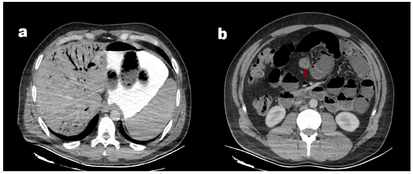
Contrast-enhanced abdominal and pelvic computed tomography (CT) with oral and intravenous contrast demonstrated dilated ileal and jejunal loops with luminal air-fluid levels and intramural gas. Air was also noted within the superior mesenteric vein, portal vein branches, and multiple intrahepatic portal venous branches Figure 1. These findings were consistent with HPVG secondary to bowel ischemia, likely resulting from mesenteric obstruction, IH, or other delayed postoperative complications. Given the imaging findings and clinical status, urgent surgical intervention was indicated. The patient was taken to the operating room within 5–6 hours of presentation to the emergency department.
Upon open laparotomy, a moderate amount of serous fluid was found in the peritoneal cavity. Scattered areas of mild ischemia were observed along the small bowel loops. A mesenteric defect adjacent to the left colon had permitted complete herniation of the entire small intestine into the right hemi-abdomen, resulting in medial displacement of the left colon Figure 2.
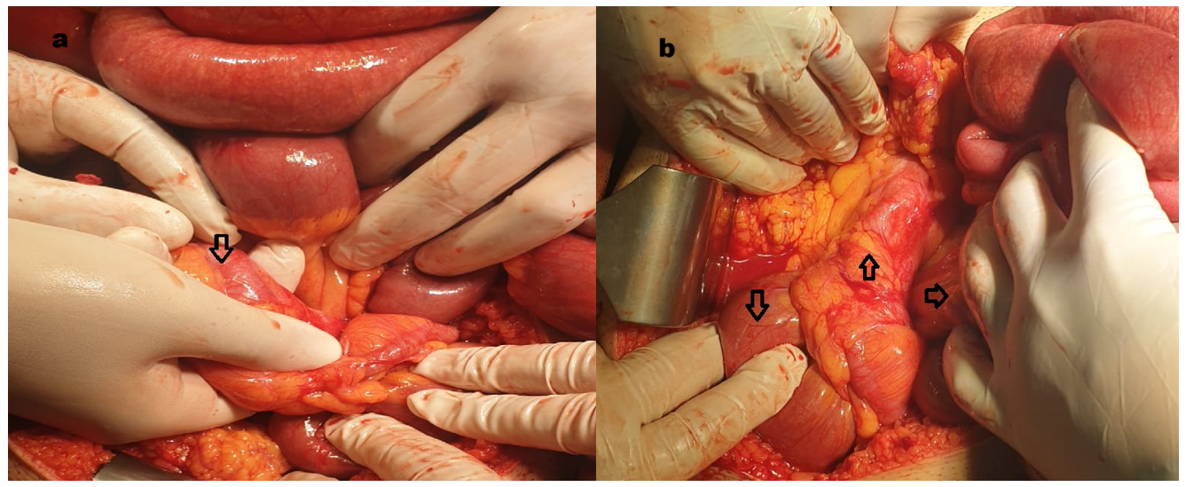
The herniated small bowel loops were manually reduced, and serosal injuries (without full-thickness compromise) were repaired primarily. Peristalsis was preserved in the ischemic segments, and the return of normal serosal colour following warm compress application indicated tissue viability. Due to the large size of the mesenteric defect and medial displacement of the left colon, the colon was secured to the lateral parietal peritoneum using interrupted sutures. The mesenteric defect was then closed in a tension-free manner at the medial retroperitoneal region to prevent stricture formation. After suturing, the viability of the colon was reassessed and confirmed. The patient was subsequently transferred to the intensive care unit for postoperative monitoring. Following consultation with the Infectious Diseases team, empiric antibiotic therapy with meropenem and vancomycin was initiated immediately after surgery. The antibiotic administration was continued over seven days, with blood cultures remaining negative throughout.
Subsequent postoperative monitoring showed a steady decline in leukocyte count and acute-phase reactants, with no significant abnormalities in vital signs. A gradual oral diet was initiated on postoperative day (POD) 3, and the patient was transferred to the surgical ward on POD 4. He was discharged on POD 7 with a normal diet and stable clinical status. At the 1-week follow-up visit, no complications were observed, and sutures were removed. No signs of recurrence or late complications were noted during the 6-month follow-up period.
Discussion
HPVG develops when gas produced in the intestinal wall due to pneumatosis—secondary to bacterial translocation—enters the portal circulation and subsequently migrates into the hepatic venous system [1]. This condition is associated with a high mortality rate and is typically detected on CT imaging, most often indicating irreversible intestinal ischemia and necrosis [1,3,2,4]. Although rare in young patients, congenital anomalies such as Ladd’s bands, as well as postoperative complications leading to bowel obstruction or injury, should be thoroughly investigated as potential secondary aetiologies [5,7,6]. IH is a serious condition that can cause complete bowel obstruction and present as an acute abdomen. It may occur congenitally or develop postoperatively, especially following colorectal surgery [9,8,7]. In patients undergoing left colon or rectal surgery, the inferior mesenteric artery (IMA) is typically ligated, and the bowel mesentery is intentionally left open to preserve perfusion to the anastomotic site [7,10]. Leaving the mesentery open may increase the risk of IH. Postoperative IH related to mesenteric defects can manifest early or several months after surgery, with the highest incidence reported around the fourth postoperative month. IH occurs in 2–3.6% of patients within three years following laparoscopic colorectal resection. Although the overall incidence after laparoscopic colorectal surgery is low (approximately 0.65%), IH carries a mortality rate of 20–50% when complicated by bowel ischemia, underscoring the critical need for early diagnosis and intervention [7,11,10].
CT is the most commonly used imaging modality for diagnosing IH; however, it may not always provide definitive confirmation. Therefore, IH should be considered in the differential diagnosis of patients presenting with unexplained abdominal pain, subileus or ileus episodes, or recurrent vomiting. Definitive diagnosis and identification of the underlying cause are typically established during surgical exploration [11].
This case is notable for several reasons: the patient’s young age, the early postoperative onset of herniation, complete displacement and transposition of the small bowel, and the presence of massive HPVG despite the absence of bowel necrosis. Unlike most cases reported in the literature where HPVG predicts irreversible necrosis, this patient achieved full recovery following successful reduction without bowel resection, culminating in complete remission. These findings underscore the critical importance of managing mesenteric defects after laparoscopic rectal surgery. However, the data remains divided on whether mesenteric defects should be routinely closed during the initial operation [12,13]. Routine closure of mesenteric defects is often avoided due to concerns about compromising anastomotic perfusion, technical complexity, and increased operative time [12]. However, some studies suggest that large mesenteric defects significantly increase the risk of IH, and that closure may help prevent this complication [10,13]. In cases where rapid weight loss or minimal postoperative adhesion formation is anticipated, careful intraoperative assessment of mesenteric defects and selective closure in high-risk patients should be considered.
Conclusions
HPVG may indicate an acute intra-abdominal event such as IH or mesenteric ischemia, particularly in elderly patients or those with a history of abdominal surgery. IH following laparoscopic colorectal surgery is a rare but serious complication that can result in HPVG. Early diagnosis and prompt surgical intervention are critical for survival in such cases. While there is no established consensus on routine closure of mesenteric defects, in our case, lateral fixation of the colon may have played a role in preventing recurrent herniation. Emergency surgical exploration remains lifesaving in suspected cases, as any delay may lead to intestinal necrosis, perforation, sepsis, or the need for extensive bowel resection (all associated with high morbidity and mortality). As a single case report, the findings are limited in generalizability. Further multicenter studies are necessary to better evaluate the risks and benefits of routine mesenteric defect closure in laparoscopic colorectal surgery.
Conflicts of Interest
The authors declare that they have no competing interests.
Funding Source
There is no specific funding related to this research.
Acknowledgments
None
Informed Consent
All necessary consent documents to publish images and clinical information relating to the cases were obtained from the patient, written in Turkish. Only accepted images were shared.
Large Language Model
The authors declare that artificial intelligence and AI-assisted technologies were not used in the article.
Authors Contribution
AT contributed to review, translation, and editing; EK was responsible for writing; UD contributed to review and editing; UK managed reference control; and DT contributed to conceptualisation (supporting).
Data Availability
Data supporting the findings of this study are available within the paper. The video of the case is available from the corresponding author upon request.
References
- Nelson A. L., Millington T. M., Sahani D., Chung R. T., Bauer C., Hertl M., Warshaw A. L., Conrad C.. Hepatic portal venous gas: the ABCs of management. Arch Surg. 2009;144(6):575-81; discussion 581. doi:10.1001/archsurg.2009.88 PMID: 19528392
- Wei H. R., Shan Y. Q., Dong F. H., Zhou L. P., Yang Y. B., Shi J., Ji C. H., Kong W. C.. Clinical characteristics and treatment of hepatic portal venous gas: case series and literature review. Front Med (Lausanne). 2025;12:1540418. doi:10.3389/fmed.2025.1540418 PMID: 40417673 PMCID: PMC12098563
- Wang C., Li Y., Yin Y., Xi C., Su M.. Hepatic Portal Venous Gas Associated with Acute Upper Gastrointestinal Hemorrhage: A Case Report and Literature Review. Curr Med Imaging. 2024;20:e15734056282877. doi:10.2174/0115734056282877240222095545 PMID: 38462831
- Zhang Y., Liu H. L., Tang M., Wang H., Jiang H. H., Lin M. B.. Clinical features and management of 20 patients with hepatic portal venous gas. Exp Ther Med. 2022;24(2):525. doi:10.3892/etm.2022.11452 PMID: 35837035 PMCID: PMC9257953
- Lanzetta M. M., Masserelli A., Addeo G., Cozzi D., Maggialetti N., Danti G., Bartolini L., Pradella S., Giovagnoni A., Miele V.. Internal hernias: a difficult diagnostic challenge. Review of CT signs and clinical findings. Acta Biomed. 2019;90(5-S):20-37. doi:10.23750/abm.v90i5-S.8344 PMID: 31085971 PMCID: PMC6625567
- Watson T. J., Moritz T.. Sliding Hernia. StatPearls. 2025:311-322. doi:10.1002/9781394322008.ch31 PMID: 29083633
- Portale G., Cipollari C., Zuin M., Spolverato Y., Fiscon V.. Prevalence of internal hernia following laparoscopic colorectal surgery: single-center report on 1300 patients. Surg Endosc. 2021;35(8):4315-4320. doi:10.1007/s00464-020-07921-w PMID: 32875409
- Mishra C., Omar K. O.. Internal Hernia: An Uncommon and Often-Missed Differential Diagnosis of Abdominal Pain. Cureus. 2024;16(12):e75801. doi:10.7759/cureus.75801 PMID: 39822468 PMCID: PMC11735237
- Gomez Corral J., Nino Rojo C., de la Fuente Olmos R.. Bowel obstruction: signs indicating the need for urgent surgery. Radiologia (Engl Ed). 2023;65 Suppl 1:S92-S98. doi:10.1016/j.rxeng.2022.09.007 PMID: 37024235
- Toh J. W., Lim R., Keshava A., Rickard M. J.. The risk of internal hernia or volvulus after laparoscopic colorectal surgery: a systematic review. Colorectal Dis. 2016;18(12):1133-1141. doi:10.1111/codi.13464 PMID: 27440227
- Svraka M., Wilhelmsen M., Bulut O.. Internal hernia following laparoscopic colorectal surgery: single center experience. Pol Przegl Chir. 2017;89(5):19-22. doi:10.5604/01.3001.0010.5405 PMID: 29154243
- Stenberg E., Szabo E., Agren G., Ottosson J., Marsk R., Lonroth H., Boman L., Magnuson A., Thorell A., Naslund I.. Closure of mesenteric defects in laparoscopic gastric bypass: a multicentre, randomised, parallel, open-label trial. Lancet. 2016;387(10026):1397-1404. doi:10.1016/S0140-6736(15)01126-5 PMID: 26895675
- Yan X., Su H., Zhang S., Zhou L., Lu J., Yang X., Li J., Xue P., He Z., Wang M., Lu A., Ma J., Zang L., Cai Z., Sun J., Hong H., Zheng M., Feng B.. Pelvic peritoneum closure reduces postoperative complications of laparoscopic abdominoperineal resection: 6-year experience in single center. Surg Endosc. 2021;35(1):406-414. doi:10.1007/s00464-020-07414-w PMID: 32086621