Bone Marrow Necrosis in A Male Patient with Anti-Phospholipid Syndrome: A Case Report with Literature Review
Maram BadraⒾ1, Mina SoryalⒾ1, Malak A. HassanⒾ2*, Nooran Soror1, Ahmed Hisham Abdulfattah3, Dalia Ahmed Nafea1
- 1Department of Internal Medicine, Faculty of Medicine, Alexandria University, Alexandria, Egypt
- 2Faculty of Medicine, Alexandria University, Alexandria, Egypt
- 3Department of Clinical Pathology, Faculty of Medicine, Alexandria University, Alexandria, Egypt
Abstract
Anti-phospholipid syndrome (APS) is a systemic autoimmune disease causing arterial and venous thrombosis, leading to macrovascular and microvascular complications. Bone marrow necrosis (BMN) is defined as the death of hematopoietic tissue and the loss of fat cells in a bone marrow biopsy. BMN is typically associated with a poor prognosis, with most patients dying within weeks. We present a case of BMN in a 32-year-old male patient with APS admitted to our hospital. The patient presented with bilateral non-healing leg ulcers and bilateral lower limb edema. The findings of the duplex ultrasound were consistent with old bilateral deep vein thromboses in the calves. The presence of acute kidney injury and proteinuria prompted a renal biopsy, which revealed chronic thrombotic microangiopathy. Bone marrow biopsy revealed BMN. Unfortunately, the patient did not respond to immunosuppressive treatment and passed away due to septic shock. The unique features in the patient’s presentation, combined with the availability of extensive data on the patient’s history and investigations, further enhance its significance. It is not possible to establish a cause-and-effect relationship or draw conclusive findings from this case report alone. More case reports from clinicians who come across BMN in patients with APS are needed to broaden our understanding of the pathogenesis, presentation, management, and outcomes.
Keywords: anti-phospholipid syndrome, bone marrow necrosis, case report
Article information
Introduction
Anti-phospholipid syndrome (APS) is a systemic autoimmune disease causing arterial and venous thrombosis, leading to macrovascular and microvascular complications [1,2]. Notably, APS also leads to pregnancy morbidity, including spontaneous abortions, preeclampsia, fetal distress, and placental insufficiency [3]. The estimated prevalence of APS is 40 to 50 cases per 100,000 people [4]. This syndrome is well-known for being more prevalent among females, with a female: male ratio of 3.5:1 [2]. The pathogenesis is primarily mediated by auto-antibodies – namely, lupus anticoagulant, anti-cardiolipin, and/or anti-2GPI antibodies [3]. The etiology may be primary isolated APS or secondary to another autoimmune disease, most commonly systemic lupus erythematosus (SLE) [3]. APS diagnosis requires the presence of clinical evidence of thrombosis or pregnancy morbidity, as well as laboratory evidence of persistently elevated titers of the previously mentioned antibodies [5]. Although thrombosis can occur anywhere, deep vein thrombosis (DVT) of the lower limbs is the most common presentation of venous thrombi [3]. Cerebral venous thrombosis is another less common manifestation [4]. Arterial thrombosis most commonly presents as transient ischemic attacks (TIAs) and ischemic strokes [3,4]. In addition, manifestations of microvascular thrombosis can also occur, most commonly affecting the skin and the kidneys [4]. Management is primarily focused on preventing thrombosis [6]. Primary prevention with low-dose aspirin is recommended for patients with a high-risk antiphospholipid profile [7]. Secondary prevention in patients with APS and a history of venous thrombosis typically involves the use of warfarin or direct oral anticoagulants (DOACs) [7]. Meanwhile, in accordance with the recommendations of the 16th International Congress on Antiphospholipid Antibodies, secondary prevention in patients with APS and a history of arterial thrombosis is either low-dose aspirin plus standard-intensity warfarin (INR range, 2 to 3) or warfarin at an INR > 3 [7]. The use of DOACs is typically limited to those who can’t tolerate warfarin or those with features of low-risk disease [7].
Bone marrow necrosis (BMN) is defined as the death of hematopoietic tissue and the loss of fat cells on bone marrow biopsy [8]. This rare entity is not only easily overlooked, but it must also be carefully distinguished from other bone marrow pathologies [8]. Underlying etiologies of BMN include hematological malignancies, infections, sickle cell disease, disseminated intravascular coagulation (DIC), and radiation [9,8,10]. Although the exact pathogenesis remains poorly understood, it is hypothesized that thrombi or emboli leading to hypoxemia is the mechanism underlying BMN [9,8]. The clinical manifestations associated with BMN include bone pain, fever, and cytopenias – all of which are non-specific, further complicating diagnosis [8]. BMN is typically associated with a poor prognosis, with most patients dying within weeks [11,9,8].
BMN is an exceedingly rare complication of APS, with only two previously reported cases in the literature [12,13]. We present a case of BMN encountered in a male patient with APS at our hospital.
Case presentation
A 32-year-old male patient presented with a two-month history of recurring attacks of fever, headache, and easy fatigability. As well as a one-month history of hypertension and bilateral painful non-healing leg ulcers. His medical history revealed left calf DVT, pulmonary embolism, and myocardial infarction three years prior to presentation. At that time, the patient was started on warfarin for a few months, then shifted to apixaban. Two months later, he developed recurrent left calf DVT two years before presentation, then bilateral calf DVTs one year prior to presentation. His past medical history was otherwise unremarkable. He has a positive family history featuring a father who developed DVT, and a sister who experienced DVT and an abortion, neither of whom was tested for APS or other autoimmune conditions. Upon presentation, physical examination revealed bilateral lower limb edema and bilateral leg ulcers. The ulcers were irregular, non-bleeding, with well-defined borders, and located in the gaiter area. Laboratory investigations at the time of admission revealed normocytic normochromic anemia, thrombocytopenia, and acute kidney injury (AKI). TTP was excluded based on normal bilirubin levels, normal LDH levels, a normal reticulocyte count, and the absence of schistocytes in the peripheral blood. More details on the lab investigations at the time of admission can be found in Table 1 and Table 2.
| Parameter | Result | Reference range |
|---|---|---|
| Hemoglobin | 9.7 g/dL | (13 – 17) |
| Hematocrit | 29.2 % | (40 – 52) |
| Mean Corpuscular Volume | 78.1 fL | (76 – 96) |
| Mean Corpuscular Hemoglobin | 26.1 pg | (26 – 32) |
| Mean Corpuscular Hemoglobin Concentration | 33.2 g/dL | (33 – 37) |
| Red blood cells | 3.74 × 10⁶/uL | (4.5 – 5.9) |
| Platelets count | 15 × 103/uL | (150 – 450) |
| White Blood Cell | 5.2 × 10³/uL | (4 - 11) |
| Basophil | 0 % | (0 – 1) |
| Eosinophil | 0 % | (1 – 6) |
| Neutrophil | 67.0 % | (40 – 70) |
| Lymphocyte | 24 % | (20 – 45) |
| Monocyte | 9 % | (2 – 8) |
| Urea | 113 mg/dL | (15 – 45) |
| Serum creatinine | 2.5 mg/dL | (0.7 – 1.4) |
| Albumin-creatinine ratio | 1233 | (< 70) |
| Glomerular filtration rate | 46 mL/min/1.73 m2 | (> 90) |
| Cystatin C | 1.68 mg/dL | (0.71 – 1.21) |
| Total serum bilirubin | 0.6 mg/dL | (0.3 – 1.2) |
| Direct serum bilirubin | 0.15 mg/dL | (0.1 – 0.3) |
| Lactate dehydrogenase | 200 units | (135 – 225) |
| Prothrombin time | 13.3 seconds | (11.3 – 13.2) |
| International normalized ratio | 1.18 | |
| Partial thromboplastin time | 53.5 seconds | (Up to 40) |
| Parameter | Result | Reference range |
|---|---|---|
| Pus cells | 2–4 | (0 – 5) |
| Red blood cells | 50–60 | (0 – 3) |
| Hyaline casts | Present | None |
| Dysmorphic RBCs | Absent | None |
| Urinary protein | 163 mg/dL | |
| Urinary creatinine | 39 mg/dL | |
| Protein-to-creatinine ratio | 4.12 | (Up to 0.2) |
Consequently, ultrasound abdomen and pelvis revealed an enlarged right lobe of liver measuring 18 cm with an echogenic pattern. Additionally, bilateral grade I nephropathy was detected. The nephrology unit recommended renal biopsy, which showed chronic thrombotic microangiopathy (CTMA), characterized by arteriolar thrombosis with marked mucinous intimal degeneration and fibrinoid necrosis. One glomerulus exhibited global necrosis, diffuse moderate interstitial fibrosis, and tubular atrophy of 40%. Diffuse moderate acute tubular necrosis with RBC casts was also observed.
Furthermore, bone marrow aspiration (BMA) revealed diluted aspirate containing only a small number of cells, including mature granulocytes, segmented neutrophils, mature lymphocytes, a few normoblasts, and occasional megakaryocytes. Blast cells were 1% and plasma cells were 0%.
Thrombophilia workup showed positivity for lupus anticoagulant, anti-cardiolipin antibodies, and anti-2GPI antibodies, consistent with triple-positive APS. Antibody levels were considered to fall within the moderate-positive titers classification. This was repeated 12 weeks later to confirm the persistent elevation in antibodies. Further details on the thrombophilia work-up can be found in Table 3. SLE was excluded based on negative antinuclear antibody (ANA), negative anti-double-stranded deoxyribonucleic acid (dsDNA) antibody, and normal C3 and C4 levels. Duplex ultrasound of the bilateral lower limbs showed the majority of the veins to be non-compressible over an iso-echoic thrombus with central canalization, consistent with the presence of old DVTs. A virology panel for hepatitis C, hepatitis B, and HIV was all negative.
| Parameter | Result | Reference range |
|---|---|---|
| PCR for factor V Leiden | Positive for heterozygosity (R506Q) | |
| PCR for MTHFR gene mutation | Positive for homozygosity (C677T) | |
| PCR for prothrombin gene mutation | Negative | |
| Protein C | 111.0 % | 70 - 140 |
| Protein S | 101.8 % | 59 - 118 |
| Antithrombin III | 97.6 % | 70 - 125 |
| Lupus anti-coagulant | 3.5 | Negative: 0.8 – 1.2 |
| Weak positive: 1.2 -1.5 | ||
| Moderate positive: 1.5 – 2.0 | ||
| Strong positive: > 2.0 | ||
| Anti-cardiolipin IgG | 39.8 U/ml | (Up to 10) |
| Anti-cardiolipin IgM | 2.2 U/ml | (Up to 7) |
| B2 glycoprotein IgG | 16.5 U/ml | Negative: < 5 |
| Equivocal: 5- 8 | ||
| Positive: > 8 | ||
| B2 glycoprotein IgM | 1.7 U/ml | Negative: < 5 |
| Equivocal: 5- 8 | ||
| Positive: > 8 |
MTHFR, methylene tetrahydrofolate reductase
The patient was administered hydroxychloroquine 200 mg twice daily, prednisolone 20 mg once daily, and cyclophosphamide 600 mg IV once weekly for two weeks, then 50 mg twice daily for two months. Regrettably, the patient did not respond to these treatments and subsequently complained of dizziness and fatigue. CBC revealed an alarming hemoglobin level of 5.7 g/dL and a WBC count of 4.7 × 10³/mm3.
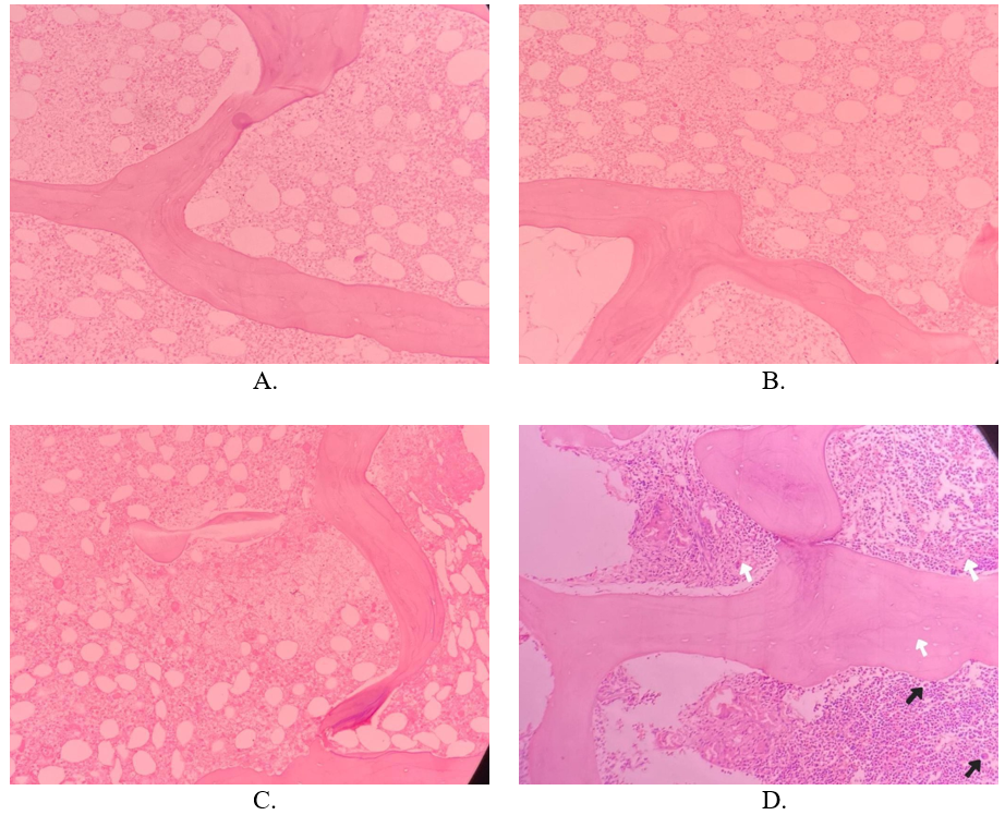
Consequently, the patient underwent bone marrow biopsy two months after presentation, which showed extensive areas of bone marrow necrosis (BMN) with a few apoptotic cells (Figure 1). Intratrabecular hematopoietic spaces were preserved, exhibiting erythroid and granulocytic hyperplasia. Bone marrow sections also demonstrated multiple small granulomas. Subsequently, the patient was administered pulse dexamethasone and rituximab 375 mg/m2 weekly for four doses. Unfortunately, the patient passed away as a result of septic shock secondary to pneumonia. This came four months after the initiation of immunosuppressive therapy and two months after being diagnosed with BMN.
Discussion
To the best of our knowledge, this is only the third case in the literature of BMN in APS, highlighting the rarity of this presentation. Interestingly, the two previous case reports were published in 1995 and 1997 [12,13]. This case report is published more than 25 years later, further underscores its significance. This male patient with a history of recurrent arterial and venous thromboembolism presented to our hospital with painful non-healing ulcers and bilateral lower limb edema. Thrombophilia work-up was consistent with APS, which was further supported by a positive family history. Upon failing to respond to treatment, a bone marrow biopsy confirmed the diagnosis of BMN. Unfortunately, the patient passed away.
There is very limited data in the literature on the epidemiology of APS in Egypt. Even when present, they typically feature small cohorts that lack diverse characteristics, making it difficult to conclude. In a study by Morad et al, one of the few studies in the literature on APS in an Egyptian cohort, 91.7% of patients were females, and 80% of patients had secondary APS [14]. Another study by Abd El-Moniem et al showed similar characteristics with 91.2% females and 71.6% secondary APS [15]. Thus, the incidence of primary APS in male patients in Egypt remains poorly studied, further emphasizing the significance of this case study. This case report highlights the urgent need for further research and country-specific data to address this knowledge gap, thereby enhancing future management. Adding to the rarity of this presentation, BMN in Egyptian patients has been rarely reported. After a thorough review of the literature, only one study has explored BMN in Egypt [16]. Elgamal et al examined 5,043 bone marrow biopsies of cancer patients at the National Cancer Institute to assess the prevalence of BMN [16]. Fifteen of the 5,043 biopsies featured BMN, resulting in a prevalence of 0.3% [16]. They reported an association between BMN and poor prognosis, with none of the patients surviving beyond 6 months from the time of BMN diagnosis [16]. No other studies have examined BMN in Egypt. Thus, this presents the first case report of BMN in Egypt in the literature. It is also the first study examining BMN due to a benign etiology in Egypt.
In 1995, Bulvik et al published the first case report of BMN in a patient with APS [12]. The patient responded to treatment with methylprednisolone [12]. Interestingly, although SLE had been previously excluded, this patient later developed positive ANA and anti-Smith antibodies [12]. Despite not meeting the diagnostic criteria for SLE, the patient was considered to be “lupus-like” [12]. The authors inferred that the high antibody titers in this patient may have had a role in the pathogenesis of BMN [12]. In 1997, a second case report was published by Paydas et al on a 27-year-old female [13]. The patient had pancytopenia refractory to treatment with steroids and plasma exchange [13]. Despite testing negative for ANA and anti-dsDNA antibodies, investigations were positive for anti-SM antibodies [13].
| Study ID | Gender/Age | Geographic location | APS type | Antibody levels | Treatment | Outcome |
|---|---|---|---|---|---|---|
| Badra 2025 (this case) | M/32 | Egypt | Triple-positive APS ANA & anti-dsDNA negative. | Moderate positive titers Anti-cardiolipin IgG: 39.8 U/ml Anti-cardiolipin IgM: 1.7 U/ml | Hydroxychloroquine, prednisolone, & cyclophosphamide. Then rituximab & dexamethasone. | No response to treatment. Death due to septic shock. |
| Paydas 1997 [13] | F/27 | Turkey | Not specified ANA & anti-dsDNA antibodies negative, but anti-SM antibody positive. | Anti-cardiolipin IgG: 215 U/I Anti-cardiolipin IgM: 48 U/I | Steroids, transfusion, & plasma exchange. | Pancytopenia did not respond. The patient discharged herself. |
| Bulvik 1995 [12] | F/23 | South Africa | Not specified Later became ANA & anti-Smith antibodies positive. | High antibody titers | Methylprednisolone | The patient responded to treatment. |
BMN, bone marrow necrosis; APS, antiphospholipid syndrome
Table 4 compares the three cases of BMN in APS, providing insight into demographics, management, and outcomes. Although our case is similar, it features a male patient. The patient’s history of bilateral DVTs, pulmonary embolism, and myocardial infarction provided valuable insight into the severity of the patient’s manifestations. The antibody titers in our patient were not as high as those reported in the two previous case reports Table 3 and Table 4, suggesting that the pathogenesis of BMN in patients with APS may not be solely explained by high antibody titers. Unfortunately, our patient did not respond to treatment and passed away shortly after being diagnosed with BMN. This is in line with the poor prognosis that has been reported with BMN due to other etiologies [16,8].
BMN may be suspected in patients with bone pain, fever, cytopenias, and elevated LDH – all of which are non-specific findings, presenting a diagnostic challenge [17]. Thus, clinical findings alone are insufficient and warrant further investigations [17]. Our patient had fever, anemia, thrombocytopenia, and elevated LDH, consistent with BMN. Bone marrow biopsy can confirm the presence of BMN [17]. It shows necrotic areas with disrupted bone marrow architecture, eosinophilic debris, and ghost cells [17]. These features were seen in our patient. Upon diagnosing BMN, thorough history-taking, clinical examination, and investigations are necessary to identify the underlying cause, if it was not already known [17]. In patients with BMN, the treatment is aimed at the underlying primary disorder. Despite treatment attempts, it carries an extremely poor prognosis and is rapidly fatal in most cases, consistent with what was seen in this case as well [18].
This case report of BMN in a male patient with APS is of significance due to the rarity of this association. However, it is not possible to establish a cause-and-effect relationship or draw conclusive findings from a single case report. Another potential limitation is the temporal association between cyclophosphamide and BMN, suggesting the possibility of cyclophosphamide-induced bone marrow toxicity. Alternative causes for bone marrow necrosis in this patient include drugs, infections, and other autoimmune diseases [17]. The poorly understood pathogenesis of both BMN and APS further hinders our ability to fully comprehend the presented case. More case reports from clinicians who come across BMN in patients with APS can help broaden our understanding of the pathogenesis, presentation, management, and outcomes. Each case report will provide a unique perspective that, together, contributes to our overall understanding of this rare complication.
Conclusions
BMN is an extremely rare complication of APS. We present the third reported case of BMN in a patient with APS. Unfortunately, the patient did not respond to treatment attempts and passed away two months after the BMN diagnosis. More case reports are needed to better understand this association.
Conflicts of Interest
The authors declare that they have no conflicts of interest.
Funding Source
This research received no external funding.
Acknowledgments
None
Informed Consent
We obtained written informed consent for publication from the patient.
Large Language Model
None
Authors Contribution
MAH, MB contributed to the original draft writing. DAN supervised the project. MS administered the project. Reviewing and editing were performed by MAH, MB, MS, and NS. All authors read and approved the final content.
Data Availability
The data supporting the findings of this case report are included within the article and its supplementary materials. Additional de-identified information may be made available upon reasonable request from the corresponding author.
References
- Sammaritano L. R.. Antiphospholipid syndrome. Best Pract Res Clin Rheumatol. 2020;34(1):101463. doi:10.1016/j.berh.2019.101463 PMID: 31866276
- Truglia S., Capozzi A., Mancuso S., Manganelli V., Rapino L., Riitano G., Recalchi S., Colafrancesco S., Ceccarelli F., Garofalo T., Alessandri C., Longo A., Misasi R., Conti F., Sorice M.. Relationship Between Gender Differences and Clinical Outcome in Patients With the Antiphospholipid Syndrome. Front Immunol. 2022;13:932181. doi:10.3389/fimmu.2022.932181 PMID: 35860235 PMCID: PMC9289158
- Bustamante J. G., Goyal A., Rout P., Singhal M.. Antiphospholipid Syndrome. StatPearls. 2025. PMID: 28613698 PMCID: PMCID
- Gaspar P., Sciascia S., Tektonidou M. G.. Epidemiology of antiphospholipid syndrome: macro- and microvascular manifestations. Rheumatology (Oxford). 2024;63(SI):SI24-SI36. doi:10.1093/rheumatology/kead571 PMID: 38320589 PMCID: PMC10846913
- Vandevelde A., Devreese K. M. J.. Laboratory Diagnosis of Antiphospholipid Syndrome: Insights and Hindrances. J Clin Med. 2022;11(8). doi:10.3390/jcm11082164 PMID: 35456258 PMCID: PMC9025581
- Garcia D., Erkan D.. Diagnosis and Management of the Antiphospholipid Syndrome. N Engl J Med. 2018;378(21):2010-2021. doi:10.1056/NEJMra1705454 PMID: 29791828
- Cohen H., Cuadrado M. J., Erkan D., Duarte-Garcia A., Isenberg D. A., Knight J. S., Ortel T. L., Rahman A., Salmon J. E., Tektonidou M. G., Williams D. J., Willis R., Woller S. C., Andrade D.. 16th International Congress on Antiphospholipid Antibodies Task Force Report on Antiphospholipid Syndrome Treatment Trends. Lupus. 2020;29(12):1571-1593. doi:10.1177/0961203320950461 PMID: 33100166 PMCID: PMC7658424
- Luan C., Zhao H., Ding Y.. Bone marrow necrosis: Facts, controversies, and perspective. Int J Lab Hematol. 2024;46(5):770-776. doi:10.1111/ijlh.14335 PMID: 38923828
- Conrad M. E.. Bone marrow necrosis. J Intensive Care Med. 1995;10(4):171-8. doi:10.1177/088506669501000403 PMID: 10155181
- Wool G. D., Deucher A.. Bone marrow necrosis: ten-year retrospective review of bone marrow biopsy specimens. Am J Clin Pathol. 2015;143(2):201-13; quiz 306. doi:10.1309/AJCP0TN1MCMOLMPK PMID: 25596246
- Badar T., Shetty A., Bueso-Ramos C., Cortes J., Konopleva M., Borthakur G., Pierce S., Huang X., Chen H. C., Kadia T., Daver N., Dinardo C., O'Brien S., Garcia-Manero G., Kantarjian H., Ravandi F.. Bone marrow necrosis in acute leukemia: Clinical characteristic and outcome. Am J Hematol. 2015;90(9):769-73. doi:10.1002/ajh.24074 PMID: 26017166 PMCID: PMC4546505
- Bulvik S., Aronson I., Ress S., Jacobs P.. Extensive bone marrow necrosis associated with antiphospholipid antibodies. Am J Med. 1995;98(6):572-4. doi:10.1016/s0002-9343(99)80016-3 PMID: 7778573
- Paydas S., Kocak R., Zorludemir S., Baslamisli F.. Bone marrow necrosis in antiphospholipid syndrome. J Clin Pathol. 1997;50(3):261-2. doi:10.1136/jcp.50.3.261 PMID: 9155683 PMCID: PMC499827
- Morad Caroline S., Fayez Dalia, Mahmoud Menna, Aboud Fatma M.. Primary and secondary antiphospholipid syndrome characteristics in an Egyptian cohort. The Egyptian Rheumatologist. 2022;44(4):373-376. doi:10.1016/j.ejr.2022.07.006
- El-Moniem G. A., El-Garf K., Sobhy N., Elmaghraby S.. Characterization of the Clinical and Laboratory Features of Primary and Secondary Antiphospholipid Syndrome in a Cohort of Egyptian Patients. Curr Rheumatol Rev. 2020;16(4):304-310. doi:10.2174/1573397116666200116095734 PMID: 31951187
- Elgamal B. M., Rashed R. A., Raslan H. N.. Prevalence of bone marrow necrosis in Egyptian cancer patients referring to the National Cancer Institute. J Egypt Natl Canc Inst. 2011;23(3):95-9. doi:10.1016/j.jnci.2011.09.007 PMID: 22776813
- Deucher A., Wool G. D.. How I investigate bone marrow necrosis. Int J Lab Hematol. 2019;41(5):585-592. doi:10.1111/ijlh.13091 PMID: 31424633
- Cabral Tamara C. S., Fernandes Carolina M., Lage Luis Alberto C., Zerbini Maria Claudia, Pereira Juliana. Bone marrow necrosis: literature review. Jornal Brasileiro de Patologia e Medicina Laboratorial. 2016. doi:10.5935/1676-2444.20160031