Near-miss Osteomyelitis in an Immunosuppressed Crohn’s Disease Patient: Diagnostic Vigilance Sparked by a Medical Student
Abbas Al BazzalⒾ1, Aya KawssanⒾ1, Atef AkoumⒾ2, Dayana Majid Kamar1, Mohammad Ali MtairekⒾ3, Hiba HamdarⒾ4*
- 1Faculty of Medical Sciences, Lebanese University, Beirut, Lebanon
- 2Internal Medicine Department, Hennepin Healthcare, Minneapolis, USA
- 3Faculty of Medicine, Damascus University, Damascus, Syria
- 4Medical Learning Skills Academy, Beirut, Lebanon
Abstract
Crohn's disease (CD) patients on immunosuppressive therapy are at increased risk of infections, including osteomyelitis, which could be diagnostically challenging. This case highlights the importance of medical student vigilance in making a near-miss diagnosis of osteomyelitis in a CD patient.
A 56-year-old CD patient developed left shoulder pain following localized muscle injection and was initially managed as a soft-tissue abscess. The condition worsened despite 21 days of antibiotics. Investigation: Cultures were positive for methicillin-sensitive Staphylococcus aureus (MSSA). Persistent pain and new neurologic complaints prompted an MRI (T1 hypointensity, T2-STIR hyperintensity, post-contrast enhancement) at 60 days, in support of osteomyelitis without bone biopsy.Management/Outcome: The patient received 6 weeks of intravenous vancomycin (1 g every 12 hours), followed by oral antibiotics, resulting in partial relief of pain (pain score: 8/10 to 4/10) and improvement in shoulder function (Constant-Murley score: 30 to 65 at 3 months). Immunotherapy (adalimumab) was restarted after infection control.
This case highlights three practical lessons: maintaining a low threshold for advanced imaging in immunosuppressed patients with persistent pain; incorporating diagnostic time-outs to invite trainee perspectives; and seeking early infectious-disease consultation when osteomyelitis is suspected.
Keywords: Crohn Disease, Osteomyelitis, Staphylococcus aureus, Diagnostic Errors, Medical Education
Article information
Introduction
Making a precise diagnosis is crucial in the challenging field of medicine, particularly for individuals with complex illnesses such as Crohn’s disease (CD). CD is a type of chronic inflammatory bowel disease (IBD) that is most commonly associated with musculoskeletal symptoms, though it can cause several other disorders as well [1,2]. The shoulder may also be affected; however, this is less common [3,4,5]. These might range from joint soreness to arthritis affecting big joints. A wide range of possibilities must be carefully considered during the diagnostic process in such circumstances. This case underscores the diagnostic pitfalls in immunosuppressed CD patients, where musculoskeletal extraintestinal manifestations (prevalence: 20–50% in IBD) can mask severe infections like osteomyelitis, particularly following injection-site complications [1,2]. The novelty lies in the role of a medical student in averting diagnostic delay through timely suspicion of osteomyelitis. This is where the new insight of trainees can be quite helpful. Because they have access to the most up-to-date academic information and frequently have more time to spend with patients, medical students can play a crucial role [6,7]. A student’s perspective can help the medical team arrive at the correct diagnosis by noticing changes in symptoms and challenging presumptions. This helps to reinforce the idea that good patient care is a team effort, in which all opinions, regardless of position, contribute to the outcome.
Case Presentation
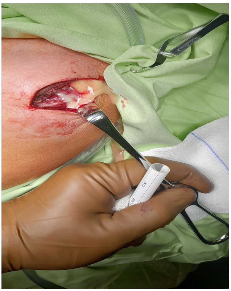
A 56-year-old man with Crohn’s disease being treated with immunotherapy (adalimumab) presented to the hospital in early September 2024 (symptoms having started in July and having worsened for the last two months) with intense left shoulder pain that progressively worsened and severely limited the range of motion. An injection was done into the peri-articular area (local muscle, deltoid) for pain relief, testing, and therapeutic reasons. An abscess developed in the injection site, as a consequence of suspected (though never formally adjudicated) poor aseptic technique and contamination, which probably made pain worse. Wound cultures taken during abscess drainage on 11.08.2024 (Day 7 from injection and abscess identification) before antibiotics yielded methicillin-sensitive Staphylococcus aureus (MSSA, MIC <1 µg/mL). The patient was started on an initial antibiotic course of vancomycin 1 g IV every 12 hours (discharge summary, approximately 21 days from 08.11.2024 to 29.11.2024) and had his Crohn’s immunotherapy drug stopped to reduce the risk of infection in the meantime.
Patient showed some improvement at 60 days after drainage (around 07.01.2025; however, pain levels increased drastically (pain score 8/10), and he developed numbness in all fingers of the left hand. Despite the worsening condition and the red flag symptoms (persistent pain greater than 72 hours duration, CRP level risen to 80 mg/L, and onset of developing neurological symptoms), the attending doctor blamed the persistence of pain on post-operative complications and reassured the patient that it would improve with time and that he was progressing well. A student doctor, observing the persistent and worsening symptoms, suggested the possibility of osteomyelitis (bone infection). Still, this opinion was initially dismissed by the attending doctor, who, in light of the initial diagnosis of a soft-tissue infection, showed confirmation bias.
Patient’s condition worsened further, necessitating an emergency room (ER) admission on 10.01.2025 with severe pain and numbness. An MRI of the left shoulder on 10.01.2025 established the diagnosis of osteomyelitis with features of increased signal intensity (T1 hypointensity, T2-STIR hyperintensity, post-contrast enhancement), tendon rupture, and fluid collection. The diagnosis relied on MRI, microbiology, and the clinical course, as a bone biopsy was postponed due to surgical risk in the immunocompromised patient. No early imaging had been done to add to the diagnostic delay.
Following diagnosis, the patient received 6 weeks of intravenous vancomycin (1 g q12h, 10.01.2025 through approximately 21.02.2025), then 4 weeks of levofloxacin orally (21.02.2025 through 21.03.2025), contingent on MSSA susceptibility and antimicrobial stewardship (local MRSA <10%; toxicity is tracked with normal CBC and BMP). MRI on 17.02.2025 showed persistent changes with rotator cuff tear and likely Hill-Sachs fracture, indicating remaining postoperative complications. Partial pain relief (pain score reduced from 8/10 to 4/10) and shoulder function (Constant-Murley score from 30 to 65) gain were attained at 3 months post-diagnosis (approximately 10.04.2025). Resumption of immunotherapy (adalimumab) was recommended by infectious disease and gastroenterology experts, in accordance with the established resolution of infection (CRP <10 mg/L, absence of fever), and occurred.
The evolution of an Injection-site abscess to osteomyelitis went through contiguous spread to the humerus and was precipitated by immunosuppression, obscuring characteristic signs. This case again brings into sharp focus the diagnostic pitfalls in immunosuppressed CD patients, in which musculoskeletal extraintestinal manifestations may masquerade or disguise infections. It reminds us of the importance of trainee contributions to preventing near-miss errors through diagnostic time-out and escalation processes.
| Date | Event |
|---|---|
| Around 09.07.2024 | Initial onset of gradual left shoulder pain, progressively worsening over two months. |
| Around 09.09.2024 | Presentation to the hospital with intense pain limiting the range of motion; peri-articular (deltoid) injection administered for pain management, testing, and treatment |
| Shortly after injection (within days, around mid-September 2024) | An abscess developed at the injection site due to infection. |
| 08.11.2024 | Abscess identified and drained; wound cultures revealed MSSA (pre-antibiotic treatment); initial vancomycin course initiated. |
| 08.11.2024–29.11.2024 | Initial IV vancomycin course; adalimumab paused. |
| Approximately 07.01.2025 | 60 days post-drainage; reported some improvement, but pain increased (8/10) with numbness in all left-hand fingers; medical student suggests osteomyelitis, dismissed. |
| 10.01.2025 | ER admission for severe pain and numbness; initial MRI confirms osteomyelitis (T1 hypointensity, T2-STIR hyperintensity, post-contrast enhancement; tendon rupture; fluid collection). |
| 10.01.2025–21.02.2025 | 6-week IV vancomycin course post-diagnosis. |
| 21.02.2025–21.03.2025 | 4-week oral levofloxacin course. |
| 17.02.2025 | Follow-up MRI shows persistent changes (rotator cuff tear, suspected Hill-Sachs fracture). |
| Approximately 10.04.2025 | 3 months post-diagnosis; pain improved to 4/10, Constant-Murley score 65; adalimumab restarted |
MSSA, Methicillin-Sensitive Staphylococcus aureus; IV, Intravenous; ER, Emergency Room; MRI, Magnetic Resonance Imaging; T1, T1-weighted MRI sequence; T2-STIR, T2-weighted Short Tau Inversion Recovery MRI sequence
.
Medical Background: Crohn’s Disease and Musculoskeletal Manifestations
Crohn’s disease (CD) can involve any portion of the gastrointestinal tract, from the mouth to the anus. CD is also linked with extraintestinal manifestations (EIMs) beyond its mere gastrointestinal symptoms, which can involve the musculoskeletal system and other organ systems [1]. Musculoskeletal symptoms are seen in more than half of the IBD patients and stand as one of the most frequent EIMs [2]. Arthralgia, i.e., pain in the joints, and peripheral arthritis, which more frequently affect large joints such as the knees and ankles, can be among them, as can axial involvement in the form of spondyloarthritis [4,5]. The shoulder can also be affected less frequently [3]. Although the exact pathophysiology of the majority of musculoskeletal EIMs is unknown, it is thought to include immunological dysregulation, genetics, and the interaction of systemic inflammation with gut microbiota [7]. In this case, the patient’s shoulder pain was initially misattributed to CD-related arthralgia, highlighting how immunosuppression can mask typical signs of infection, thereby delaying suspicion of osteomyelitis. Musculoskeletal manifestations in CD patients can have a significant negative impact on their quality of life and typically require a multidisciplinary management strategy.
Osteomyelitis: Diagnosis and Treatment
Osteomyelitis is an infectious disease that results in an inflammatory bone and bone marrow condition induced by bacterial infection, leading to progressive bone destruction [6]. It can be either acute or chronic, and it is typically diagnosed based on a constellation of clinical symptoms, imaging studies (such as X-rays, MRIs, and CT scans), and laboratory results (including elevated inflammatory markers and positive blood cultures) [8]. In this case, diagnosis relied on MRI (T1 hypointensity, T2-STIR hyperintensity, post-contrast enhancement) and clinical course, as bone biopsy was deferred due to surgical risk in an immunocompromised patient [8,9]. A definitive diagnosis is the gold standard and typically includes a bone biopsy, histological study, and microbiological culture [9]. Osteomyelitis treatment is typically long-term and involves surgical debridement to remove infected or necrotic bone, along with long-term antibiotic treatment [10]. The antibiotics are chosen based on culture findings, as well as the patient’s specific characteristics. Here, vancomycin (1 g IV every 12 hours) was administered for 6 weeks based on MSSA susceptibility (MIC < 1 µg/mL), followed by oral levofloxacin for 4 weeks. In some instances, particularly in chronic osteomyelitis, surgery is necessary for the definitive removal of the infection [11]. The duration of antibiotic treatment may vary, but it typically lasts weeks to months, depending on the seriousness and type of infection [12].
Staphylococcus aureus and Osteomyelitis
Staphylococcus aureus (S. aureus) is a common skin and nasal bacterium that is the leading cause of osteomyelitis [13]. S. aureus can produce infection of the bone by a variety of mechanisms, including hematogenous spread, direct inoculation (like trauma, surgery, or, in our patient, an unsterile injection), and contiguous spread from an infective overlying soft tissue infection [14]. Here, a contiguous spread from an injection-site abscess (MSSA, as indicated by wound culture collected prior to antibiotics) was likely, given the low local MRSA prevalence per hospital epidemiology. The ability of S. aureus to form biofilms on bone and medical devices, as well as its capacity to survive intracellularly within host cells, contributes to the recurrence and chronicity of osteomyelitis, making treatment challenging [15,16]. Initial antibiotic failure was likely due to inadequate duration and bone penetration, underscoring the need for early imaging. The presence of S. aureus in an abscess, especially in close proximity to bone, should raise a high suspicion of underlying osteomyelitis, particularly if this does not respond to standard treatment or if it is recurrent.
Immunotherapy and Infection Risk in Crohn’s Disease
Patients with Crohn’s disease frequently require immunosuppressive medications, such as biologics and immunomodulators, to control their chronic inflammation [17]. While these medications are quite efficient at controlling disease activity, they do increase the patient’s vulnerability to infections from bacterial, viral, and fungal pathogens [18]. The nature and severity of immunosuppression vary depending on the drug and combination therapy used. For example, anti-tumor necrosis factor (TNF) medications, which are routinely used in CD, can impede the immune system’s ability to generate an effective response to infections, including those caused by S. aureus [19]. In this case, adalimumab was paused after the diagnosis of an abscess and restarted 3 months after antibiotic completion, following consultations with infectious disease and gastroenterology specialists who confirmed resolution of the infection (CRP <10 mg/L, no fever). This increased risk of infection necessitates careful monitoring and a heightened awareness of potential infectious complications, as infections in immunocompromised patients can present atypically and progress rapidly. The interruption of immunotherapy, as occurred in this case, is a common practice when an infection is suspected or confirmed. Still, it also highlights the delicate balance between controlling the underlying disease and mitigating the risk of infection.
| Condition | Symptoms and features |
|---|---|
| Osteomyelitis | Focal pain, numbness |
| Septic Arthritis | Joint effusion, warmth |
| Bursitis | Localized tenderness |
| Rotator Cuff Tear | Weakness, limited ROM |
| SIRVA | Post-injection pain |
| Neuropathic Pain | Burning, no focal signs |
SIRVA, Shoulder Injury Related to Vaccine Administration; ROM, Range of Motion.
Progression from Abscess to Osteomyelitis
The spread from soft tissue infection, i.e., abscess, to osteomyelitis is a well-documented process when infection is not managed or is managed improperly, or if it occurs in very close proximity to bone [20]. An abscess at the deltoid injection site likely seeded the humerus via contiguous spread, exacerbated by immunosuppression. In immunocompromised individuals, this can be even quicker and insidious due to a weakened immune response. An abscess, which is a localized collection of pus, can exert pressure on adjacent bone, leading to local ischemia and creating an environment conducive to bacterial invasion of the bone tissue. Furthermore, bacteria from the abscess can directly spread into the bone through microtrauma or through pre-existing vascular channels. The acute management of the abscess, although mandatory, may not always eradicate all of the bacteria, especially if the infection has already begun to involve the bone. Red flags (persistent pain for more than 72 hours, CRP 80 mg/L, and new numbness) were ignored, delaying the MRI; a safety-net checklist should have prompted faster imaging [21] (Table 3).
| Checklist Item |
|---|
| Persistent focused pain after more than 72 hours of antibiotics |
| Fever or increasing CRP (more than 20 mg/L) |
| New neurological symptoms (e.g., numbness) |
| Unresolved soft-tissue infection near the bone |
CRP, C-reactive protein.
Persistent pain and new neurological symptoms of numbness are critical red flag signs that should be further investigated with further workup for deeper, more serious infections such as osteomyelitis, even if imaging was initially negative or inconclusive. This case exemplifies how a seemingly localized infection can escalate to a severe bone infection, especially in a vulnerable patient population.
Clinical Reasoning and Medical Education
This case exemplifies the complexity of clinical reasoning, as well as the crucial role of medical education in teaching critical thinking and a willingness to question assumptions. Clinical reasoning is an interactive process of data collection, hypothesis generation, and hypothesis testing, which is typically conducted under conditions of uncertainty [22]. Although experience is beneficial, it can also lead to cognitive biases, such as anchoring bias (overreliance on initial information) and confirmation bias (seeking information that confirms preconceived views) [23]. In this case, the attending physician, perhaps fixated on the initial diagnosis of a soft tissue infection and subsequent abscess drainage, may have overlooked the possibility of a deeper, more severe infection, blaming the patient’s persistent symptoms on expected post-surgical complications. Anchoring on post-operative complications and confirmation bias delayed MRI; diagnostic time-outs and trainee escalation pathways could mitigate such errors [23]. This demonstrates a typical weakness in medical practice: the tendency to prematurely stop a diagnostic investigation when a plausible explanation is available, even if it does not fully account for all of the patient’s symptoms. The medical student with a fresh perspective was able to synthesize the evolving clinical picture—the patient’s immunocompromised state, the S. aureus infection, the persistent and worsening pain despite antibiotic treatment, and the new neurological symptoms—to propose an alternative, more severe diagnosis. This highlights the importance of creating an environment in medical school where students feel empowered to express their findings and hypotheses, even if they disagree with those of their superiors. Effective medical education entails more than just imparting knowledge; it also includes cultivating diagnostic curiosity, supporting a systematic approach to problem solving, and developing the courage to challenge. Although the student’s suggestion was initially rejected, it ultimately proved accurate, highlighting the possibility that insightful information can come from all levels of the medical hierarchy. This instance supports the ideas that responsiveness to different viewpoints is crucial for the best possible patient outcomes and that learning in medicine is an ongoing, team-based process.
Conclusions
This case highlights the crucial importance of a comprehensive differential diagnosis, especially in challenging cases involving immunocompromised patients. While experience is necessary for clinical intuition, it must be combined with a continual openness to new information and a willingness to alter preliminary diagnostic impressions. Despite initial dismissal, the medical student’s correct anticipation of the possibility of osteomyelitis demonstrates the absolute necessity of multiple perspectives in clinical decision-making. This incident serves as a forceful reminder to all healthcare professionals, regardless of their level of experience, to attentively listen to and take seriously the opinions of every member of the healthcare team. The generation of an atmosphere of intellectual humility and shared inquiry is not an academic goal, but a practical imperative for the delivery of optimum patient outcomes. Learning Points: 1) In immunosuppressed patients with persistent focal pain after soft-tissue infection, maintain a low threshold for advanced imaging (MRI/CT). 2) MRI findings (marrow edema, enhancement) should prompt early infectious disease/orthopedics consultation. 3) Formal trainee-voice mechanisms (e.g., diagnostic time-outs, escalation pathways) mitigate anchoring bias. 4) Persistent pain (>72 hours) or new neurological symptoms warrant urgent re-imaging. 5) Patient outcomes (pain score 8/10 to 4/10, Constant-Murley score 30 to 65) highlight the value of timely diagnosis. The patient’s journey from a seemingly simple infection to a severe bone infection, and the subsequent delay in diagnosis, highlights the potential consequences of ignoring warning signs and dismissing valid clinical hypotheses. Ultimately, this case reinforces the timeless lesson that in medicine, continuous learning, critical thinking, and a commitment to thoroughness are paramount.
Conflicts of Interest
The authors declare no competing interests that could have influenced the objectivity or outcome of this research
Funding Source
The authors declare that no specific grant or funding was received for this research from any public, commercial, or not-for-profit funding agency.
Acknowledgments
None
Informed Consent
Consent for publication was obtained from the patient involved in this case report. All clinical photographs have been de-identified, and written informed consent for their publication was obtained from the patient’s legal guardian.
Large Language Model
The authors declare that generative AI was used solely for language editing and grammar correction during the preparation of the manuscript. No part of the scientific content, data interpretation, analysis, conclusions, or author responses to peer review was generated by AI. The authors take full responsibility for the integrity, originality, and accuracy of all content presented.
Authors Contribution
AB conceptualization and writing of the original manuscript, AK data collection and writing the original manuscript, AA writing the original manuscript, DK writing the original manuscript, MM writing the original manuscript, and HH drafted the manuscript, provided critical revisions, and clinical expertise.
Data Availability
Patient data related to this study are not publicly available but can be obtained upon request from the corresponding author.
References
- Bourikas L. A., Papadakis K. A.. Musculoskeletal manifestations of inflammatory bowel disease. Inflamm Bowel Dis. 2009;15(12):1915-24. doi:10.1002/ibd.20942 PMID: 19408334
- Salvarani C., Fornaciari G., Beltrami M., Macchioni P. L.. Musculoskeletal manifestations in inflammatory bowel disease. Eur J Intern Med. 2000;11(4):210-214. doi:10.1016/s0953-6205(00)00093-5 PMID: 10967509
- Rogler G., Singh A., Kavanaugh A., Rubin D. T.. Extraintestinal Manifestations of Inflammatory Bowel Disease: Current Concepts, Treatment, and Implications for Disease Management. Gastroenterology. 2021;161(4):1118-1132. doi:10.1053/j.gastro.2021.07.042 PMID: 34358489 PMCID: PMC8564770
- Sheth T., Pitchumoni C. S., Das K. M.. Musculoskeletal manifestations in inflammatory bowel disease: a revisit in search of immunopathophysiological mechanisms. J Clin Gastroenterol. 2014;48(4):308-17. doi:10.1097/MCG.0000000000000067 PMID: 24492406
- Sheth T., Pitchumoni C. S., Das K. M.. Management of Musculoskeletal Manifestations in Inflammatory Bowel Disease. Gastroenterol Res Pract. 2015;2015:387891. doi:10.1155/2015/387891 PMID: 26170832 PMCID: PMC4478299
- Hatzenbuehler J., Pulling T. J.. Diagnosis and management of osteomyelitis. Am Fam Physician. 2011;84(9):1027-33. PMID: 22046943
- Hountondji L., Rudler F., Blanc P.. An Atypical Cause of Musculoskeletal Pain in a Patient With Chronic Inflammatory Bowel Disease. Gastroenterology. 2022;163(6):e8-e10. doi:10.1053/j.gastro.2022.06.065 PMID: 35777475
- Lee Y. J., Sadigh S., Mankad K., Kapse N., Rajeswaran G.. The imaging of osteomyelitis. Quant Imaging Med Surg. 2016;6(2):184-98. doi:10.21037/qims.2016.04.01 PMID: 27190771 PMCID: PMC4858469
- Lew D. P., Waldvogel F. A.. Osteomyelitis. N Engl J Med. 1997;336(14):999-1007. doi:10.1056/NEJM199704033361406 PMID: 9077380
- Besal R., Adamic P., Beovic B., Papst L.. Systemic Antimicrobial Treatment of Chronic Osteomyelitis in Adults: A Narrative Review. Antibiotics (Basel). 2023;12(6). doi:10.3390/antibiotics12060944 PMID: 37370263 PMCID: PMC10294961
- UCHealth. Osteomyelitis: Diagnosis & treatment [Internet]. 2025.
- Schmitt S. K.. Osteomyelitis. Infect Dis Clin North Am. 2017;31(2):325-338. doi:10.1016/j.idc.2017.01.010 PMID: 28483044
- Urish K. L., Cassat J. E.. Staphylococcus aureus Osteomyelitis: Bone, Bugs, and Surgery. Infect Immun. 2020;88(7). doi:10.1128/IAI.00932-19 PMID: 32094258 PMCID: PMC7309607
- Nasser A., Azimi T., Ostadmohammadi S., Ostadmohammadi S.. A comprehensive review of bacterial osteomyelitis with emphasis on Staphylococcus aureus. Microb Pathog. 2020;148:104431. doi:10.1016/j.micpath.2020.104431 PMID: 32801004
- Mouton W., Josse J., Jacqueline C., Abad L., Trouillet-Assant S., Caillon J., Bouvard D., Bouchet M., Laurent F., Diot A.. Staphylococcus aureus internalization impairs osteoblastic activity and early differentiation process. Sci Rep. 2021;11(1):17685. doi:10.1038/s41598-021-97246-y PMID: 34480054 PMCID: PMC8417294
- Wu S., Wu B., Liu Y., Deng S., Lei L., Zhang H.. Mini Review Therapeutic Strategies Targeting for Biofilm and Bone Infections. Front Microbiol. 2022;13:936285. doi:10.3389/fmicb.2022.936285 PMID: 35774451 PMCID: PMC9238355
- Catalan-Serra I., Brenna O.. Immunotherapy in inflammatory bowel disease: Novel and emerging treatments. Hum Vaccin Immunother. 2018;14(11):2597-2611. doi:10.1080/21645515.2018.1461297 PMID: 29624476 PMCID: PMC6314405
- Irving P. M., de Lusignan S., Tang D., Nijher M., Barrett K.. Risk of common infections in people with inflammatory bowel disease in primary care: a population-based cohort study. BMJ Open Gastroenterol. 2021;8(1). doi:10.1136/bmjgast-2020-000573 PMID: 33597152 PMCID: PMC7893652
- Lichtenstein G. R., Panaccione R., Mallarkey G.. Efficacy and safety of adalimumab in Crohn's disease. Therap Adv Gastroenterol. 2008;1(1):43-50. doi:10.1177/1756283X08092548 PMID: 21180513 PMCID: PMC3002485
- Johnston S. L.. Clinical immunology review series: an approach to the patient with recurrent superficial abscesses. Clin Exp Immunol. 2008;152(3):397-405. doi:10.1111/j.1365-2249.2008.03640.x PMID: 18422735 PMCID: PMC2453199
- Hamdar Hiba, Petrova Atanaska, Paunov Lubomir, Murdjeva Mariana. Microbiological Aspects of Joint and Bone Infections. Acta Microbiologica Bulgarica:45.
- Marcum James A. The Bloomsbury companion to contemporary philosophy of medicine. 2016. doi:10.5040/9781474233033
- Norman G. R., Monteiro S. D., Sherbino J., Ilgen J. S., Schmidt H. G., Mamede S.. The Causes of Errors in Clinical Reasoning: Cognitive Biases, Knowledge Deficits, and Dual Process Thinking. Acad Med. 2017;92(1):23-30. doi:10.1097/ACM.0000000000001421 PMID: 27782919