Aqueous Basella alba Mitigates Cyclosporine-Induced Nephrotoxicity in Wistar Rats: Relevance in Adjuvant Therapy
Christian Eseigbe ImafidonⒾ1*, Oluwamayowa Gracious OkeⒾ2,3, Esther Opeyemi Ojo-AyangokeⒾ1, Aderonke Kehinde AdemoyeⒾ4
- 1Physiology Programme, College of Health Sciences, Bowen University, Iwo, Osun State, Nigeria
- 2Department of Anatomy, Faculty of Basic Medical Sciences, Ladoke Akintola University of Technology, Ogbomoso, Oyo State, Nigeria
- 3Department of Nursing Science, Faculty of Allied Health Sciences, Kings University, Ode-Omu, Osun State, Nigeria
- 4Department of Physiological Sciences, Faculty of Basic Medical Sciences, Obafemi Awolowo University, Ile-Ife, Osun State, Nigeria
Abstract
Introduction: Cyclosporine A (CsA) is an immunosuppressant agent that is usually considered as a first-line therapy against organ rejection after a transplant procedure. However, its administration is often associated with nephrotoxicity and a compromise of kidney function. There is a paucity of literature on the effects of aqueous Basella alba leaf extract (ABALE) in this condition. This study aimed to bridge the knowledge gap.
Methods: Thirty male Wistar rats were divided into 6 groups of 5 rats each, such that the experimental groups received graded doses of ABALE at 100mg/kg, 200mg/kg, and 400mg/kg for 21 consecutive days, after inducing nephrotocity with CsA at 20mg/kg/day (i.p).
Results: Treatment with ABALE resulted in a dose-dependent reduction of oxidative stress, inflammation, and elevated plasma markers of kidney dysfunction, with the highest dose showing the greatest protective effect (p < 0.05). Histological analysis of the kidneys also revealed near-normal architecture following ABALE treatment, while CsA administration was associated with marked vacuolation of the kidney interstitium and glomerular atrophy. However, no significant difference was observed between the untreated recovery group and the nephrotoxicity model group.
Conclusion: ABALE mitigated cyclosporine-induced nephrotoxicity by suppressing plasma pro-inflammatory cytokines and restoring antioxidant balance. These findings suggest that the extract may serve as a promising adjuvant therapy in CsA-induced nephrotoxicity.
Keywords: Cyclosporine A, Nephrotoxicity, Basella alba, Pro-inflammatory pathway, Oxidative stress
Article information
Introduction
The kidney, recognized as the principal organ of homeostasis, serves both excretory and regulatory functions, primarily through its capacity to produce urine [1,2,3]. Impairment of normal kidney function can lead to the accumulation of metabolic waste products, which may have harmful effects on other organs [1,2]. Progressively, such dysfunction manifests as chronic kidney disease (CKD), a condition with rising global prevalence that, if left unaddressed, can significantly reduce life expectancy [4].
CKD affects approximately 10% of the global population, predominantly adults, and contributes to millions of premature deaths due to its associated health risks [1,4]. Current treatment strategies focus on slowing disease progression and preventing related complications, with the primary approaches being pharmacological intervention and adherence to a regulated dietary plan.
Cyclosporine is an immunosuppressive agent commonly used as a first-line treatment to prevent organ rejection following transplantation and to manage various immunologically mediated disorders [5,6]. However, its use is frequently associated with adverse side effects, most notably nephrotoxicity, which can impair the kidneys’ essential excretory and regulatory functions. The treatment of such complications carries a significant financial burden worldwide, particularly in low- and middle-income countries. Given these challenges, there is growing interest in exploring plant-based or natural products as potential adjuvant therapies. In this context, the present study investigates the medicinal potential of Basella alba in mitigating cyclosporine-induced nephrotoxicity.
Basella alba is an annual and perennial leafy vegetable that is native to Africa and tropical Asia [7,8,9]. Commonly known as Malabar spinach, its pharmacological properties have been widely documented, demonstrating notable health benefits, including anti-diabetic, antioxidant, anti-inflammatory, antimicrobial, digestive, and wound-healing effects [8,10,9]. These therapeutic effects are largely attributed to its rich phytochemical composition, including tannins, flavonoids, polyphenols, phytosterols, alkaloids, and triterpenoids [11,12]. Despite advances in medical research and these documented benefits, there remains a scarcity of studies investigating the effects of aqueous Basella alba extract on cyclosporine-induced nephrotoxicity. Most science indexing databases, including PubMed, scopus, and web of science, have journals with publications that seem to focus on the anti-diabetic, antimicrobial, anti-stress, cardio-protective, and anti-inflammatory potential of different alcohol fractions of the extract of Basella alba [13,14,8,15,10,9], other than the effects of its aqueous extract in an experimental model of CsA-induced nephrotoxicity. To mimic the natural mode of ingestion in humans, this study aims to address the existing literature gap by investigating the potential nephroprotective effects of its aqueous extract in a Wistar rat model of cyclosporine-induced kidney injury.
Methods
Animal Management and Experimental Protocol
Thirty (30) male Wistar rats of about 6 to 8 weeks old and weighing 130 to 145g were recruited for this study. The rats were housed in standard, conventional plastic cages under a natural light-dark cycle, as well as natural temperature and humidity conditions during the harmattan season. The study was conducted in a hygienic laboratory with a natural ecosystem at Obafemi Awolowo University, Ile-Ife, Osun State, Nigeria. The rats were divided into six (6) groups of five (5) rats each as follows: Groups 1 were allowed ad libitum feeding on normal rat chow throughout the study period, after which they were euthanized. Group 2 served as the model for CsA-induced nephrotoxicity. They were administered intraperitoneal CsA administration at 20 mg/kg/day for 7 consecutive days using olive oil as a vehicle, after which they were euthanized. This model was as delineated by Murray et. al [16] and validated in our pilot study. Group 3 was pre-treated as group 2 and, thereafter, left for a recovery period of 21 days before they were euthanized. Groups 4, 5, and 6 were also pre-treated as group 2, after which they received graded doses of aqueous Basella alba leaf extract (ABALE) at 100mg/kg, 200mg/kg, and 400mg/kg, respectively, for 21 consecutive days before they were euthanized Table 1. The rats were euthanized under ketamine anesthesia (60mg/kg i.m.) and blood samples were collected into separate lithium heparinized tubes. These samples were centrifuged at 4000 rpm for 15 minutes using a cold centrifuge (Centrium Scientific, model 8881) at -4 °C. The obtained plasma was decanted into separate plain bottles for biochemical assays. Thereafter, the kidney of each rat was carefully excised and fixed in 10% formal saline solution for histological examinations.
| N = 30 | Description | Duration |
|---|---|---|
| Group 1 (n = 5) | Normal rat chow and water | 28 days |
| Group 2 (n = 5) | Cyclosporine A (CsA) at 20 mg/kg/day (i.p.), in olive oil as vehicle | 7 days |
| Group 3 (n = 5) | CsA (7 days) + recovery period (21 days) | 28 days |
| Group 4 (n = 5) | CsA (7 days) + oral 100 mg/kg ABALE (21 days) | 28 days |
| Group 5 (n = 5) | CsA (7 days) + oral 200 mg/kg ABALE (21 days) | 28 days |
| Group 6 (n = 5) | CsA (7 days) + oral 400 mg/kg ABALE (21 days) | 28 days |
CsA, Cyclosporine A; i.p., Intraperitoneal; ABALE, Aqueous Basella alba leaf extract; ^* point at which rats were sacrificed; N, total number of rats recruited for the study; n, number of rats in a group.
Dose Regimen and Stock Solution of Basella alba
According to Krishna [17], Basella alba has an oral LD50 of > 2,000 mg/kg. This indicates that the plant is of very low toxicity. The adopted graded doses for this study were 100mg/kg, 200mg/kg, and 400mg/kg.
A stock solution for 100mg/kg of ABALE was prepared by dissolving 1g of the extract in 20 mL of normal saline. This was done to prevent overloading, such that each 100g rat received 0.2 mL of ABALE. On the other hand, both 200 and 400 mg/kg of ABALE were prepared by dissolving 2g and 4g of the extract in separate 20ml of normal saline, respectively.
Each stock solution was refrigerated after use at 5°C, while fresh samples were prepared every 48 hours throughout the study period.
Assessment of Plasma Concentrations of Kidney Function Biomarkers
Plasma concentrations of creatinine and urea were assayed using the Randox standard laboratory test kit (Randox Lab. Ltd., County Antrim, United Kingdom). At the same time, the level of kidney injury molecule-1 (KIM-1) was determined using the ELK Biotechnology (Rat) KIM-1 ELISA kit (China), catalog number ELK11099, with a species sensitivity range of 0.32 to 20 ng/mL.
Assessment of Plasma Concentrations of Pro-inflammatory Cytokines and Oxidative Stress Biomarkers
Standard biochemical kits for systemic inflammatory profiling were procured from Elabscience (China); hs-CRP (rat) ELISA kit with catalog number E-EL-R3002 and species sensitivity of 4.69 to 500 pg/mL, IL-6 (rat) ELISA kit with catalog number E-EL-R0015 and species sensitivity of 7.5 to 800 pg/mL, and TNF- (rat) ELISA kit with catalog number E-HSEL-R0006 and species sensitivity of 0.94 to 100 pg/mL. The protocols were performed as delineated by the manufacturer.
The plasma GSH concentration was determined according to the method of Beutler and Kelly [18], as described as follows: To 1 mL of the sample, 0.5 mL of Ellman’s reagent (10 mM) and 2 mL of phosphate buffer (0.2 M, pH 8.0) were added. The yellow precipitate developed was read at 412 nm against a blank containing 3.5 mL of phosphate buffer. A series of standards was treated similarly, and the amount of GSH was expressed in g/mg protein. Meanwhile, SOD activity was determined by the method of McCord and Fridovich [19]. In contrast, the TBARS level was determined by the method of Ohkawa and co-workers [20], as described as follows: To 0.5 mL of the sample, 0.5 mL of phosphate buffer (0.1 M, pH 8.0) and 0.5 mL of 24% TCA were added. The resulting mixture was incubated at room temperature for 10 min, followed by centrifugation at 2000 rpm for 20 min. To 1 mL of the supernatant, 0.25 mL of 0.33% TBA in 20% acetic acid was added, and the resulting mixture was boiled at 95 C for 1 h. The resulting pink-coloured product was cooled, and the absorbance was read at 532 nm.
Histological Examination
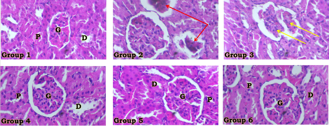
The excised kidneys, fixed in a 10% formalin-saline solution, were subjected to histological examination using conventional hematoxylin and eosin (H&E) staining techniques. Tissue preparation on the slides was captured for photomicrograph assessment using a Leica DM750 microscope with a camera at a magnification of ×400.
Statistical Analysis
Data were expressed as mean ± standard error of mean at p < 0.05. These were subjected to the Newman-Keuls post hoc test using GraphPad Prism 5.03 (GraphPad Software Inc., CA, USA).
Results
Effects of ABALE on Plasma Concentrations of Kidney Function Indices of Wistar Rats with Cyclosporin-induced Nephrotoxicity
CsA administration induced significantly elevated levels of plasma creatinine (mg/dL), urea (mg/dL), and Kim-1 (ng/mL) (p<0.0001). While there was no significant difference between the recovery and toxic group 2, ABALE administration was associated with a dose-dependent improvement in these kidney function indices, compared with the toxic CsA group 2 (p < 0.0001) Table 2.
| Group | Creatinine (mg/dl) | Urea (mg/dl) | KIM-1 (ng/ml) |
|---|---|---|---|
| Group 1 | 0.62 ± 0.04 | 35.46 ± 0.35 | 7.36 ± 2.35 |
| Group 2 | 0.87 ± 0.03 | 50.22 ± 0.37 | 17.62 ± 2.14 |
| Group 3 | 0.84 ± 0.04 | 49.84 ± 0.51 | 16.37 ± 2.01 |
| Group 4 | 0.69 ± 0.02 | 39.75 ± 0.40 | 9.45 ± 1.87 |
| Group 5 | 0.65 ± 0.03 | 36.61 ± 0.50 | 8.58 ± 2.17 |
| Group 6 | 0.64 ± 0.02 | 36.00 ± 0.44 | 7.69 ± 2.46 |
KIM-1, Kidney injury molecule 1; a, significant difference compared with Group 1 (Negative control); b, significant difference compared with Group 2 (CsA); c, significant difference compared with Group 3 (CsA + Recovery); d, significant difference compared with Group 4 (CsA + 100 mg/kg ABALE); e, significant difference compared with Group 5 (CsA + 200 mg/kg ABALE); Values represent mean ± SEM, p < 0.05.
Effects of ABALE on Plasma Concentrations of Oxidative Stress Biomarkers of Wistar Rats with Cyclosporin-induced Nephrotoxicity
While plasma concentrations of GSH (g/mg protein) and SOD (mM) were significantly reduced following CsA administration, these were dose-dependently restored by ABALE administration, with the highest dose having the most beneficial effect Table 3. These biomarkers were not significantly reversed in the recovery group compared with control group 1 (). However, the plasma TBARS level (nmol/mg protein) was significantly elevated in the toxic group 2 compared with the ABALE-treated groups, with the highest dose (400 mg/kg) also exhibiting the most risk-averse profile () Table 3.
| Group | GSH (g/mg protein) | SOD (U/mL) | TBARS (nmol/mg protein) |
|---|---|---|---|
| Group 1 | 3.62 ± 0.28 | 186.00 ± 14.50 | 29.31 ± 0.52 |
| Group 2 | 2.04 ± 0.30 | 57.84 ± 12.35 | 78.75 ± 0.30 |
| Group 3 | 2.15 ± 0.25 | 72.37 ± 12.40 | 77.15 ± 0.44 |
| Group 4 | 3.21 ± 0.32 | 173.40 ± 14.32 | 34.55 ± 0.37 |
| Group 5 | 3.38 ± 0.32 | 181.10 ± 13.72 | 29.82 ± 0.41 |
| Group 6 | 3.44 ± 0.21 | 182.50 ± 14.24 | 29.02 ± 0.35 |
GSH, Reduced glutathione; SOD, Superoxide dismutase; TBARS, Thiobarbituric acid reactive substances; a, significant difference compared with Group 1 (Negative control); b, significant difference compared with Group 2 (CsA); c, significant difference compared with Group 3 (CsA + Recovery); d, significant difference compared with Group 4 (CsA + 100 mg/kg ABALE); e, significant difference compared with Group 5 (CsA + 200 mg/kg ABALE); Values represent mean ± SEM, p < 0.05.
Effects of ABALE on Plasma Concentrations of Pro-inflammatory Cytokines of Wistar Rats with Cyclosporin-induced Nephrotoxicity
The pro-inflammatory cytokines (IL-6, CRP, and TNF-) were significantly elevated in the plasma compared to the control group (). However, following administration of ABALE, there was a dose-dependent amelioration of inflammation, with the highest dose also expressing the highest risk-averse potential Table 4.
Effects of ABALE on Kidney Histology of Wistar Rats with Cyclosporin-induced Nephrotoxicity
CsA administration was associated with distortion of the glomerulus, expressed as both atrophic and shrunken histo-architecture. However, the ameliorative effects of graded doses of ABALE were associated with an improved histoarchitecture of the kidney compared to that of the toxic CsA group Figure 1.
| Group | IL-6 (pg/ml) | CRP (pg/ml) | TNF- (pg/ml) |
|---|---|---|---|
| Group 1 | 69.21 ± 2.54 | 54.00 ± 2.00 | 33.54 ± 2.67 |
| Group 2 | 102.54 ± 3.55 | 86.00 ± 2.00 | 57.44 ± 2.18 |
| Group 3 | 100.63 ± 2.30 | 84.00 ± 3.00 | 55.25 ± 2.59 |
| Group 4 | 76.66 ± 3.21 | 61.00 ± 2.00 | 40.21 ± 2.36 |
| Group 5 | 72.54 ± 3.00 | 57.00 ± 1.00 | 36.45 ± 2.61 |
| Group 6 | 70.89 ± 3.73 | 56.00 ± 2.00 | 35.07 ± 2.42 |
TNF-α, Tumor necrosis factor-alpha; IL-6, Interleukin-6; CRP, C-Reactive protein; a, significant difference compared with Group 1 (Negative control); b, significant difference compared with Group 2 (CsA); c, significant difference compared with Group 3 (CsA + Recovery); d, significant difference compared with Group 4 (CsA + 100 mg/kg ABALE); Values represent mean ± SEM, p < 0.05.
Discussion
This study demonstrated that CsA-induced nephrotoxicity is implicated by oxidative stress, as well as an imbalance of plasma pro-inflammatory cytokines, in its pathogenesis. This is also associated with histo-architectural distortion of the glomerulus / glomerular filtration barrier (with a downstream effect on glomerular filtration), which culminated in significantly elevated plasma levels of kidney function biomarkers. If this condition is left unchecked, the resultant bio-accumulation of metabolic wastes may spiral into a compromise of body homeostasis and consequent expression of health risks. However, administration of graded doses of ABALE was associated with the amelioration of this nephrotoxicity, presenting a potential therapeutic choice in adjuvant therapy against CsA-induced kidney injury or, possibly, immunologically mediated clinical conditions. Generally, for the indices assessed in this study, the highest dose of ABALE (400mg/kg) showed the most risk-averse potential against CsA-induced nephrotoxicity, even though the extract demonstrated a dose-dependent therapeutic effect.
The use of both enzymatic (SOD) and non-enzymatic (GSH) antioxidant biomarkers [21], along with their significantly reduced plasma levels observed in this study, supports the role of oxidative stress in the development of CsA-induced nephrotoxicity. This indicates an overwhelming of the systemic antioxidant defenses, exceeding the body’s capacity to neutralize free radicals. The marked increase in lipid peroxidation, measured by TBARS, provides clear evidence of heightened systemic cellular and organ damage. This oxidative damage is further corroborated by pronounced histological and architectural disruption of the glomerulus, resulting in impairment of the glomerular filtration barrier and elevated plasma kidney function biomarkers.
Both classical (creatinine and urea) and novel (KIM-1) kidney function biomarkers were significantly elevated in the plasma of experimental groups following CsA administration. These harmful effects were notably improved by ABALE treatment in a dose-dependent manner. While the recovery group failed to restore kidney excretory and regulatory functions fully (as depicted by the assayed plasma levels of kidney function biomarkers), the highest dose of ABALE demonstrated the most significant protective effect. This underscores the extract’s potential as a valuable adjuvant therapeutic agent for CsA-induced nephrotoxicity. A limitation of this study, deserving further exploration, is the absence of KIM-1 measurement in urine samples, which could have been facilitated through the use of metabolic cages for clean urine collection within a Wistar rat experimental model. Additionally, circulatory CsA levels were not assessed; determining these would help clarify the relationship between ABALE administration and CsA half-life, as per its clearance by the kidney. Both aspects warrant further scientific investigation.
Administration of ABALE was linked to a significant decrease in plasma pro-inflammatory cytokines in the experimental groups. The extract’s ability to reduce systemic inflammation, evidenced by notable declines in plasma CRP, IL-6, and TNF- levels, highlights its potent anti-inflammatory properties. Nevertheless, the underlying pro-inflammatory pathways merit further detailed investigation.
Conclusion
Aqueous Basella alba leaf extract (ABALE) mitigated cyclosporine-induced nephrotoxicity by suppressing plasma pro-inflammatory cytokines and restoring antioxidant balance. The highest dose (400mg/kg) demonstrated the most risk-averse potential, while a recovery period (without medication) is associated with significant features of nephrotoxicity. This presents ABALE as a potential therapeutic choice in adjuvant therapy for cyclosporine-induced nephrotoxicity.
Conflicts of Interest
The authors declare that they have no competing interests that could have influenced the objectives or outcome of this research.
Funding Source
This research did not receive any specific grant from any funding agency in the public, commercial, or not-for-profit sector(s).
Acknowledgments
The authors acknowledge the team of Physiology Laboratory Technologists of the College of Health Sciences, Bowen University, Iwo, Osun State, Nigeria, as well as the Animal attendants of the Animal Holding Units of the Physiology Programme, College of Health Sciences, Bowen University, for their kind support and technical assistance.
Institutional Review Board (IRB)
The experimental protocols were in strict compliance with the guidelines for animal research, as contained in the NIH guidelines for the care and use of laboratory animals and approved by the local institutional research committee (Institute of Public Health of the Obafemi Awolowo University, Ile-Ife, Osun State, Nigeria) with ethics reference IPH/OAU/12/110A.
Large Language Model
None
Authors Contribution
Conceptualization of the study was performed by ICE. Methodology was developed by ICE and OOG, and data analysis was conducted by ICE, OOG, and OAEO. Investigation was carried out by ICE, OOG, OAEO, and AAK, while manuscript writing was undertaken by ICE, OOG, and OAEO. Funding acquisition was managed by ICE, OOG, and OAEO, and supervision was provided by ICE and OOG. All authors contributed to data collection, interpretation of results, enhancement of the intellectual content, and approval of the final manuscript.
Data Availability
The datasets generated and/or analyzed during the current study are not publicly available, as the work was based solely on animal experiments and no additional data were created. Further details are available from the corresponding author on reasonable request.
References
- Adekunle I. A., Imafidon C. E., Oladele A. A., Ayoka A. O.. Ginger polyphenols attenuate cyclosporine-induced disturbances in kidney function: Potential application in adjuvant transplant therapy. Pathophysiology. 2018;25(2):101-115. doi:10.1016/j.pathophys.2018.02.001 PMID: 29433768
- Fox Stuart Ira. Human physiology. 2011.
- Scanlon Valerie C, Sanders Tina. Essentials of anatomy and physiology. 2018.
- World Kidney Day. Chronic Kidney Disease. 2025.
- Elsayed Ata, Bayomy Mohamed, Azab Azab. Effect of acute and chronic treatment of Cyclosporin A on liver and kidney functions in rats. Journal of Applied Pharmaceutical Science. 2016;6(3):116-119. doi:10.7324/japs.2016.60320
- Faulds D., Goa K. L., Benfield P.. Cyclosporin. A review of its pharmacodynamic and pharmacokinetic properties, and therapeutic use in immunoregulatory disorders. Drugs. 1993;45(6):953-1040. doi:10.2165/00003495-199345060-00007 PMID: 7691501
- Chaurasiya Ajay, Pal Rajesh Kumar, Verma Pradeep Kumar, Katiyar Avineet, Razauddin, Kumar Narendra. An updated review on Malabar spinach (Basella alba and Basella rubra) and their importance. Journal of Pharmacognosy and Phytochemistry. 2021;10(2):1201-1207. doi:10.22271/phyto.2021.v10.i2p.13974
- D'Souza O. J., Gasti T., Hiremani V. D., Pinto J. P., Contractor S. S., Shettar A. K., Olivia D., Arakera S. B., Masti S. P., Chougale R. B.. Basella alba stem extract integrated poly (vinyl alcohol)/chitosan composite films: A promising bio-material for wound healing. Int J Biol Macromol. 2023;225:673-686. doi:10.1016/j.ijbiomac.2022.11.130 PMID: 36403767
- Sheik A., Kim E., Adepelly U., Alhammadi M., Huh Y. S.. Antioxidant and antiproliferative activity of Basella alba against colorectal cancer. Saudi J Biol Sci. 2023;30(4):103609. doi:10.1016/j.sjbs.2023.103609 PMID: 36970255 PMCID: PMC10036729
- Shantha T. R., Patchaimal P., Reddy M. P., Kumar R. K., Tewari D., Bharti V., Venkateshwarlu G., Mangal A. K., Padhi M. M., Dhiman K. S.. Pharmacognostical Standardization of Upodika- Basella alba L.: An Important Ayurvedic Antidiabetic Plant. Anc Sci Life. 2016;36(1):35-41. doi:10.4103/0257-7941.195411 PMID: 28182032 PMCID: PMC5255969
- Halayal Rekha Y., Bagewadi Zabin K., Khan T. M. Yunus, Shamsudeen Shaik Mohamed. Investigating compounds from Basella alba for their antioxidant, anti-inflammatory, and anticancer properties through in vitro and network pharmacology, molecular simulation approach. Green Chemistry Letters and Reviews. 2025;18(1). doi:10.1080/17518253.2025.2481945
- Omotoso D., Olubowale V., Aina M., Daramola O. O.. Phytochemical Profiling of Basella alba Using Gas chromatography-Mass Spectrometry. Tropical Journal of Natural Product Research. 2024;8(6). doi:10.26538/tjnpr/v8i6.36
- Bamidele O., Okeke N. C., Adedeji T. G., Adedayo L. D., Akinnuga A. M.. Methanol extracts of Basella alba leaves alleviate stress in rats. Chin Herb Med. 2020;12(2):163-170. doi:10.1016/j.chmed.2019.11.004 PMID: 36119801 PMCID: PMC9476701
- Bamidele Olubayode, Fasipebi Oyinkansola, Tomoloju Eniola E., Adedayo Lawrence D., Ajayi Ayodeji F., Afolabi Ayobami O.. Protective activity of Basella alba l. Leaf extract on cardiac injury markers following intestinal ischemia-reperfusion: potential for cardiovascular therapy. Pharmaceutical Science Advances. 2025;3. doi:10.1016/j.pscia.2025.100083
- Kumari Reshma, Goldar Waquar Ahmed, Mondal Subhodeep, Patra Susmita, Bhattacharya Sanjib, Haldar Pallab Kanti. Protective effect of Basella alba leaf against diabetic nephropathy in rats. Advances in Traditional Medicine. 2020;21(1):111-119. doi:10.1007/s13596-020-00458-2
- Murray B. M., Paller M. S., Ferris T. F.. Effect of cyclosporine administration on renal hemodynamics in conscious rats. Kidney Int. 1985;28(5):767-74. doi:10.1038/ki.1985.196 PMID: 3910916
- Chaitanya B. Krishna. Anti Inflammatory Activity of Basella Alba Linn. in Albino Rats. Journal of Applied Pharmaceutical Science. 2012;2(4):87-89. doi:10.7324/japs.2012.2413
- Beutler E., Duron O., Kelly B. M.. Improved method for the determination of blood glutathione. J Lab Clin Med. 1963;61:882-8. PMID: 13967893
- McCord Joe M., Fridovich Irwin. Superoxide Dismutase. Journal of Biological Chemistry. 1969;244(22):6049-6055. doi:10.1016/s0021-9258(18)63504-5
- Ohkawa H., Ohishi N., Yagi K.. Assay for lipid peroxides in animal tissues by thiobarbituric acid reaction. Anal Biochem. 1979;95(2):351-8. doi:10.1016/0003-2697(79)90738-3 PMID: 36810
- Imafidon C. E., Akomolafe R. O., Abubakar S. A., Ogundipe O. J., Olukiran O. S., Ayowole O. A.. Amelioration of Cadmium-Induced Nephropathy using Polyphenol-rich Extract of Vernonia amygdalina (Del.) Leaves in Rat Model. Open Access Maced J Med Sci. 2015;3(4):567-77. doi:10.3889/oamjms.2015.120 PMID: 27275289 PMCID: PMC4877889