Adult Meckel's Diverticulum Presenting with Massive Lower Gastrointestinal Bleeding and Hemorrhagic Shock: A Case Report
Yusuf HallakⒾ1, Jad FakhouryⒾ2*, Ajay KatwalaⒾ2, Umberto Battistin1, Lea El-Sayyad3, Bisher SawafⒾ1, Keith Burns4, Sara Stanley5
- 1Department of Internal Medicine, University of Toledo Medical Center, Toledo, OH, United States
- 2University of Toledo College of Medicine and Life Sciences, Toledo, OH, United States
- 3University of Toledo, Toledo, OH, United States
- 4Department of Gastroenterology, University of Toledo Medical Center, Toledo, OH, United States
- 5ProMedica Gastroenterology, Toledo, OH, United States
Abstract
Meckel's diverticulum (MD) is a congenital ileal outpouching that may contain ectopic gastric mucosa. Although typically asymptomatic, it may cause significant lower gastrointestinal (GI) bleeding in adults and, rarely, hemorrhagic shock. We report a case of a 31-year-old man who presented with sudden-onset painless hematochezia, syncope, and eventual hemorrhagic shock. Despite negative CT angiography and upper endoscopy, colonoscopy revealed active bleeding originating in the terminal ileum and entering the cecum, suggesting a small-bowel source and providing partial localization. The patient required substantial blood transfusion support, including 3 units of packed red blood cells, 2 units of platelets, and 2 units of cryoprecipitate. Ongoing hemorrhage and hemodynamic instability prompted emergent laparotomy, which identified and allowed for resection of an MD. Histopathologic examination confirmed MD containing ectopic gastric mucosa with an ulcerated bleeding focus. This case highlights the clinical challenges of MD in adults. It demonstrates that it may present with life-threatening lower gastrointestinal bleeding despite initially non-diagnostic imaging and the absence of definitive endoscopic identification of the lesion. Although uncommon, this case supports maintaining clinical suspicion for MD in adults with unexplained severe gastrointestinal hemorrhage and further characterizes its potential presentation in the adult population.
Keywords: Meckel’s diverticulum, Gastrointestinal hemorrhage, Hemorrhagic shock, Lower gastrointestinal bleeding, Small bowel bleeding
Article information
Introduction
Acute gastrointestinal (GI) hemorrhage is a common emergency that may manifest as hematemesis, melena, or hematochezia and, in severe cases, hypovolemic shock [1]. Massive GI bleeding is often defined by transfusion requirements (e.g., greater than 4 units of RBCs), clinical indicators such as systolic blood pressure less than 90 mmHg or heart rate more than 110 beats/minute, and is associated with significant mortality [2]. Thus, rapid localization and control of the bleeding source are critical for patient survival. In patients with acute, unstable lower GI bleeding, initial management focuses on hemodynamic stabilization and localization of the bleeding source using endoscopy, radiographic, and/or angiographic modalities as clinically appropriate. CTA may aid in identifying active bleeding, while endoscopy and angiographic embolization can provide both diagnostic and therapeutic benefit in certain patients, achieving technical success rates of 93-100% in some studies [3]. However, when bleeding cannot be localized or controlled, and hemodynamic instability persists despite resuscitative efforts, surgical intervention may be required for definitive diagnosis and management.
Meckel’s diverticulum (MD) is the most common congenital GI malformation (~2% prevalence), arising from incomplete obliteration of the primitive vitelline duct [4]. It is a true diverticulum on the anti-mesenteric border of the ileum, typically 3 cm in length, and located around 60 cm from the ileocecal junction [4]. Although typically asymptomatic, 4-7% of MDs will develop complications: most commonly bleeding, obstruction, and inflammation, leading to their diagnosis [4]. Notably, more than half of MDs harbor heterotopic mucosa (most often gastric) that secretes acid, leading to adjacent ulceration and bleeding [4,5]. While MD classically presents as painless hematochezia in children, it is rare and often overlooked in adults [4].
Diagnosis of MD in adults is challenging as the symptoms are nonspecific, and standard imaging or endoscopy often do not reveal a lesion. Furthermore, Technetium-99m pertechnetate (Meckel’s scan) is less sensitive in adults (60% in adults versus 95% in children) [6]. In practice, unexplained massive lower GI bleeding in an adult should prompt consideration of MD despite a negative workup. Definitive treatment of bleeding MD is surgical resection, with small-bowel resection and anastomosis preferred over other surgical techniques to ensure removal of ectopic mucosa; however, the approach may vary from case to case depending on risk factors and complications [4].
We present the case of a young adult with massive lower gastrointestinal bleeding and hemorrhagic shock in whom CTA and upper endoscopic evaluation failed to identify a definitive source. At the same time, colonoscopy localized bleeding to the terminal ileum. Definitive diagnosis was achieved only after emergent surgical exploration and pathological confirmation of MD with ectopic gastric mucosa. This case highlights the challenges of diagnosing Meckel’s diverticulum in adults. It underscores the importance of maintaining suspicion for a small bowel source when standard evaluations are non-diagnostic or only provide partial localization, particularly in patients with ongoing hemorrhage and hemodynamic instability, in whom surgery may ultimately serve as both the diagnostic and definitive therapeutic intervention.
Case Presentation
A 31-year-old previously healthy man initially presented to an outside hospital on April 10 with a one-day history of acute painless hematochezia and transient syncope and presyncope. The day before the presentation, he reported having a normal bowel movement. However, on the day of presentation, he experienced 4-5 episodes of bloody stools, which were initially dark (maroon) and later bright red. He denied any abdominal pain, cramping, nausea, vomiting, or fever. He reported intermittent syncopal episodes coinciding with bleeding, once at home and twice after arrival at an outside hospital, where he struck his head. Before being transferred to our institution later that day, his systolic blood pressure decreased to 80-90 mmHg. The patient denied any anticoagulant or excessive NSAID use, but upon further questioning, did report taking daily Excedrin (formulation with acetaminophen, aspirin, and caffeine). Family history was significant for a grandmother with rectal cancer.
Upon arrival at our emergency department on April 10, the patient remained hypotensive and tachycardic, requiring intravenous fluids and transfusion of 1 unit of packed red blood cells (pRBCs). The patient underwent CT angiography of the abdomen and pelvis, which did not demonstrate active extravasation or identify a definitive source of bleeding, and non-contrast CTs of the head and neck, considering the fall, which revealed no acute abnormalities. His hemoglobin fell rapidly from 12.5 g/dL to 8.7 g/dL and further to 7.4 g/dL after another syncopal episode, for which he received an additional 2 units of pRBCs. Physical examination revealed a pale, diaphoretic male with transient improvement in vital signs after resuscitation efforts. Abdominal examination was benign and non-tender. Rectal examination confirmed the presence of gross blood without a palpable source and revealed no masses or hemorrhoids. Given ongoing bright red blood per rectum with persistent tachycardia and hypotension despite resuscitation, he was admitted to the intensive care unit (ICU).
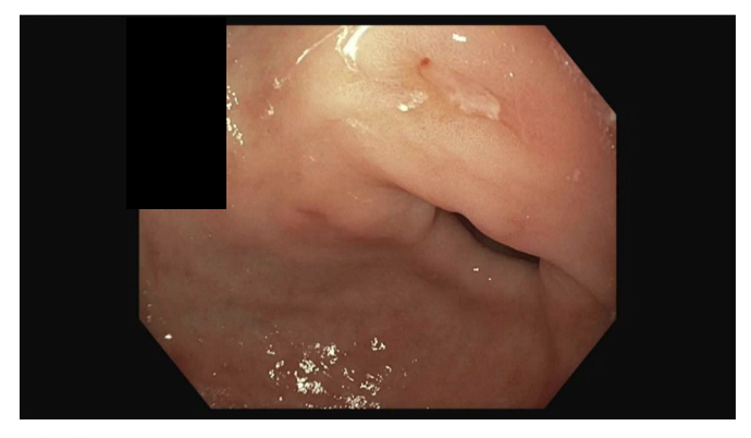
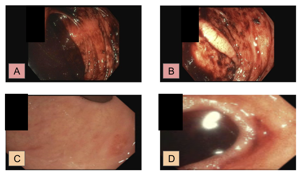
Given continued bleeding, gastroenterology was consulted. The patient was started on a proton pump inhibitor (PPI), and subsequently underwent emergent esophagogastroduodenoscopy (EGD) and colonoscopy later that day (April 10). EGD revealed a normal esophagus and stomach except for a single 5-mm superficial gastric ulcer with a clean base (Forrest III) in the antrum without evidence of active bleeding Figure 1. However, the small ulcer was judged unlikely to account for his hemorrhage. Colonoscopy was limited by poor bowel preparation and copious blood. Fresh and old blood filled the colon, with no obvious colonic lesion or source of bleeding identified. With aggressive suctioning, fresh blood was seen draining into the cecum, suggesting a small bowel source of hemorrhage, and further intubation of the terminal ileum revealed active bleeding in the small bowel Figure 2. There was high suspicion for a small bowel source, specifically MD, given the clinical picture. During colonoscopy, the patient developed worsening hemodynamic instability, with a nadir blood pressure of 88/48 mmHg and a peak heart rate of 146 beats/min despite ongoing transfusion support, prompting immediate general surgery consultation.
Despite aggressive intravenous fluid resuscitation and blood product administration (total of 3 units of pRBCs, 2 units of platelets, and 2 units of cryoprecipitate), the patient remained tachycardic and hypotensive, consistent with hemorrhagic shock. Although vasopressor support was not required, the combination of ongoing massive gastrointestinal blood loss, hypotension, marked tachycardia, transfusion requirements, and need for surgical intervention was considered consistent with hemorrhagic shock. Additionally, given the severity of these factors and the need for urgent source control, additional diagnostic studies—including repeat CT angiography, tagged red blood cell scintigraphy, mesenteric angiography, capsule endoscopy, deep enteroscopy, and technetium-99m pertechnetate (Meckel’s scan)—were not pursued, as they were not considered feasible in the setting of active hemorrhage requiring emergent surgical intervention. The patient was therefore intubated and transferred directly from the endoscopy suite to the operating room for exploratory laparotomy.
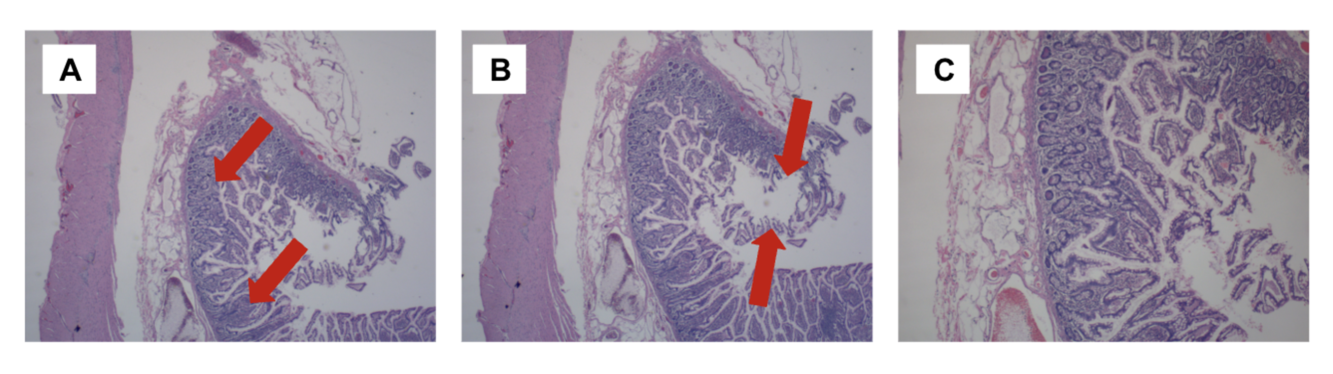
At exploratory laparotomy, an MD was identified on the anti-mesenteric border of the distal ileum. The involved segment of ileum containing the diverticulum was resected, and bowel continuity was restored with an 80 mm stapled side-to-side functional end-to-end anastomosis. The procedure was completed without complications, and the patient was admitted to the surgical intensive care unit (SICU). No additional sources of GI bleeding were identified. Gross pathologic examination demonstrated a 4.8 cm × 2.2 cm intact Meckel’s diverticulum arising from a 3.2 cm segment of resected ileum. The diverticular wall measured approximately 0.4 cm in thickness with a tan-pink, glistening serosal surface and prominent dilated vessels. On sectioning, the mucosa was tan-pink, smooth, and glistening without evidence of polyps, masses, or nodules. Representative sections from the proximal margin, distal margin, central diverticulum, and diverticular tip were submitted for histopathologic evaluation. Histopathological examination confirmed MD with gastric-type mucosa and an ulcerated bleeding focus Figure 3.
Postoperatively, the patient’s hemoglobin remained stable with no further hematochezia. He was successfully extubated on postoperative day 1, remained hemodynamically stable, and was transferred out of the SICU later that day. Serial abdominal examinations and hemoglobin monitoring continued throughout his hospitalization. On April 14, he received an additional 1 unit of pRBCs for a hemoglobin nadir of 6.9 g/dL, after which his hemoglobin recovered appropriately. His diet was gradually advanced as tolerated with return of normal bowel function. No further hematochezia or syncopal episodes occurred. By hospital day 4, he was ambulating, passing flatus and stool, and pain was controlled with oral medications. He was discharged home in stable condition on hospital day 5 with outpatient surgical follow-up planned for staple removal; additionally, avoiding NSAIDs post-discharge was emphasized.
At early post-discharge follow-up on April 16, the patient reported continued clinical improvement without chest pain, shortness of breath, dizziness, nausea, vomiting, or recurrent hematochezia. He reported normal bowel function and adherence to his prescribed medication and treatment regimen. Laboratory evaluation obtained on April 22 showed an increase in hemoglobin to 11.3 g/dL, supporting hematologic recovery following operative management and resolution of the acute bleeding episode. At surgical follow-up on April 27, the patient continued to do well without complications. He was tolerating a regular diet, maintaining normal bowel function, and denied recurrent gastrointestinal bleeding, fever, chills, nausea, vomiting, diarrhea, chest pain, or shortness of breath. Examination demonstrated satisfactory postoperative wound healing, and surgical staples were removed without issue. Hemoglobin and hematocrit levels were observed to improve progressively. The patient had completed his postoperative pain regimen and required only intermittent acetaminophen as needed. No further surgical intervention was required, and follow-up was recommended on an as-needed basis.
Discussion
This case illustrates that MD, although uncommon in adults, should be considered in cases of severe, unexplained GI hemorrhage. Our patient presented with painless, massive hematochezia, rapid hemoglobin decline, and hemorrhagic shock, features consistent with previously reported cases of bleeding MD. In adults, MD-related bleeding can be life-threatening and often evades initial detection [7]. Moreover, the current literature suggests that MD-related bleeding is most commonly attributed to ulceration from ectopic gastric acid secretion [4].
Diagnosis of MD in adults remains challenging due to nonspecific clinical presentation and the limited sensitivity of diagnostic modalities. For hemodynamically unstable patients, the 2019 British Society of Gastroenterology guidelines recommended CTA to localize active bleeding [3]; however, as demonstrated in our case and in others reported in the literature, both imaging and endoscopic evaluation may fail to establish a definitive diagnosis. Moreover, angiographic embolization may temporize active bleeding but often fails without definitive resection, as in the case report by Zheng et al. [7]. Zheng et al. described the case of a 41-year-old man whose colonoscopy and CTA were initially non-diagnostic; only combined laparoscopic exploration and selective angiography revealed the MD as the bleeding source [7]. Similarly, a pediatric case presented by Shewaye et al. [8] reported a 15-year-old boy with massive bleeding, negative EGD/colonoscopy, and negative Meckel’s scan. MD with ectopic gastric mucosa was ultimately found on laparoscopic resection after CTA suggested the lesion [8]. These cases, along with our case, underscore the limitations of noninvasive diagnostics in establishing a definitive diagnosis of MD-related bleeding even when they provide clinically useful partial localization of the bleeding source.
Considering that surgical resection of symptomatic MD-related bleeding is the standard of care, early surgical intervention is appropriate once resuscitation is underway [4]. In adults, guidelines suggest segmental small-bowel resection with anastomosis rather than simple diverticulectomy to ensure complete removal of the heterotopic mucosa [4]. In our case, we performed an ileal resection containing the MD with an 80 mm stapled side-to-side functional end-to-end anastomosis. Other reports, such as that by Haro et al., described successful laparoscopic resection after bleeding persisted despite embolization [9]. Additionally, our patient required multiple blood product transfusions before surgery, underscoring the severity of his hemorrhage and clinical instability. Delays in achieving definitive source control in severe gastrointestinal bleeding are associated with worse outcomes, highlighting the importance of timely surgical intervention when ongoing hemorrhage persists despite resuscitative efforts [2].
In the discussion of adult GI bleeding, MD is a rare but important etiology that clinicians should not overlook when standard evaluations are unrevealing, as emphasized by nearly all studies in the literature. Indeed, more than a dozen adult cases of MD-related bleeding have been reported in recent years, many of which required ICU support and emergent surgery, such as the study by Ahmad et al. [10]. In addition to echoing these findings, our case illustrates the key role of coordinated multidisciplinary care in the evaluation and management of severe gastrointestinal hemorrhage. This includes gastroenterology to localize the bleeding source and surgery for definitive management. Finally, our case highlights potential considerations regarding aspirin-containing medications in patients with MD. While the patient endorsed daily use of aspirin-containing Excedrin and pathology demonstrated an ulcerated bleeding focus associated with ectopic gastric mucosa, the extent to which aspirin contributed to the bleeding event cannot be determined. Nevertheless, aspirin use may have represented an additional factor in a patient already predisposed to bleeding.
This report has several limitations. The diagnosis of MD was established intraoperatively, limiting our ability to evaluate the effectiveness of imaging and endoscopic modalities in definitively diagnosing the lesion. Additionally, although operative and pathologic findings are available, the operative report did not document the exact distance of the diverticulum from the ileocecal valve, and the pathology report did not specify the ulcer location or the extent of ectopic gastric mucosa. Furthermore, serum lactate and base deficit measurements were not obtained during the acute resuscitation period, limiting the availability of additional objective markers of hemorrhagic shock. Finally, while the patient endorsed daily use of aspirin-containing Excedrin, its contribution to the bleeding events described cannot be determined.
Conclusion
This report describes a 31-year-old man who developed hemorrhagic shock from massive lower GI bleeding due to MD. Despite non-diagnostic CT imaging and the absence of a definitive endoscopic diagnosis, colonoscopy localized active bleeding to the terminal ileum, supporting suspicion for a small bowel bleeding source and prompting exploratory surgery and resection. The case underscores that, although rare in adults, MD can present with life-threatening hemorrhage. In such patients, prompt aggressive resuscitation and definitive surgical intervention are crucial. It is important for clinicians to be suspicious of MD when encountering severe, unexplained GI bleeding after common etiologies have been ruled out. Early recognition and surgical treatment can be lifesaving in these atypical presentations.
Conflicts of Interest
All authors report no disclosures or conflicts of interest.
Funding Source
No funding was received for this study.
Acknowledgments
None.
Institutional Review Board (IRB)
Institutional Review Board approval was not required for this single de-identified case report. The report was prepared in accordance with the principles of the Declaration of Helsinki.
Informed Consent
Written informed consent was obtained from the patient for publication of this case report and the accompanying clinical images.
Large Language Model
We have employed an advanced Large Language Model (LLM) to enhance and refine the English-language writing. This process focused solely on improving the text’s clarity and style, without generating or adding any new information to the content.
Authors Contribution
All authors contributed to the conception of the report, the acquisition and interpretation of the clinical data, and the drafting and critical revision of the manuscript. All authors read and approved the final manuscript and agree to be accountable for all aspects of the work.
Data Availability
All relevant data are within the manuscript.
References
- Nable J. V., Graham A. C.. Gastrointestinal bleeding. Emerg Med Clin North Am. 2016;34(2):309-325. doi:10.1016/j.emc.2015.12.001
- Natarajan M. U. V., O'Brien J. W., Rogers M., Gallagher M., Rockall T.. Management of massive gastrointestinal haemorrhage. Surgery (Oxford). 2025;43(9):580-592. doi:10.1016/j.mpsur.2025.07.006
- Oakland K., Chadwick G., East J. E., et al. Diagnosis and management of acute lower gastrointestinal bleeding: guidelines from the British Society of Gastroenterology. Gut. 2019;68(5):776-789. doi:10.1136/gutjnl-2018-317807
- Lequet J., Menahem B., Alves A., Fohlen A., Mulliri A.. Meckel's diverticulum in the adult. J Visc Surg. 2017;154(4):253-259. doi:10.1016/j.jviscsurg.2017.06.006
- Carlioz P.. Le diverticule de Meckel, de l'embryologie à la chirurgie. e-mémoires de l'Académie Nationale de Chirurgie. 2014;13(2):1-6.
- Anand S. S., Handa R., Chauhan M. S.. Nuclear imaging in Meckel's diverticulum. Med J Armed Forces India. 2004;60(2):198-199. doi:10.1016/S0377-1237(04)80124-2
- Zheng L., Li L., Huo Y., Su H.. Massive gastrointestinal hemorrhage in an adult caused by Meckel's diverticulum: a case report. Int Med Case Rep J. 2025;18:1253-1259. doi:10.2147/IMCRJ.S541384
- Shewaye A. B., Ramchandani M., Berhane K. A., Endalew D. T., Minyilshewa A. T., Elias T. P.. Meckel's diverticulum causing massive lower intestinal bleeding in a teenager: a case report. J Pediatr Surg Case Rep. 2025;116:102986. doi:10.1016/j.epsc.2025.102986
- Haro C., Duffau A., Muniz N., Mansilla S., Sanguinetti M., Viola M.. Shock hemorrágico como presentación inicial de divertículo de Meckel. Rev Argent Coloproctol. 2021;32(3):102-104. doi:10.46768/racp.v32i03.81
- Ahmad I., Lakshman S., Estevez A.. Hemorrhagic shock: where is the bleed?. Chest. 2021;160(4):A925. doi:10.1016/j.chest.2021.07.861