An Unusual Case of Disseminated Hydatid Disease: A Laparoscopic Wonder
Gajendra Bhati1, Raghav BansalⒾ2, Vanessa Pamela Salolin VargasⒾ3*, Ripanbir Singh KahlonⒾ4
- 1Medical Intern, Department of Surgical Gastroenterology, Adesh Institute of Medical Sciences and Research, Bathinda, Punjab, India
- 2Department of General Surgery, Adesh Institute of Medical Sciences and Research, Bathinda, Punjab, India
- 3Facultad de Medicina, Universidad Westhill, CDMX, Mexico
- 4Government Medical College, Punjab, India
Abstract
Human echinococcosis, caused by Echinococcus tapeworms, is a zoonotic disease in which ingested eggs of the tapeworm form cysts in the organs known as Hydatid cysts. These cysts are commonly found in the Liver and lungs, but in rare cases, they can also be found in the spleen, ovaries, round ligament, and peritoneal cavity. Treatment typically involves surgery. This is a case of a 32-year-old female who presented with chief complaints of pain in the abdomen for 2 months, which was associated with nausea, vomiting, reduced appetite, and increased size of mass per abdomen. Further investigations revealed the presence of multiple disseminated hydatid cysts in the upper abdomen and the peritoneal cavity. This was a rare & unusual entity that was meticulously managed by minimally invasive laparoscopic surgery. Echinococcosis can occur anywhere in the body, with concurrent localizations in the liver, spleen, round ligament, ovaries, and peritoneal cavity, which are rare and pose a diagnostic as well as a surgical management challenge. The inconvenience of multiple localization and the risk of contamination usually lead to open surgery in these cases. The presented case is unique due to the laparoscopic approach to the multiple localizations of hydatid disease, advocating for a minimally invasive first-line approach even in these particular localizations.
Keywords: Echinococcus, Human echinococcosis, Disseminated hydatid disease, Laparoscopic surgery, Case report
Article information
Introduction
Human echinococcosis is a zoonotic disease caused by Echinococcus tapeworms. Transmission occurs through ingesting eggs, releasing oncospheres in the intestine that migrate and form hydatid cysts in organs. Rarely, cyst rupture spreads protoscolices, causing secondary cyst formation, known as secondary or disseminated echinococcosis. Disseminated hydatid disease is uncommon, even in endemic areas, with an incidence of 1% to 8% [1]. The spleen, kidney, peritoneal cavity, skin, and muscles each have about a 2% involvement rate, while the heart, brain, vertebral column, ovaries, pancreas, gallbladder, thyroid gland, breast, and bones each have about a 1% involvement rate [2]. Management typically involves open or laparoscopic surgery. Surgical treatment depends on the cysts’ size and spread, requiring either an open or laparoscopic approach. Laparoscopic surgery offers shorter operative duration, fewer intraoperative complications, less pain, shorter hospital stays, and better cosmesis [3]. This report aims to enhance understanding of the atypical presentation and management of disseminated hydatid disease involving multiple tissues like the liver, spleen, ovaries, round ligament, and peritoneal cavity. This case report seeks to contribute valuable insights to the knowledge on managing disseminated hydatidoses, ultimately guiding future clinical interventions.
Case Presentation
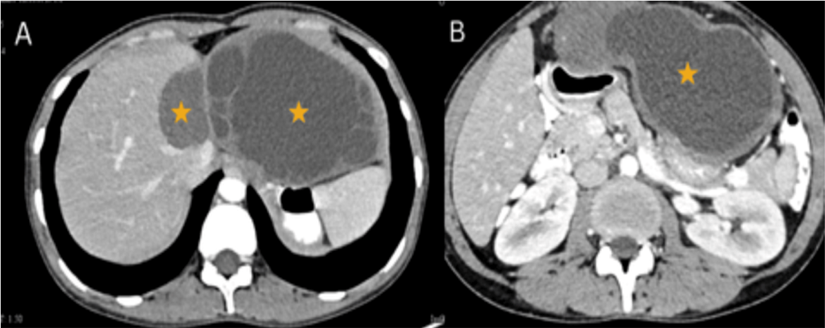
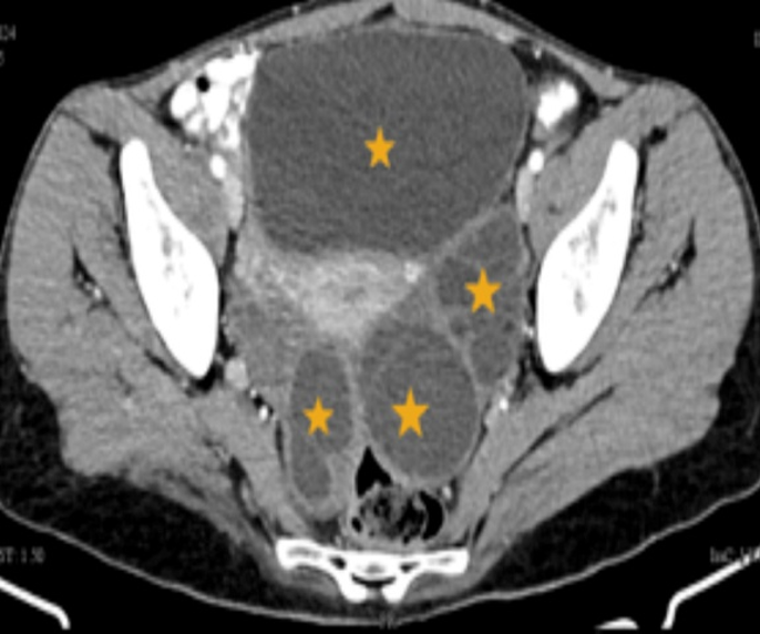
A 32-year-old woman presented to the outpatient clinic with a primary complaint of abdominal pain persisting for two months. The pain had a sudden onset, was severe in intensity, radiated to the flanks, and was accompanied by a progressively enlarging abdominal mass located in the epigastric region, which had been present for six years. Additionally, the patient reported experiencing nausea, vomiting, and anorexia over the past month. She denied any history of fever, significant weight loss, melena, or rectal bleeding. There was no prior use of medications before the presentation. The patient had no known comorbidities and no history of diabetes mellitus, hypertension, tuberculosis, bronchial asthma, epilepsy, or any significant medical or surgical conditions. She also reported no known allergies to food or medications. There was no relevant family history or any similar complaints in the past. She denied having any domestic animals, including dogs. On physical examination, a round, tender mass of approximately 10 × 10 cm was palpated in the epigastric region, with associated guarding and rigidity. The patient had remained asymptomatic until two months prior, when the onset of severe abdominal pain likely delayed medical consultation. There was no prior history of diagnostic imaging or medical treatment. Initial investigations, summarized in Table 1, revealed mildly decreased hemoglobin levels, a normal white blood cell count, slightly elevated liver enzymes, and renal function tests within normal limits. Viral markers, including HCV, HBsAg, and HIV, were non-reactive. CT imaging revealed multiple characteristic features consistent with disseminated hydatid disease. As shown in Figure 1 1A, a cystic lesion was observed in segment IV of the liver, classified as WHO Stage CE-4. Adjacent to this, a large exophytic hypodense cyst was visualized, consistent with WHO Stage CE-2, and containing multiple peripheral daughter cysts. Figure 1 1B demonstrated a focal loculated extension of this hepatic cyst along its infero-medial wall. Figure 2 displays axial CT sections revealing well-defined cysts in the recto-uterine pouch and left adnexa, in addition to the infero-medial extension of the hepatic cyst.
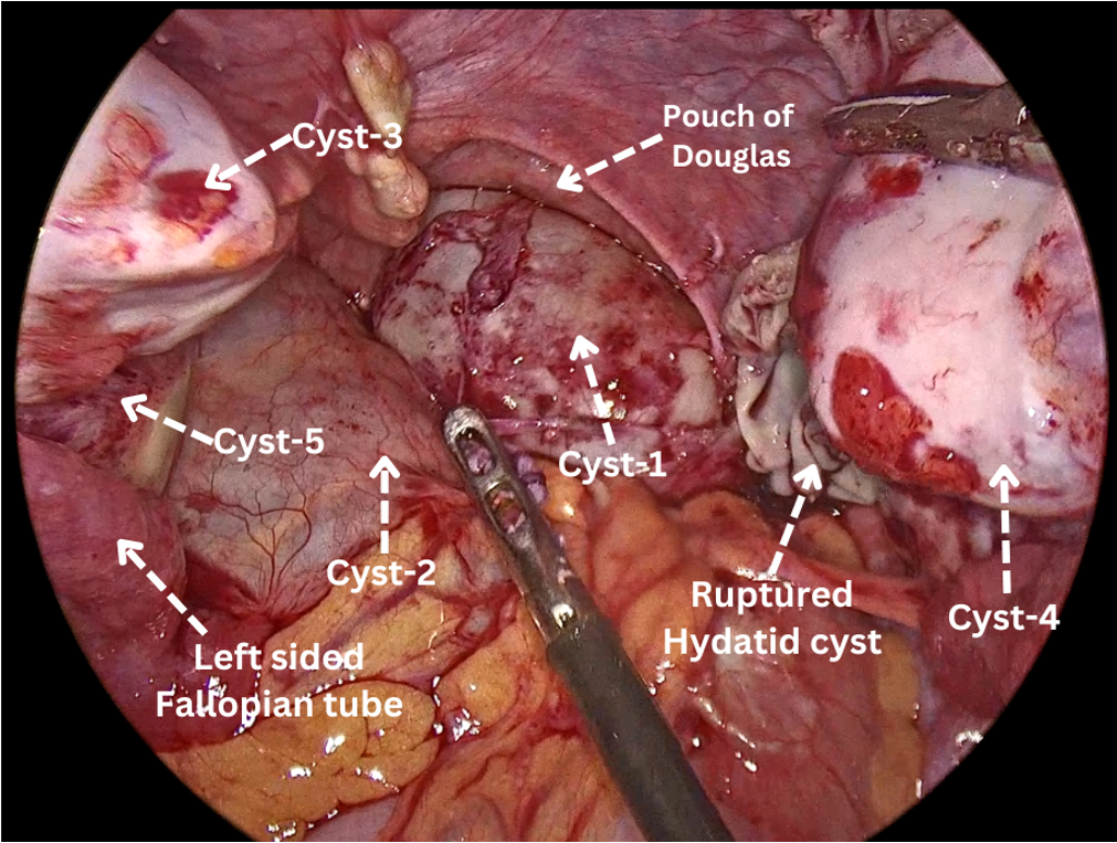
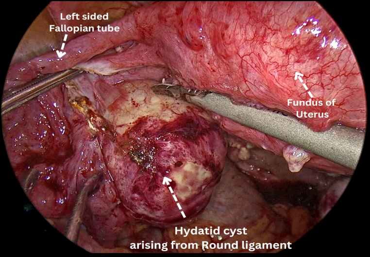
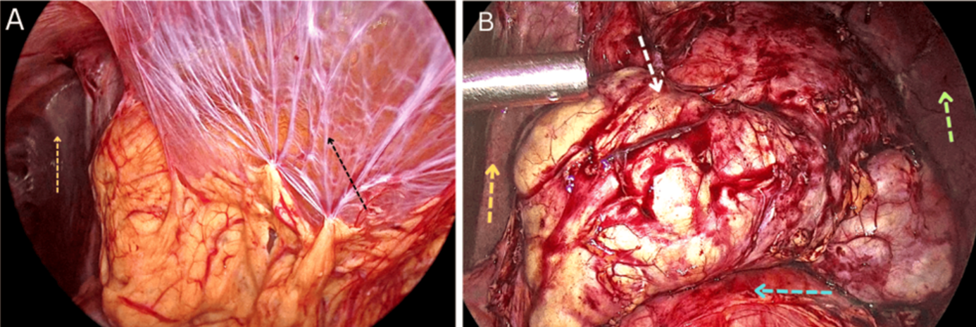
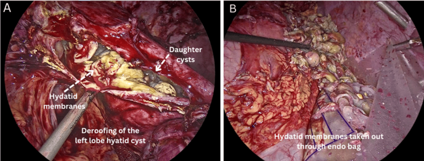
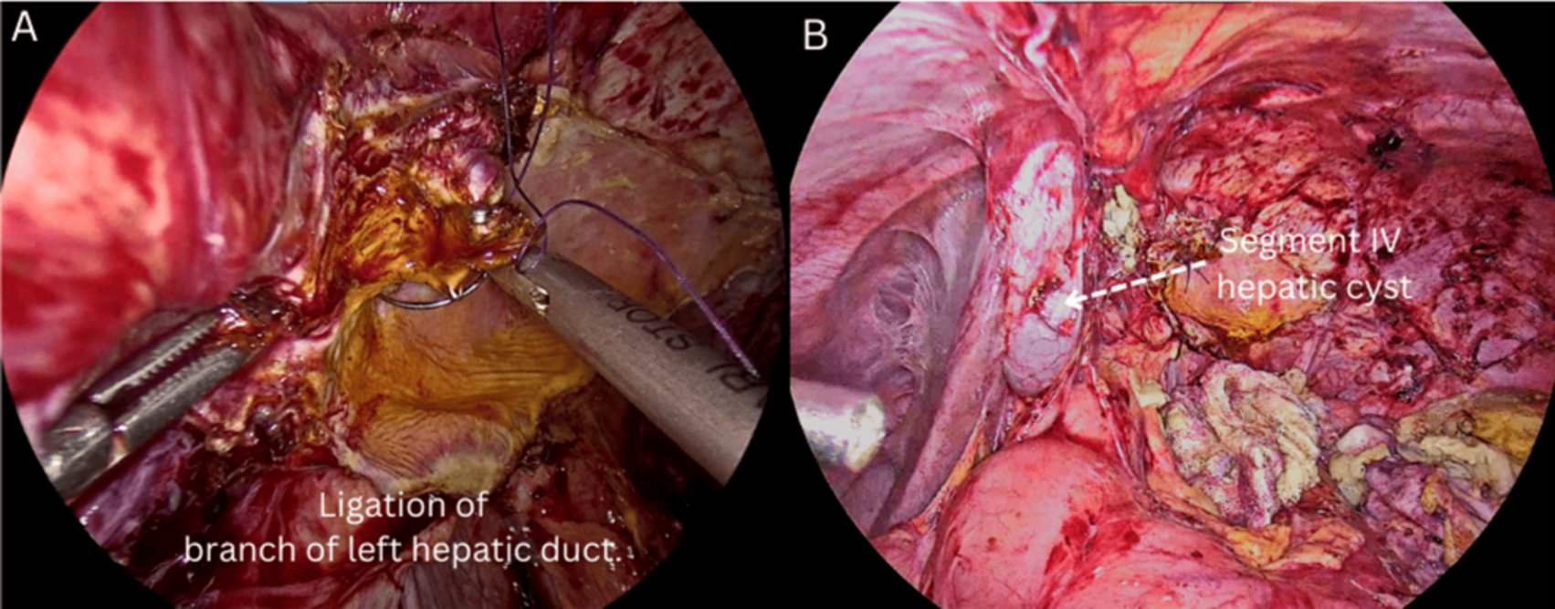
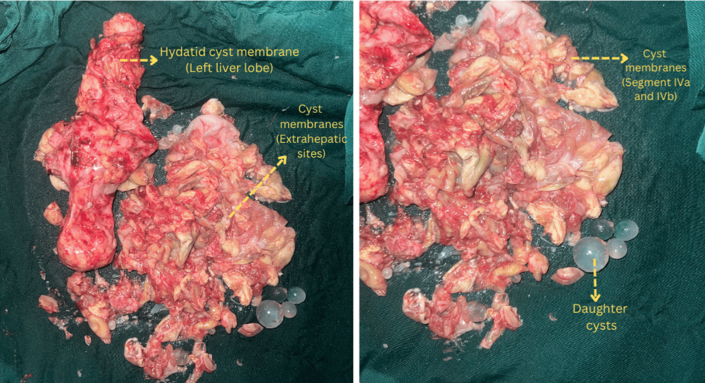
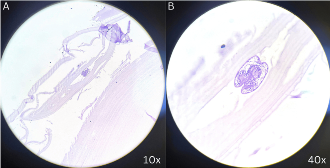
Surgery was planned based on these findings. Under General anesthesia, the patient was painted and draped. An infra-umbilical curvilinear incision was given, and Carbon dioxide pneumoperitoneum was created. Laparoscopic exploration (using a 30° laparoscope) was carried out through trocar placement, with a 12 mm trocar infra-umbilically and two 5mm working ports in the right and left mid-clavicular line. Approx. 5 intact cysts (measuring approx. 3-5cm in size) were identified in the cul-de-sac (anterior to the rectum), right fornix, and left fornix. One ruptured hydatid cyst was also identified in the right fornix with intact membranes Figure 3. Another cyst, which was arising from the round ligament on the left side (approx. 4cm in size), was identified. All the cysts were meticulously dissected and excised intact using harmonic shears Figure 4. After pelvic clearance, the upper abdomen was examined, which showed extensive omental adhesions that were adhered to the anterior abdominal wall as well as the underlying cyst. It was found out that the omentum was wrapped around a large liver hydatid cyst (measuring approx. 15cm), replacing the entire left liver lobe and causing pressure atrophy of the lobe. Adhesiolysis was done, and it was found that this cyst was also adhered to the surface of the spleen Figure 5. After meticulous adhesiolysis, the cyst was carefully punctured, and the fluid inside the cavity was aspirated before keeping povidone iodine-soaked gauze around the cyst to prevent recurrence due to spillage. The cyst was then de-roofed, along with the instillation of 3% hypertonic saline in the cavity, and was excised using a harmonic scalpel. The cystic content was taken out in an endo bag along with the excised pelvic cysts Figure 6. A Bile leak was identified over the edge of the left lobe (possibly a cysto-biliary fistula), the source was identified (Branch of left hepatic duct) and was ligated using ligaclip 400 & 4-0 vicryl suture. Biliostasis was achieved. Another cyst (measuring approx. 3cm) was identified over segment IV of the liver (behind the gall bladder), which was de-roofed, and all the contents were aspirated out Figure 7. Romovac drain was placed in the upper left quadrant through the 5mm port, and for postoperative pain management, a local anesthetic agent, Bupivacaine, was administered in the subcutaneous plane. Closure of 12mm ports was performed using polyglactin 910 (vicryl) sutures, and the skin of the port entry site was approximated with a skin stapler Figure 8. The patient was extubated successfully and was shifted to the ward. The post-op period was uneventful, and the patient was ambulatory & accepting diet per oral within post-op Day 1. The patient was monitored for another 3 days for any anaphylactic reactions, and the patient was discharged on Day 4 as the patient became asymptomatic and fit for discharge. The patient was put on 4-week anthelminthic medication of 400mg Albendazole, and the follow-up ultrasound in the 6th week showed no residual disease. Histopathological features were consistent with a clinical diagnosis of Hydatid disease. Examination of the cyst revealed acellular laminated membranes with debris, scolex, and protoscolices. Cyst wall tissue was lined by cuboidal lining epithelium, with mixed inflammatory infiltrates comprising lymphocytes, eosinophils, and plasma cells underneath. Areas of fibrosis, calcification, congested blood vessels, and necrosis were discernible Figure 9.
| Investigations | Value | Normal Range |
|---|---|---|
| Haemoglobin | 11.2 g/dl | 13.0-17.0 |
| Total Leukocyte Count (TLC) | 7.2 103/uL | 4.0-10.0 |
| Platelet | 285 103/uL | 150.0-410.0 |
| Liver Function Tests | ||
| Serum Glutamic-Oxaloacetic Transaminase (SGOT) | 37 IU/L | 0.0-35.0 |
| Serum Glutamic Pyruvic Transaminase (SGPT) | 46 IU/L | 0.0-41.0 |
| Alkaline Phosphatase (ALP) | 127 IU/L | 40.0-129.0 |
| Albumin | 5.5 g/dl | 3.5-5.2 |
| Bilirubin | 0.2 mg/dl | 0.1-1.2 |
| Renal Function Tests | ||
| Serum urea | 13 mg/dl | 17.0-43.0 |
| Serum Creatinine | 0.7 mg/dl | 0.6-1.1 |
| International Normalized Ratio (INR) | 1.05 | 0.8-1.2 |
| Serum Sodium (Na+) | 140 mmol/L | 136.0-145.0 |
| Serum Potassium (K+) | 3.9 mmol/L | 3.5-5.0 |
| Viral Markers | ||
| Hepatitis C virus (HCV) | Non-Reactive | |
| Hepatitis B surface antigen (HBsAg) | Non-Reactive | |
| Human Immunodeficiency Virus (HIV) | Non-Reactive | |
Discussion
Disseminated hydatid disease is a rare condition, with an incidence ranging from 1% to 8% [1]. Involvement of sites other than the liver and lungs is uncommon. Clinical presentation varies depending on the size, location, and presence of complications related to the cysts. Involvement of organs such as the spleen, ovaries, round ligament, and peritoneum typically occurs secondary to the rupture of a primary hepatic hydatid cyst. Ultrasound serves as the first-line diagnostic modality for hydatid disease [4]; however, computed tomography (CT) has significantly enhanced both diagnostic accuracy and treatment planning. Surgical intervention remains the cornerstone of treatment and may be performed via open or laparoscopic approaches. Laparoscopic surgery offers several advantages, including reduced operative time, fewer intraoperative complications, diminished postoperative pain, shorter hospital stays, and superior cosmetic outcomes.
Preoperative and postoperative administration of albendazole for one month is recommended to sterilize the cyst and reduce the risk of recurrence. Nonetheless, laparoscopic procedures have inherent limitations, such as dependence on the surgeon’s expertise and the potential risk of cyst content spillage, which may lead to intraoperative anaphylaxis and recurrence.
The strength of this case report lies in its documentation of a rare and atypical manifestation of hydatid disease, successfully managed through a laparoscopic approach. Initial diagnostic laparoscopy confirmed the feasibility of a minimally invasive surgical strategy. This approach offered superior intraoperative visualization, facilitated by advanced 4K imaging technology, which allowed for enhanced anatomical detail beyond the capabilities of direct visualization in open surgery. As a result, a large incision was avoided in a young patient, leading to improved surgical outcomes such as minimal blood loss, reduced postoperative pain, a shorter hospital stay—which lowers the risk of hospital-acquired infections—and improved cosmetic results due to minimal scarring. Collectively, these factors contributed to an overall enhanced surgical experience. An extensive literature review indicates that, to our knowledge, this is among the first documented cases of its kind globally, underscoring the uniqueness of the cysts’ size and location and the laparoscopic approach used in management. However, a key limitation of this report is that it describes a single case, which restricts the generalizability of its findings.
A comprehensive review of documented cases involving disseminated and atypical sites of hydatid disease was conducted. Mihetiu A. et al. [5] reported 49 cases with unusual abdominal localizations, among which only one involved the round ligament [6]. Of these 49 cases, merely two were managed laparoscopically, reinforcing our case report’s significance. El Bakaouri et al. [7] described a rare case involving the liver, spleen, and peritoneum, managed through cystectomy combined with splenectomy; the patient was discharged on the sixth postoperative day, suggesting a relatively extended hospital stay.
Ziad F. et al. [1] also reported a similar case with hydatid cysts localized to comparable sites, with cyst sizes ranging from 3 to 10 cm. That case was managed via a median laparotomy and had an uneventful postoperative course. In contrast, our case demonstrates the successful application of a minimally invasive laparoscopic approach to similar anatomical sites despite the presence of even larger cysts. This highlights the feasibility and advantages of laparoscopy in managing such complex presentations.
A rare case presented by Achraf S et al. [8] shows how a patient with multiple hepatic as well as extrahepatic hydatid cysts was managed by resection of the cysts but died on postoperative day 15, further strengthening our case, which was managed laparoscopically.
A recent case reported by Kandel et al. [9] described multiple primary intraperitoneal cysts ranging in size from 2 to 15 cm, which were managed through an open surgical approach. In contrast, our report offers valuable insights into managing similarly complex cases of hydatid disease using a minimally invasive laparoscopic technique.
A comparable case by Zayati M. et al. [10] also demonstrated similar localization of hydatid cysts, with sizes ranging from 3 to 6 cm—closely resembling those in our case. Notably, they reported the presence of a cysto-biliary fistula, a complication also identified in our patient. However, while their case was managed via open surgery, our case highlights the successful laparoscopic management of the same, further reinforcing our approach’s strength.
A comprehensive literature review also identified a rare case of disseminated hydatid disease reported by Babiker et al. [11], involving cysts localized to the lungs, liver (5 cm), and spleen (7.5 cm). Although this case was successfully managed laparoscopically, the degree of dissemination and the cyst sizes were notably smaller than those observed in our report.
The literature review indicates that only a limited number of cases have utilized laparoscopy as the primary modality for managing hydatid disease. One such case was reported by Obeid M. et al. [12], where hydatid cysts were localized to the liver and the small intestine mesentery, with the largest measuring 10 cm. This case was successfully managed laparoscopically. Similarly, Jangjoo et al. [13] described a large cyst measuring 10 × 5 cm located in the greater omentum, which was also treated via a laparoscopic approach. These reports further support the uniqueness of our case, both in terms of presentation and the successful use of minimally invasive management.
In contrast, Baimakhanov et al. [14] documented a rare instance of disseminated hydatid disease involving multiple organs—including the liver, spleen, kidneys, and pancreas—with cyst sizes ranging from 1 to 6 cm. This case was managed using open surgery. Likewise, Ranjan R. et al. [15] reported a mesenteric hydatid cyst measuring 12 × 10 cm, which was also treated through an open surgical approach. Delis SG et al. [16] reported a case series of patients presenting with unusual localization of hydatid cysts for which complete cyst excision in most patients was performed, along with omentoplasty in a few cases of hydatid extension into vertebrae.
G. Ozturk et al. [17] conducted a study involving twenty patients with post-traumatic ruptured liver hydatid cysts, all of whom were managed surgically. The authors advocated for a conservative surgical approach—primarily involving de-roofing and various techniques to manage the residual cavity—over more radical procedures such as hepatic resection or pericystectomy. This study also highlights the pivotal role of computed tomography (CT) in transforming the diagnosis and management of hydatid disease.
Mushtaque M. et al. [18] further emphasized that surgery remains the cornerstone of hydatid disease treatment. Among the surgical options available, laparoscopic intervention is generally preferred over traditional open surgery. The PAIR technique (Puncture, Aspiration, Injection, and Reaspiration/Removal) is commonly used in laparoscopic procedures, as it facilitates the conversion of the cyst into a non-dependent cavity. This approach offers several advantages, including a shorter hospital stay, reduced postoperative pain, and improved cosmetic outcomes.
Evidence indicates that surgical intervention remains the cornerstone in managing complex cases of hydatid disease. While the optimal surgical approach continues to be a topic of discussion, multiple studies have demonstrated that laparoscopic techniques offer superior outcomes, including reduced hospital stays, lower complication rates, and improved prognoses compared to open surgery. Our case underscores that even rare and atypical presentations of hydatid disease can be effectively managed through minimally invasive methods. This supports the growing body of evidence advocating for laparoscopic surgery as a transformative approach in treatment guidelines, ultimately enhancing patient care. We advocate for adopting minimally invasive techniques, even in cases involving large cysts or multiple localizations, to optimize patient outcomes and avoid unnecessary open procedures when not explicitly indicated. In managing sizable cysts, de-roofing combined with the instillation of sporicidal agents such as hypertonic saline have improved surgical outcomes. Additionally, using povidone-iodine-soaked gauze during surgery can effectively prevent spillage of cystic contents, thereby reducing the risk of recurrence. Laparoscopy, as a novel surgical modality, offers numerous advantages, including shorter hospital stays, enhanced cosmetic results, and the avoidance of large incisions associated with open surgery. These benefits contribute to improved overall patient experiences and outcomes.
Conclusions
Echinococcosis can occur anywhere in the body, with concurrent localizations in the liver, spleen, round ligament, peritoneum, mesentery, and omentum. It is rare and poses a diagnostic as well as a surgical management challenge. The inconvenience of multiple localization and the risk of contamination usually lead to open surgery in these cases. A thorough literature review suggested that our case was unique in various aspects, including variable localization of the cysts, cyst size, and the laparoscopic approach to such complex cases of hydatid disease, advocating for a minimally invasive first-line approach even in these types of cases.
Conflicts of Interest
GB, RB, VS, and RK declare that they have no financial or non-financial competing interests related to the content of this article. No conflicts of interest, financial ties, or funding sources have influenced the results or interpretations presented in this manuscript.
Funding Source
This research did not receive any specific grant from funding agencies in the public, commercial, or not-for-profit sectors.
Acknowledgments
None
Informed Consent
This study was conducted in accordance with the Declaration of Helsinki. Ethical approval was waived by the Institutional Review Board (IRB) for the case reports/case series. Written informed consent was obtained from the patient to participate in the study and publish their clinical information and images. No identifiable patient information is included in this publication.
Large Language Model
None
Authors Contribution
GB and RB supervised, conceptualized, designed methodology, provided resources, investigated, drafted the original manuscript, and reviewed and edited the draft; VS and RK reviewed and edited the draft. All authors contributed to the manuscript’s text and content, approved the final version, and agreed to be accountable for the work.
Data Availability
All data are included in this published article.
References
- Ziad F., Zouaoui I., El Mamoune M., Aoufi S.. Disseminated hydatidosis an unusual presentation: a case report. Access Microbiol. 2024;6(9). doi:10.1099/acmi.0.000803.v3 PMID: 39314747 PMCID: PMC11418919
- McManus D. P., Zhang W., Li J., Bartley P. B.. Echinococcosis. Lancet. 2003;362(9392):1295-304. doi:10.1016/S0140-6736(03)14573-4 PMID: 14575976
- Sharma Dr Dhawal, Wadhawan Dr Gaurav, Vyas Dr K. C.. Unusual locations, presentations of hydatid cyst and their laparoscopic management. International Journal of Surgery Science. 2020;4(3):214-218. doi:10.33545/surgery.2020.v4.i3d.494
- Badi M., Arifi M., Kaddouri N., Abdelhak M., Benhmamouch N., Barahioui M.. [Peritoneal hydatidosis in children. Report of a historical case]. Arch Pediatr. 2003;10(10):895-7. doi:10.1016/s0929-693x(03)00460-3 PMID: 14550979
- Mihetiu A., Bratu D. G., Tanasescu C., Vintila B. I., Sandu A., Sandu M., Serban D., Sabau D., Hasegan A.. Laparoscopic Management of Multiple Liver, Omental, Mesenteric, Peritoneal, and Round Ligament Hydatid Cysts-A Rare Report of a Case and a Systematic Literature Review. J Pers Med. 2024;14(2):10-3390 14020205. doi:10.3390/jpm14020205 PMID: 38392638 PMCID: PMC10890065
- Sachar S., Goyal S., Goyal S., Sangwan S.. Uncommon locations and presentations of hydatid cyst. Ann Med Health Sci Res. 2014;4(3):447-52. doi:10.4103/2141-9248.133476 PMID: 24971224 PMCID: PMC4071749
- El Bakouri A., Fatine A., Eddaoudi Y., Bouali M., El Hattabi K., Bensardi F., Fadil A.. Peritoneal hydatidosis: An exceptional case report. Ann Med Surg (Lond). 2022;83:104606. doi:10.1016/j.amsu.2022.104606 PMID: 36389191 PMCID: PMC9661632
- Achref Sarraj, Yassine Tlili, Sahir Omrani, Rached Bayar. Disseminated hydatidosis: a case report. Journal of Infectious Diseases & Case Reports. SRC/JIDSCR-201. 2023;4(1):2-3.
- Kandel B. P., Luitel P.. Primary intraperitoneal hydatid cyst presenting with abdominal distension: A case report. Int J Surg Case Rep. 2025;126:110789. doi:10.1016/j.ijscr.2024.110789 PMID: 39731794 PMCID: PMC11741092
- Zayati M., Chaouch M. A., Mokni S., Maaref M., Gafsi B., Noomen F.. Peritoneal hydatidosis secondary to an asymptomatic liver hydatid cyst rupture: A case report. Int J Surg Case Rep. 2024;123:110220. doi:10.1016/j.ijscr.2024.110220 PMID: 39216290 PMCID: PMC11402151
- Mohamed Babiker A., Abdelrahman A., Abdalkarim A., Algaly G., Sanosi A., Mohamed Abdalla E., Zaki H. A., Elgassim M., Abdeen M., Elgassim M.. Case Report: Disseminated hydatid cyst: Unusual presentation and therapeutic challenges. F1000Res. 2023;12(1492):1492. doi:10.12688/f1000research.142072.2 PMID: 39050979 PMCID: PMC11267145
- Obeid M., Mansour S., Damouny M., Farah A., Halloun K., Marjiyeh R., Ghalia J., Khuri S.. A Conservative Management of Spontaneously Ruptured Liver Hydatid Cyst. Gastroenterology Res. 2021;14(2):125-128. doi:10.14740/gr1373 PMID: 34007355 PMCID: PMC8110234
- Jangjoo A., Norouzi Asl S., Azadmanesh Y., Zandbaf T.. Isolated greater omentum solid mass, a rare manifestation of hydatid disease; A Case Report and Review of Literature. Caspian J Intern Med. 2023;14(2):386-390. doi:10.22088/cjim.14.2.386 PMID: 37223303 PMCID: PMC10201126
- Baimakhanov B. B., Harandi M. F., Kaniyev S. A., Ismailova G. N., Nurlanbayev E. K., Sadykov C. T., Muratkyzy G.. Disseminated Abdominal Cystic Echinococcosis After Blunt Abdominal Trauma: A Case Report. Iran J Med Sci. 2024;49(11):741-747. doi:10.30476/ijms.2024.102545.3553 PMID: 39678530 PMCID: PMC11645415
- Ranjan R., Kumari K., Milan K. C., Awale L., Khaniya B.. Mesenteric hydatid cyst mimicking an ovarian cyst - A case report. Int J Surg Case Rep. 2023;106(108260):108260. doi:10.1016/j.ijscr.2023.108260 PMID: 37105028 PMCID: PMC10165032
- Delis S. G., Bakoyiannis A., Exintabelones T., Triantopoulou C., Papailiou J., Dervenis C.. Rare localizations of the hydatid disease. Experience from a single center. J Gastrointest Surg. 2007;11(2):195-8. doi:10.1007/s11605-006-0036-4 PMID: 17390172
- Ozturk G., Aydinli B., Yildirgan M. I., Basoglu M., Atamanalp S. S., Polat K. Y., Alper F., Guvendi B., Akcay M. N., Oren D.. Posttraumatic free intraperitoneal rupture of liver cystic echinococcosis: a case series and review of literature. Am J Surg. 2007;194(3):313-6. doi:10.1016/j.amjsurg.2006.11.014 PMID: 17693274
- Mushtaque M., Mir M. F., Malik A. A., Arif S. H., Khanday S. A., Dar R. A.. Atypical localizations of hydatid disease: experience from a single institute. Niger J Surg. 2012;18(1):2-7. doi:10.4103/1117-6806.95466 PMID: 24027383 PMCID: PMC3716240