Comparative Outcomes of Esophageal Stent Placement in Esophageal Cancer Patients: A Prospective Study of 183 Cases
Ahmed E. SalemⒾ1*, Hazem AbosheaishaaⒾ2, Sarah MeriboutⒾ1, Morad ZaayaⒾ1, Carla Barberan ParragaⒾ1, Kantash KumarⒾ1, Aye CherryⒾ1, Mohammed AbusulimanⒾ3, Mohamed Eldesouki4, Omar Abdelhalim2, Yuriy TsirlinⒾ5, Aaron Z. Tokayer5
- 1Department of Medicine, Maimonides Medical Center, Brooklyn, New York, USA
- 2Department of Medicine, Mount Sinai Queens, Queens, New York, USA
- 3Department of Medicine, Henry Ford Hospital, Michigan, USA
- 4Department of Internal Medicine, St Micheal Hospital, New Jersey, USA
- 5Department of Gastroenterology and Hepatology, Maimonides Medical Center, Brooklyn, New York, USA
Abstract
Introduction: Esophageal stent placement is a vital, minimally invasive procedure for alleviating dysphagia and enabling smoother food passage. It offers immediate relief, reduces hospital stays, and enhances quality of life for high-risk patients. Effective for both malignant and benign conditions, stenting is cost-efficient and requires skilled professionals for optimal outcomes. This study examines outcomes of various stent types in esophageal cancer patients, evaluating efficacy, safety, and quality of life impacts.
Methods: Between January and December 2022, 183 patients with esophageal cancer were enrolled from three tertiary healthcare facilities. Patients were divided into three groups based on the esophageal stent used: self-expanding metallic stents (SEMS, n = 84), self-expanding plastic stents (SEPS, n = 61), and fully covered self-expanding metallic stents (FCSEMS, n = 38). All stent placements were performed under endoscopic guidance, with follow-up assessments at 1 week, 1 month, 3 months, and 6 months to evaluate stent patency, dysphagia relief, and complications.
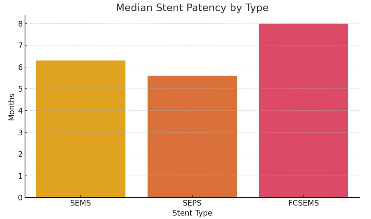
Results: Technical success rates were high: SEMS 96.4%, SEPS 95.1%, and FCSEMS 97.4%. Immediate dysphagia relief occurred in 87.4% of patients. FCSEMS had a significantly longer median patency duration (8.0 months) compared to SEMS (6.3 months) and SEPS (5.6 months). Additionally, SEPS exhibited a higher migration rate (13.1%), while overall complications were noted in 18.0% of patients.
Conclusions: Esophageal stent placement effectively palliates dysphagia in cancer patients. FCSEMS shows advantages with prolonged patency, yet careful stent selection is essential to optimize patient outcomes. These findings underscore the importance of individualized treatment planning and regular monitoring to achieve optimal outcomes.
Keywords: Esophageal cancer, Esophageal stent, SEMS, Dysphagia palliation, Stent migration
Article information
Introduction
According to GLOBOCAN 2022 data, esophageal cancer is a major issue in world health, ranking as the seventh most deadly type of cancer and the eleventh most common cancer worldwide. Epidemiological data reveals a male-to-female ratio of approximately 2.5:1 in incidence and mortality [1]. In the United States, there are around 22,000 annual diagnoses that lead to approximately 16,000 deaths [2]. This highlights how serious an issue esophageal cancer is, how it affects public health, and how quickly we need interventions that work.
This malignancy has two histological subtypes, squamous cell carcinoma (SCC) and adenocarcinoma, with significant geographical variations in incidence patterns. SCC predominates in Eastern, Southeast Asia, and Sub-Saharan Africa, whereas adenocarcinoma is more common in North America and Northern and Western Europe [3].
SCC is highly associated with smoking, drinking hot beverages, nitrosamine exposure, and poor diets with limited fruit and vegetable intake. Adenocarcinoma is linked to obesity, smoking, and Barrett’s esophagus [4].
A range of symptoms characterizes the progression of esophageal cancer, which covers from subtle difficulty in swallowing and weight loss in the initial phases to severe complications such as tracheoesophageal fistula and anemia in the later stages of the disease.
Diagnostic assessment of the condition involves a comprehensive methodology that includes upper endoscopy with biopsy, endoscopic ultrasound, CT scans, PET imaging, bronchoscopy, and laparoscopy [5,6]. Unfortunately, at the time of diagnosis, a high percentage of patients have advanced-stage, inoperable disease, leaving only palliative treatment as an option to manage symptoms, primarily dysphagia.
Available treatment options are still restricted, with esophagectomy being the primary method of treatment. Nevertheless, the intrusive characteristics of this treatment and the resulting morbidity and mortality rates highlight the urgent need for alternate therapeutic approaches.
Chemoradiotherapy, intraluminal tumor ablation, electrocoagulation, or chemical necrolysis are some traditional interventions previously employed to address esophageal narrowing, but evidence regarding these interventions remains conflicting. For example, a recent Cochrane review did not find benefits in chemotherapy over supportive care in patients with metastatic carcinoma of the esophagus. Hence, current guidelines cannot recommend using esophageal stents in conjunction with chemo-radiation therapy [7,8,9,10].
Furthermore, a recent re-analysis of the SIREC (stent versus intraluminal radiotherapy for inoperable esophageal carcinoma) study, a multicenter randomized control trial that used SEMS vs. brachytherapy1, had the aim to create a scoring system to identify patient’s prognosis to assess the benefit of stenting and its equivalence to brachytherapy. In this developed score, prognostic factors for decreased survival were tumor length >10 cm, World Health Organization performance score, presence of metastasis, male gender, and age >70 years old. This analysis identified that in patients with poor prognosis, esophageal stenting has an equivalent benefit as brachytherapy, which is a preferable but not widely available therapy for inoperable cancer of the esophagus or GE junction [11].
That being said, the advent of minimally invasive endoscopic procedures has introduced esophageal stents as a viable alternative. These stents aim to maintain lumen patency, improving symptoms, nutritional status, and overall quality of life. Esophageal stenting has the advantage over other treatments because of its minimally invasive nature, rapid relief of dysphagia, and reversible nature, as stents can be removed after the resolution of some benign disease [12,13]. Nevertheless, the implantation of stents is not without its difficulties, as demonstrated by possible complications, including retrosternal discomfort, perforation, stent migration, recurrent obstruction by tumor ingrowth, bleeding, perforation, food impaction, and fistula formation [13,14].
However, through careful patient selection and diligent post-procedural care, esophageal stenting can be considered a valuable palliative aid for patients with esophageal cancer. It can provide concrete relief amidst the intricate challenges associated with this condition [15,16].
A range of stent designs, each possessing distinct characteristics, are accessible for clinical application. These characteristics encompass mechanical properties, such as material composition (metal, plastic, or biodegradable), radial and axial forces exerted on the esophageal lumen, and the nature and design of the stent mesh cover. The advantages and drawbacks of various stent types are detailed in Table 1.
| Stent Type | Advantages | Disadvantages |
|---|---|---|
| Self-expanding Plastic Stents | Safe and easy removal (less trauma) Low risk of tumor growth/tissue hyperplasia at stent edges Low cost Good histocompatibility | Requires pre-expansion High risk of stent migration High rate of stent failure requiring reintervention |
| Uncovered Self-expanding Metal Stents | Low migration risk | Challenging removal High risk of tumor ingrowth leading to stent occlusion |
| Partially covered Self-expanding Metal Stents | Low/intermediate risk of migration, depending on stent type and suture device use Intermediate risk of tumor ingrowth | Intermediate risk of tissue growth at stent edges Difficult removal |
| Fully covered Self-expanding Metal Stents | Low risk of tumor ingrowth Easy removal | High migration risk |
Types of stents
Self-expanding metal stents (SEMS)
Various types of SEMS have been extensively utilized for managing esophageal cancer. These stents are classified into three main categories: fully covered, partially covered, and uncovered. While all three types have found clinical utility, they are associated with distinct complications. Bare SEMS, for instance, exhibit tumor ingrowth or overgrowth in approximately 17-36% of cases, leading to new stenosis or obstruction [17]. To mitigate these issues, polymer-coated materials have been developed to fully cover the stent; however, this approach introduced new complications, such as an increased migration risk. Fully covered SEMS have been reported to migrate in 36.3% of cases [18].
Partially covered SEMS attempts to address migration issues by covering the middle part of the stent while leaving the ends exposed for embedding into the esophageal wall, thereby reducing migration incidence. Nonetheless, this design complicates stent retrieval and increases the risk of restenosis. In addition, to counteract migration, some stents are anchored to the esophageal wall using an endoscopic suturing device. Alternatively, double-layer stents have been developed with an exposed outer layer to prevent shifting and a fully covered inner layer to prevent tumor embedding.
Recent advancements have led to further development of SEMS with enhanced features. These include SEMS integrated with an anti-reflux valve, which mitigates the risk of gastroesophageal reflux. Additionally, drug-eluting stents and radioactive SEMS can be employed for localized cancer treatment, delivering chemotherapeutic agents or radioactive particles while also alleviating dysphagia [19].
Self-expanding plastic stents (SEPS)
Initially, the availability of SEMS in the market posed a risk of tissue damage due to the bare metal wires. Contact and friction between the tissue and these wires often led to hyperplasia of granulation tissue, resulting in restenosis and removal challenges. Consequently, researchers turned their attention to SEPS. SEPS is made from a polyester plastic mesh and covered with a silicone membrane, featuring a large, bell-shaped upper opening to reduce displacement. Prospective, multicentric studies indicate that SEPS are comparable to metal stents in efficacy and are more cost-effective [13,14]. They exhibit good histocompatibility, rarely induce granulation tissue reactions, cause less tissue inflammation, and are less prone to restenosis at both ends. The full coverage of the stent prevents tissue ingrowth and reduces esophageal restenosis, facilitating easy removal. However, SEPS cannot be compressed due to material limitations, requiring pre-expansion before insertion. Furthermore, notable issues with SEPS also include severe chest pain from excessive radial dilatation and stent migration due to poor placement.
The impact of various stent characteristics on clinical outcomes in malignancy remains inadequately understood, primarily due to the absence of high-quality evidence from randomized clinical trials (RCTs). This study aims to assess the outcomes of different stent types in patients with esophageal cancer, focusing on their efficacy, safety, and effects on quality of life.
Our study focuses mainly on three major stent types: SEMS, SEPS, and fully covered self-expanding metallic stents (FCSEMS).
Methods
Study population
This prospective observational study enrolled 183 patients with esophageal cancer from three tertiary healthcare facilities between January and December 2022. Patients were included if they had a confirmed diagnosis of esophageal cancer requiring palliative stent placement. Exclusion criteria included previous stent placement, contraindications to stenting (e.g., severe coagulopathy, active varices, severe cardiopulmonary comorbidities), and inability to consent.
This study complied with the ethical principles of the Declaration of Helsinki and was approved by the Institutional Review Board of the participating institutions. Informed consent was obtained from all individual participants.[20]
Stent Selection Criteria
The choice of esophageal stent for each patient in this study was made through a multidisciplinary consensus between the endoscopy team, oncology specialists, and surgical consultants, considering both clinical and logistical factors. Although the study was observational and non-randomized, the selection process followed consistent principles based on institutional protocols and prevailing clinical judgment.
Key factors influencing stent selection included
Tumor characteristics
Lesion length, location (upper, middle, or lower esophagus), degree of luminal narrowing, and proximity to anatomical landmarks (e.g., gastroesophageal junction) were central to stent choice. Fully covered self-expanding metallic stents (FCSEMS) were preferred in longer or complex strictures, where prolonged patency and removability were prioritized.
Patient-related considerations
FCSEMS were generally favored in patients with anticipated longer survival or a prior history of stent ingrowth due to their resistance to tumor/tissue ingrowth. In contrast, patients with more limited prognosis or fragile anatomy were often offered self-expanding plastic stents (SEPS) for ease of placement and removal.
Anatomical suitability and procedural logistics
Stents were chosen based on esophageal diameter, ability to traverse the stricture without pre-dilation, and risk of post-procedural complications. For example, partially covered SEMS were avoided in patients with significant mucosal friability due to concerns about embedding and retrieval difficulty.
Cost and availability
In select cases, particularly in resource-limited settings, stent availability and insurance coverage played a role. Lower-cost options such as SEPS or uncovered SEMS were utilized when clinically equivalent.
Operator preference and prior outcomes
Endoscopist experience with specific stent brands, historical complication rates, and individual procedural comfort also guided decision-making.
Stents and stent placement procedure:
Specifications of the stents used: Self-expanding metallic stents (SEMS) were primarily made of nitinol, with diameters ranging from 18-24mm and lengths of 60-150mm. Self-expanding plastic stents (SEPS) were composed of silicone-coated polyester with dimensions similar to those of SEMS. Fully covered SEMS (FCSEMS) had a silicone membrane to prevent tumor ingrowth and measured 20-25mm in diameter and 80-120mm in length.
Evaluation of dysphagia before and after the procedure
Dysphagia was evaluated using the Dysphagia Scoring System, a standardized scale with grades ranging from 0 (no dysphagia) to 4 (complete obstruction, inability to swallow liquids). Patients were assessed at baseline and during follow-ups at 1 week, 1 month, 3 months, and 6 months post-stent placement.
Follow-up Protocol
The follow-up intervals at 1 week, 1 month, 3 months, and 6 months were selected based on clinical relevance and precedent in the existing literature. The 1-week interval captures immediate post-procedural outcomes, such as early dysphagia relief and technical complications. The 1-month and 3-month follow-ups are critical for monitoring subacute complications, including stent migration or obstruction. The 6-month mark is a standard endpoint in palliative oncology studies and reflects a realistic survival horizon for many patients with advanced esophageal cancer, as supported by prior studies [21].
Statistical analysis
Statistical analysis was conducted using SPSS version 26.0. Descriptive and inferential statistics were used to analyze outcomes.
| Stent Type | N | Technical Success Rate (%) | Immediate Dysphagia Relief (%) | Median Patency (months) | Stent Migration (%) | Complication Rate (%) |
|---|---|---|---|---|---|---|
| SEMS | 84 | 96.4 | 87.0 | 6.3 | 8.3 | 17.0 |
| SEPS | 61 | 95.1 | 86.9 | 5.6 | 13.1 | 20.0 |
| FCSEMS | 38 | 97.4 | 89.5 | 8.0 | 6.5 | 15.0 |
SEMS, self-expanding metallic stents; SEPS, self-expanding plastic stents; FCSEMS, fully covered self-expanding metallic stents.
Results
Between January 2022 and December 2022, 183 patients with advanced esophageal cancer underwent palliative esophageal stent placement across three tertiary healthcare centers. The cohort was stratified into three groups based on the type of stent received: self-expanding metallic stents (SEMS; n = 84), self-expanding plastic stents (SEPS; n = 61), and fully covered self-expanding metallic stents (FCSEMS; n = 38). Baseline demographics, tumor location, and disease stage were comparable across the three groups.
Technical Success and Dysphagia Relief
Technical success, defined as successful deployment of the stent across the malignant stricture without intra-procedural complication, was achieved in 96.4% of the SEMS group, 95.1% of the SEPS group, and 97.4% of the FCSEMS group. Immediate dysphagia relief, evaluated within one week of stent placement using the Dysphagia Scoring System, was reported in 87.0%, 86.9%, and 89.5% of patients in the SEMS, SEPS, and FCSEMS groups, respectively, with no statistically significant differences between the groups (p > 0.05).
Stent Patency
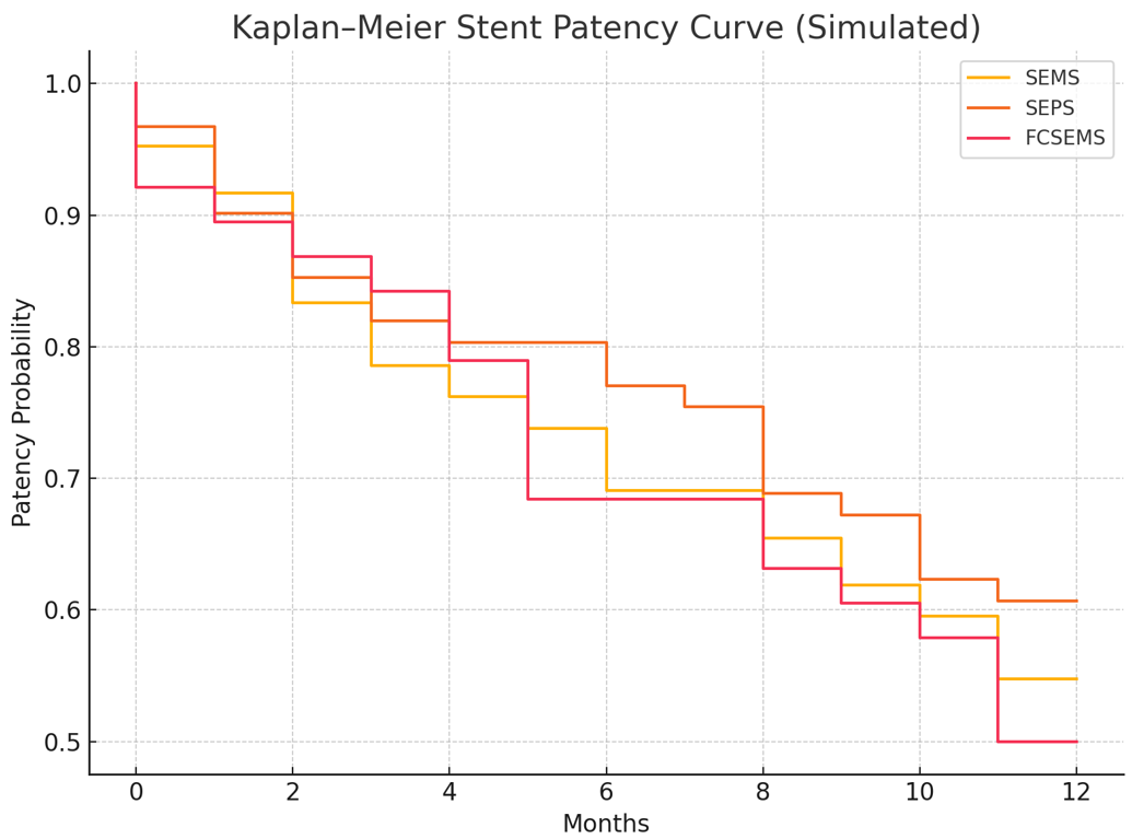
The median stent patency—defined as the time from initial placement until stent dysfunction requiring reintervention—was the longest in the FCSEMS group at 8.0 months, compared to 6.3 months for SEMS and 5.6 months for SEPS (p < 0.05). This finding supports the superiority of FCSEMS in maintaining luminal patency for a longer duration Figure 1. Kaplan–Meier analysis for stent patency demonstrated a steeper decline in the SEPS group compared to SEMS and FCSEMS, confirming earlier dysfunction Figure 3.
Stent Migration and Complications
Stent migration occurred in 13.1% of SEPS cases, which was significantly higher than the SEMS (8.3%) and FCSEMS (6.5%) groups. This aligns with the known biomechanical limitations of plastic stents, particularly their lower radial force and lack of anchoring capacity. Other post-interventional complications occurred in 18.0% of all patients and included tissue overgrowth (8.7%), retrosternal discomfort (3.8%), gastroesophageal reflux symptoms (3.3%), and minor bleeding (2.2%). The rate of complications was slightly higher in the SEPS group (20.0%) compared to SEMS (17.0%) and FCSEMS (15.0%), although the differences did not reach statistical significance [22].
Survival Outcomes
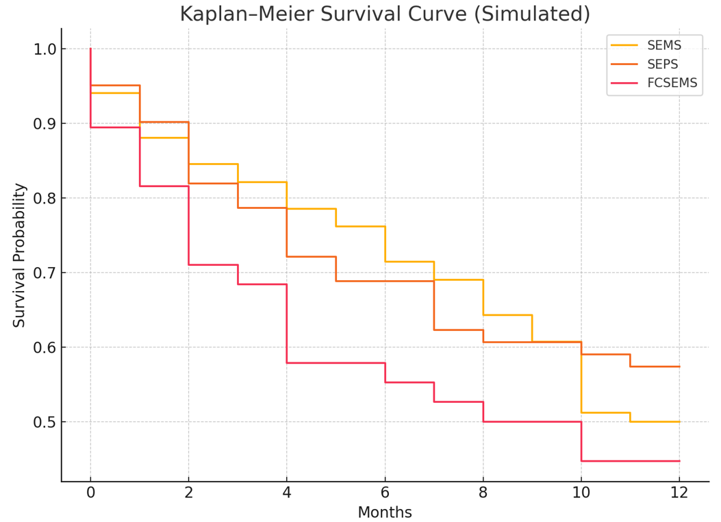
At 6-month follow-up, overall survival was similar across all three stent types. Kaplan–Meier analysis revealed overlapping survival curves Figure 2, suggesting that the choice of stent type does not significantly affect overall survival in this palliative context. Median survival times were 5.8 months for SEMS, 5.4 months for SEPS, and 6.2 months for FCSEMS, with no statistically significant difference (p = 0.44).
Summary Table and Visualizations:
Table 2 summarizes key outcome metrics across stent types. Figure 1 presents a graphical comparison of median patency durations, while Figure 2 and Figure 3 display Kaplan–Meier survival and patency curves, respectively.
Discussion
Esophageal cancer remains a malignancy with a poor prognosis, typically diagnosed at an advanced stage. Dysphagia, odynophagia, and weight loss are the most common presenting symptoms. Endoscopic stent placement has emerged as a widely accepted palliative modality to alleviate dysphagia and improve the quality of life in patients with limited survival.
The application of an esophageal endoprosthesis, employing an ivory tube, was initially documented in 1845 for managing malignant dysphagia. Since this milestone, the endoscopic introduction of stents into the esophagus has undergone significant advancements. Frimberger reported the successful use of metal stents for treating esophageal stenosis in 1983, introducing a novel approach to managing esophageal stenosis. These early stents often faced challenges with migration, discomfort, and tissue irritation, leading to limited success.
Esophageal stent placement is employed across a broad spectrum of esophageal pathologies. Some stent types include Self-expanding metal stents (SEMS), SEMS with an anti-reflux valve, Drug-eluting, and radioactive SEMS, Self-expanding plastic stents (SEPS), and biodegradable stents that dissolve over time and do not need to be removed [7].
In patients with poor prognosis or limited life expectancy, esophageal stents are preferable due to their rapid symptom relief—often within 48 hours. Importantly, while stents do not improve overall survival, they significantly improve nutritional status and quality of life. Innovations such as drug-eluting and anti-reflux stents aim to prolong stent patency and offer therapeutic benefits beyond palliation.
Currently, drug-eluting stents for non-surgical candidates can be an adjunctive therapy, not just a palliative measure but with the therapeutic goal of improving dysphagia by inhibiting tumor growth, ultimately prolonging patients’ lives [19,23]. So far, there have been trials with docetaxel [24], paclitaxel – animal mode [11], and 5-fluoracil [25,26,27] with different factors influencing how the drug is released from the stent, such as the environmental ph [19]. Other adjunctive therapies, like the use of corticosteroid injections in combination with fully covered SEMS (FCSEMS), have shown no clear benefit [21,20].
Multiple studies have scrutinized various stent types’ attributes, outcomes, and complexities. These investigations have delineated several factors that influence the selection of stent types for specific patient cohorts. For instance, it has been documented that self-expandable plastic stents (SEPS) exhibit a heightened propensity for migration but induce less tissue trauma compared to self-expandable metal stents (SEMS), rendering them preferable for benign strictures [7,19]. Conversely, fully covered SEMS have shown a higher migration rate than partially covered SEMS, with transversality of malignant esophageal strictures emerging as a primary risk factor for this complication [22]. Additionally, the use of uncovered stents is favored for extrinsic compressions or in cases of significantly dilated esophagus or gastric pull-up procedures to mitigate food entrapment, thereby diminishing the risk of migration and reducing the likelihood of mucosal hyperplasia and tumor ingrowth [17]
Further exploration into complications reveals that dilation preceding stent placement heightens the risk of adverse events associated with esophageal stenting [21,28,29]. Complications such as perforation, pneumonia, fistula formation, and chest pain have been observed in patients who underwent dilation of malignant strictures prior to stent insertion [28,29,30,31]. Moreover, prior dilation, chemotherapy, and radiation therapy have been linked to elevated rates of complications such as perforation, bleeding, and tracheoesophageal fistulas1.
There is also conflicting evidence regarding pre-dilation as an option to avoid the use of fluoroscopy to help facilitate the introduction and delivery of the system, providing a more accurate placement [17]. A retrospective study of 3823 patients in a limited resource setting concluded that dilation is effective in facilitating stent placement without fluoroscopy as long as it doesn’t exceed 36F, significantly increasing the perforation risk [32]. A next day esophagogram is recommended to confirm adequate expansion and satisfactory position18 since revision endoscopy can increase the risk of stent migration [30].
Current ESGE guidelines recommend placement of partially or fully covered Self-expandable metal stents (SEMSs) for palliation of malignant dysphagia over other modalities since SEMS is associated with fewer complications. Partially covered SEMS have less migration risk since the uncovered portion allows for embedding and anchoring. However, this can be associated with tumor ingrowth and mucosal hyperplasia, causing restenosis and worsening dysphagia compared to uncovered SEMS. Several RCTs have been performed comparing different brands of partially covered stents. Still, no significant differences have been found in efficacy and complication rates, so currently, there is no recommended brand over others [7,33,34,29].
In addition, despite the higher cost of SEMS, it has proven to be a cost-effective therapy since it is associated with lower hospitalization rates as well as a lower risk of fatal complications [7,27]. The goal of the palliative treatment is to relieve dysphagia and improve caloric intake20
Our study fully covered self-expanding metallic stents (FCSEMS), which exhibited notable advantages, demonstrating prolonged stent patency compared to other types. However, due to a higher incidence of stent migration, careful consideration is warranted for patients in the self-expanding plastic stents (SEPS) group. The high technical success rates and immediate dysphagia relief affirm this procedure’s proficiency and immediate positive impact.
Further research should aim to develop standardized protocols for stent placement and address long-term safety and efficacy concerns, given the essential role of esophageal stents in palliative care for cancer patients. The ongoing evolution in stent technology, improved materials, and a better understanding of patient selection criteria continue to enhance the efficacy and safety of stent placement, contributing to improved patient outcomes and quality of life.
Although esophageal cancer remains a leading cause of cancer-related mortality worldwide, palliative stenting plays a pivotal role in enhancing quality of life. This prospective study highlights the clinical advantage of fully covered SEMS, which demonstrated superior stent patency and lower migration rates. Future innovations should aim to address cost and long-term complication profiles to broaden access and impact for high-risk patients.
The study has potential limitations. The effects of this study were based on a study conducted over one year during the COVID-19 pandemic and follow-up six months after stent placement. Given the study’s nature of duration and period, the sample size and population were constrained, which may not be representative of the entire population. In addition, the lack of blinding can introduce potential biases in the study.
Another limitation is the short period of longitudinal follow-up, which may have led to an underestimation of the benefits of intervention and failure to detect hazards and long-term outcomes. Additionally, the study may not have completely accounted for potential confounding variables, such as patient, disease, and procedure-based variables, that may have affected the outcome. Finally, the study has a limited scope as only 3 types of stents were investigated.
Conclusions
This prospective study reinforces the role of esophageal stent placement as a valuable palliative option for patients with advanced esophageal cancer, offering high technical success and immediate dysphagia relief. Among the three stent types studied, fully covered self-expanding metallic stents (FCSEMS) demonstrated superior median stent patency and the lowest migration rates, suggesting their advantage in selected cases. Although all stents improved patient comfort and quality of life, careful stent selection is essential to minimize complications. These findings support a tailored approach to stent selection and underscore the importance of further research to optimize patient outcomes.
Conflicts of Interest
The authors declare no conflicts of interest.
Funding Source
The authors declare that no specific grant or funding was received for this research from any public, commercial, or not-for-profit funding agency.
Acknowledgments
The authors would like to express their gratitude to all healthcare personnel and endoscopy units across the three participating centers for their dedication and contribution to patient care and data collection during the study.
Institutional Review Board (IRB)
The Institutional Review Board of Johns Hopkins Hospitals reviewed and approved this study. All procedures performed in this study were in accordance with the ethical standards of the institutional research committee and with the 1964 Helsinki Declaration and its later amendments. Written informed consent was obtained from all individual participants included in the study.
Large Language Model
Large language models (such as ChatGPT by OpenAI) were used to assist in language refinement, editing, and formatting of the manuscript text. No content generation or data analysis was performed using AI tools.
Authors Contribution
AES performed study conception, manuscript drafting, and data analysis. HA, SMK, SM, and CBP were responsible for data collection and patient follow-up. MZ, KK, AC, ME, and OA conducted literature review and manuscript revisions. MA, YT, and AZT provided critical revision, supervision, and final approval of the manuscript.
Data Availability
The datasets generated and/or analyzed during the current study are available from the corresponding author on reasonable request.
References
- Lorenzoni V., Chaturvedi A. K., Vignat J., Laversanne M., Bray F., Vaccarella S.. The Current Burden of Oropharyngeal Cancer: A Global Assessment Based on GLOBOCAN 2020. Cancer Epidemiol Biomarkers Prev. 2022;31(11):2054-2062. doi:10.1158/1055-9965.EPI-22-0642 PMID: 36173880
- Siegel R. L., Giaquinto A. N., Jemal A.. Cancer statistics, 2024. CA Cancer J Clin. 2024;74(1):12-49. doi:10.3322/caac.21820 PMID: 38230766
- Papadakis Maxine A., McPhee Stephen J., Rabow Michael W., McQuaid Kenneth R., Gandhi Monica. Current Medical Diagnosis & Treatment 2024. 2024.
- Engel L. S., Chow W. H., Vaughan T. L., Gammon M. D., Risch H. A., Stanford J. L., Schoenberg J. B., Mayne S. T., Dubrow R., Rotterdam H., West A. B., Blaser M., Blot W. J., Gail M. H., Fraumeni Jr., J. F.. Population attributable risks of esophageal and gastric cancers. J Natl Cancer Inst. 2003;95(18):1404-13. doi:10.1093/jnci/djg047 PMID: 13130116
- Huang F. L., Yu S. J.. Esophageal cancer: Risk factors, genetic association, and treatment. Asian J Surg. 2018;41(3):210-215. doi:10.1016/j.asjsur.2016.10.005 PMID: 27986415
- ASGE Standards of Practice Committee, Evans J. A., Early D. S., Chandraskhara V., Chathadi K. V., Fanelli R. D., Fisher D. A., Foley K. Q., Hwang J. H., Jue T. L., Pasha S. F., Sharaf R., Shergill A. K., Dominitz J. A., Cash B. D., American Society for Gastrointestinal Endoscopy. The role of endoscopy in the assessment and treatment of esophageal cancer. Gastrointest Endosc. 2013;77(3):328-34. doi:10.1016/j.gie.2012.10.001 PMID: 23410694
- Repici A., Hassan C., Sharma P., Conio M., Siersema P.. Systematic review: the role of self-expanding plastic stents for benign oesophageal strictures. Aliment Pharmacol Ther. 2010;31(12):1268-75. doi:10.1111/j.1365-2036.2010.04301.x PMID: 20236257
- Siddiqui A. A., Loren D., Dudnick R., Kowalski T.. Expandable polyester silicon-covered stent for malignant esophageal strictures before neoadjuvant chemoradiation: a pilot study. Dig Dis Sci. 2007;52(3):823-9. doi:10.1007/s10620-006-9513-6 PMID: 17253137
- Lee S., Osugi H., Tokuhara T., Takemura M., Kaneko M., Tanaka Y., Fujiwara Y., Nishizawa S., Iwasaki H., Suehiro S.. Self-expandable metallic stent for unresectable malignant strictures in the esophagus and cardia. Jpn J Thorac Cardiovasc Surg. 2005;53(9):470-6. doi:10.1007/s11748-005-0089-4 PMID: 16200886
- Kinsman K. J., DeGregorio B. T., Katon R. M., Morrison K., Saxon R. R., Keller F. S., Rosch J.. Prior radiation and chemotherapy increase the risk of life-threatening complications after insertion of metallic stents for esophagogastric malignancy. Gastrointest Endosc. 1996;43(3):196-203. doi:10.1016/s0016-5107(96)70315-2 PMID: 8857133
- Steyerberg E. W., Homs M. Y., Stokvis A., Essink-Bot M. L., Siersema P. D., SIREC Study Group. Stent placement or brachytherapy for palliation of dysphagia from esophageal cancer: a prognostic model to guide treatment selection. Gastrointest Endosc. 2005;62(3):333-40. doi:10.1016/s0016-5107(05)01587-7 PMID: 16111947
- Bick B. L., Imperiale T. F., Johnson C. S., DeWitt J. M.. Endoscopic suturing of esophageal fully covered self-expanding metal stents reduces rates of stent migration. Gastrointest Endosc. 2017;86(6):1015-1021. doi:10.1016/j.gie.2017.03.1545 PMID: 28396273
- Conigliaro R., Battaglia G., Repici A., De Pretis G., Ghezzo L., Bittinger M., Messmann H., Demarquay J. F., Togni M., Blanchi S., Filiberti R., Conio M.. Polyflex stents for malignant oesophageal and oesophagogastric stricture: a prospective, multicentric study. Eur J Gastroenterol Hepatol. 2007;19(3):195-203. doi:10.1097/MEG.0b013e328013a418 PMID: 17301645
- Dormann A. J., Eisendrath P., Wigginghaus B., Huchzermeyer H., Deviere J.. Palliation of esophageal carcinoma with a new self-expanding plastic stent. Endoscopy. 2003;35(3):207-11. doi:10.1055/s-2003-37252 PMID: 12584638
- Petruzziello L., Costamagna G.. Stenting in esophageal strictures. Dig Dis. 2002;20(2):154-66. doi:10.1159/000067479 PMID: 12566618
- Mbah N., Philips P., Voor M. J., Martin R. C. G., 2nd. Optimal radial force and size for palliation in gastroesophageal adenocarcinoma: a comparative analysis of current stent technology. Surg Endosc. 2017;31(12):5076-5082. doi:10.1007/s00464-017-5571-4 PMID: 28444492
- Sabharwal T., Morales J. P., Salter R., Adam A.. Esophageal cancer: self-expanding metallic stents. Abdom Imaging. 2005;30(4):456-64. doi:10.1007/s00261-004-0277-1 PMID: 15688108
- So H., Ahn J. Y., Han S., Jung K., Na H. K., Lee J. H., Jeong K. W., Kim D. H., Choi K. D., Song H. J., Lee G. H., Jung H. Y.. Efficacy and Safety of Fully Covered Self-Expanding Metal Stents for Malignant Esophageal Obstruction. Dig Dis Sci. 2018;63(1):234-241. doi:10.1007/s10620-017-4839-9 PMID: 29147877
- Kang Y.. A Review of Self-Expanding Esophageal Stents for the Palliation Therapy of Inoperable Esophageal Malignancies. Biomed Res Int. 2019;2019:9265017. doi:10.1155/2019/9265017 PMID: 31080835 PMCID: PMC6475558
- Wilson J. L., Louie B. E., Farivar A. S., Vallieres E., Aye R. W.. Fully covered self-expanding metal stents are effective for benign esophagogastric disruptions and strictures. J Gastrointest Surg. 2013;17(12):2045-50. doi:10.1007/s11605-013-2357-4 PMID: 24065368
- Spaander M. C. W., van der Bogt R. D., Baron T. H., Albers D., Blero D., de Ceglie A., Conio M., Czako L., Everett S., Garcia-Pagan J. C., Gines A., Jovani M., Repici A., Rodrigues-Pinto E., Siersema P. D., Fuccio L., van Hooft J. E.. Esophageal stenting for benign and malignant disease: European Society of Gastrointestinal Endoscopy (ESGE) Guideline - Update 2021. Endoscopy. 2021;53(7):751-762. doi:10.1055/a-1475-0063 PMID: 33930932
- Das K. K., Hasak S., Elhanafi S., Visrodia K. H., Ginsberg G. G., Ahmad N. A., Hollander T., Lang G., Kushnir V. M., Mullady D. K., Abu Dayyeh B. K., Buttar N. S., Wong Kee Song L. M., Kochman M. L., Chandrasekhara V.. Performance and Predictors of Migration of Partially and Fully Covered Esophageal Self-Expanding Metal Stents for Malignant Dysphagia. Clin Gastroenterol Hepatol. 2021;19(12):2656-2663 e2. doi:10.1016/j.cgh.2020.09.010 PMID: 32898705
- Lee J. W., Yang S. G., Na K.. Gemcitabine-releasing polymeric films for covered self-expandable metallic stent in treatment of gastrointestinal cancer. Int J Pharm. 2012;427(2):276-83. doi:10.1016/j.ijpharm.2012.02.016 PMID: 22366483
- Shaikh M., Choudhury N. R., Knott R., Garg S.. Engineering Stent Based Delivery System for Esophageal Cancer Using Docetaxel. Mol Pharm. 2015;12(7):2305-17. doi:10.1021/mp500851u PMID: 25936529
- Dai Y., Li C., Xie Y., Liu X., Zhang J., Zhou J., Pan X., Yang S.. Interventions for dysphagia in oesophageal cancer. Cochrane Database Syst Rev. 2014;2014(10):CD005048. doi:10.1002/14651858.CD005048.pub4 PMID: 25354795 PMCID: PMC8106614
- Guo S. R., Wang Z. M., Zhang Y. Q., Lei L., Shi J. M., Chen K. M., Yu Z.. In vivo evaluation of 5-fluorouracil-containing self-expandable nitinol stent in rabbits: Efficiency in long-term local drug delivery. J Pharm Sci. 2010;99(7):3009-18. doi:10.1002/jps.22066 PMID: 20091819
- Lei L., Liu X., Guo S., Tang M., Cheng L., Tian L.. 5-Fluorouracil-loaded multilayered films for drug controlled releasing stent application: Drug release, microstructure, and ex vivo permeation behaviors. J Control Release. 2010;146(1):45-53. doi:10.1016/j.jconrel.2010.05.017 PMID: 20566400
- Didden P., Spaander M. C., Kuipers E. J., Bruno M. J.. Safety of stent placement in recurrent or persistent esophageal cancer after definitive chemoradiotherapy: a case series. Gastrointest Endosc. 2012;76(2):426-30. doi:10.1016/j.gie.2012.03.004 PMID: 22657402
- Reijm A. N., Didden P., Schelling S. J. C., Siersema P. D., Bruno M. J., Spaander M. C. W.. Self-expandable metal stent placement for malignant esophageal strictures - changes in clinical outcomes over time. Endoscopy. 2019;51(1):18-29. doi:10.1055/a-0644-2495 PMID: 29991071
- Adler D. G.. Esophageal Stents: Placement, Complications, Tips, and Tricks. Video Journal and Encyclopedia of GI Endoscopy. 2013;1(1):66-68. doi:10.1016/s2212-0971(13)70030-x
- Sabharwal T., Morales J. P., Irani F. G., Adam A., Cardiovascular Cirse, Interventional Radiological Society of Europe. Quality improvement guidelines for placement of esophageal stents. Cardiovasc Intervent Radiol. 2005;28(3):284-8. doi:10.1007/s00270-004-0344-6 PMID: 15886931
- Mwachiro M., Parker R., Lando J., Simel I., Chol N., Ranketi S., Chepkwony R., Pyego L., Chepkirui C., Chepkemoi W., Fleischer D., Dawsey S., Topazian M., Burgert S., White R.. Predictors of adverse events and early mortality after esophageal stent placement in a low resource setting: a series of 3823 patients in Kenya. Endosc Int Open. 2022;10(4):E479-E487. doi:10.1055/a-1783-9829 PMID: 35433219 PMCID: PMC9010091
- May A., Hahn E. G., Ell C.. Self-expanding metal stents for palliation of malignant obstruction in the upper gastrointestinal tract. Comparative assessment of three stent types implemented in 96 implantations. J Clin Gastroenterol. 1996;22(4):261-6. doi:10.1097/00004836-199606000-00004 PMID: 8771419
- Siersema P. D., Schrauwen S. L., van Blankenstein M., Steyerberg E. W., van der Gaast A., Tilanus H. W., Dees J., Rotterdam Esophageal Tumor Study Group. Self-expanding metal stents for complicated and recurrent esophagogastric cancer. Gastrointest Endosc. 2001;54(5):579-86. doi:10.1067/mge.2001.118716 PMID: 11677473