Annular pancreas in adults presenting as chronic pancreatitis and duodenal obstruction: A review of literature and a rare case report
Gajendra BhatiⒾ1, Shruti PandeyⒾ2, Ripanbir Singh KahlonⒾ3, Raghav BansalⒾ4
- 1Department of Surgical Gastroenterology, Adesh Institute of Medical Sciences and Research, Bathinda, Punjab, India
- 2Department of Anaesthesiology and Critical Care, Dayanand Medical College, Ludhiana, Punjab, India
- 3Government Medical College, Amritsar, Punjab, India
- 4Department of General Surgery, Adesh Institute of Medical Sciences and Research, Bathinda, Punjab, India
Abstract
Background: Annular pancreas is a rare congenital anomaly with an estimated incidence of 3.4 per 1,00,000 live births. It is more commonly diagnosed in infants. Its occurrence in adults is exceptionally rare but clinically significant, as seen in our case, where it presented as a duodenal obstruction.
Case presentation: We present a case of an adult male who presented with a 3-week history of projectile bilious vomiting associated with postprandial abdominal pain and early satiety. Imaging studies revealed a segment of the pancreas encircling the second part of the duodenum, along with pancreatolithiasis. Based on the diagnosis of annular pancreas with chronic calcific pancreatitis, the patient underwent pylorus-preserving pancreatoduodenectomy.
Materials and Methods: A structured literature review was performed using PubMed and Scopus databases, covering the period from 2018 to 2024. A total of 24 case reports were analyzed.
Results: The review confirms that annular pancreas remains a rare condition, often undiagnosed in asymptomatic individuals with abdominal pain (83.3%) and vomiting (41.6%) as the most common symptoms. The most common diagnostic modality is the CT scan (computed tomography), at 64.1%, and surgical procedures, such as gastrojejunostomy, are the most common treatment modality, at 46.5%.
Conclusions: It is a rare congenital condition that usually remains undiagnosed in asymptomatic individuals but typically presents with abdominal pain and vomiting when symptomatic. Diagnosis relies primarily on CT imaging. Conservative management is preferred for asymptomatic cases, and surgery is for symptomatic patients. Further research is needed to develop standardized management protocols.
Keywords: Congenital pancreatic anomaly, Acute Intestinal Obstruction, Annular pancreas, Chronic Calcific Pancreatitis, Pancreatoduodenectomy, Whipple's procedure
Article information
Introduction
The annular pancreas (AP) is a rare congenital anomaly characterized by pancreatic tissue partially or completely encircling the second part of the duodenum. This results from the aberrant migration of the ventral pancreatic bud during embryogenesis. This condition was first described by Tiedemann in 1818 and later termed “annular pancreas” by Ecker [1]. Between the fourth and eighth week of embryonic growth, the pancreas typically forms through the rotation and merging of the dorsal and ventral pancreatic buds, driven by the duodenum’s expansion. Thereafter, the ventral bud gives rise to the lower portion of the pancreatic head and the uncinate process, while the dorsal bud develops into the body and tail of the pancreas [2]. The majority of cases of AP are identified in the pediatric population, exhibiting signs of gastric outlet obstruction [3]. Common symptoms in adults include abdominal discomfort, vomiting, and hematemesis in patients [4]. A recent population-based study in the United States analyzed data from 6,162,600 patients who underwent abdominal imaging, identifying 210 cases of AP. This finding suggests an estimated prevalence rate of 3.4 per 100,000 individuals [5]. It impacts both sexes, with a modest predominance towards males [6]. The current standard for diagnosing AP relies on abdominal imaging techniques, including ultrasound, computed tomography (CT) scans, barium studies, endoscopic retrograde cholangiopancreatography (ERCP), and magnetic resonance cholangiopancreatography (MRCP) [7]. Approximately 40% of cases necessitate surgical interventions, such as duodenoduodenostomy or duodeno-jejunostomy, to circumvent the blocked duodenal section. Pancreatoduodenectomy is advised only in instances where the AP coexists with pancreatolithiasis and is further complicated by chronic pancreatitis [4]. This report condenses a meticulous review of 24 recently published case reports (2018–2024), providing a contemporary overview of symptomatic adult AP, along with a case report that further contributes to its clinical significance.
Objective
This paper aims to advance the understanding of AP presentation in adults, a rare condition with limited published research. Through a case report and a comprehensive literature review, we seek to provide valuable insights that enhance the current knowledge of this pathology.
Case Description
A 50-year-old male presented to the outpatient clinic with chief complaints of vomiting and abdominal pain for 3 weeks. The pain was intermittent, moderate to severe in intensity, poorly localized in the upper abdomen, radiating to the back, and was relieved by vomiting. There was a history of projectile bilious vomiting associated with post-prandial abdominal pain and fullness for 3 weeks. The patient also had a history of chronic smoking and alcohol intake for 20 years. There was no history of weight loss, fever, or melena. There were no co-morbidities associated, including no history of diabetes mellitus, hypertension, tuberculosis, bronchial asthma, or epilepsy, and no significant past medical or surgical history was reported. There was no history of similar complaints in the past. On examination, the abdomen was distended and tender, along with guarding and rigidity. No mass was palpable per abdomen. No significant findings in per rectal examination.
Initial investigations, as detailed in Table 1, revealed slightly decreased hemoglobin levels, an elevated white blood cell count, elevated random blood sugar, and slightly deranged liver enzymes. Renal function tests were within normal limits. Electrolytes were slightly deranged, and pancreatic enzymes were highly elevated. Viral markers, including HCV, HBsAg, and HIV, were non-reactive.
| Investigations | Value | Normal Range |
|---|---|---|
| Haemoglobin | 9.5 g/dl | 13.0–17.0 |
| TLC | 18 /uL | 4.0–10.0 |
| RBS | 155 mg/dL | 80.0–140.0 |
| Liver Function Tests | ||
| SGOT | 89 IU/L | 0.0–35.0 |
| SGPT | 92 IU/L | 0.0–41.0 |
| Alkaline Phosphatase (ALP) | 109 IU/L | 40.0–129.0 |
| Albumin | 3.2 g/dl | 3.5–5.2 |
| Bilirubin | 2.5 mg/dl | 0.1–1.2 |
| Renal Function Tests | ||
| Serum urea | 42 mg/dl | 17.0–43.0 |
| S. Creatinine | 1.4 mg/dl | 0.6–1.1 |
| INR | 0.91 | 0.8–1.2 |
| Electrolytes | ||
| S. Na | 135 mmol/L | 136.0–145.0 |
| S. K | 4.1 mmol/L | 3.5–5.0 |
| S. Ca | 6.2 mg/dl | 8.6–10.3 |
| Pancreatic Enzymes | ||
| S. Lipase | 2850 U/L | 13.0–60.0 |
| S. Amylase | 1549 U/L | 22.0–80.0 |
| Viral Markers | ||
| HCV | Non-Reactive | – |
| HBsAg | Non-Reactive | – |
| HIV | Non-Reactive | – |
Hb, Haemoglobin; TLC, Total Leukocyte Count; RBS, Random Blood Sugar; SGOT, Serum Glutamic Oxaloacetic Transaminase (AST); SGPT, Serum Glutamic Pyruvic Transaminase (ALT); ALP, Alkaline Phosphatase; INR, International Normalized Ratio; S. Na⁺, Serum Sodium; S. K⁺, Serum Potassium; S. Ca²⁺, Serum Calcium; S. Lipase, Serum Lipase; S. Amylase, Serum Amylase; HCV, Hepatitis C Virus; HBsAg, Hepatitis B Surface Antigen; HIV, Human Immunodeficiency Virus.
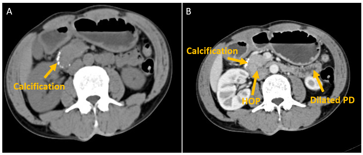
Further, the patient was investigated, and a CT scan of the abdomen revealed an abnormal configuration of the head of the pancreas with features suggestive of (F/S/O) chronic calcific pancreatitis (CCP) and a triangular segment of the pancreatic tissue encircling the second part of the duodenum (D2), causing complete obstruction. A few foci of calcification were also noted in the head of the pancreas (HOP) along with stranding of peripancreatic fat and dilated pancreatic duct with no evidence of malignancy or necrosis Figure 1.
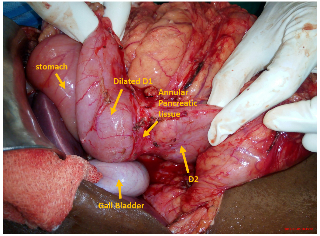
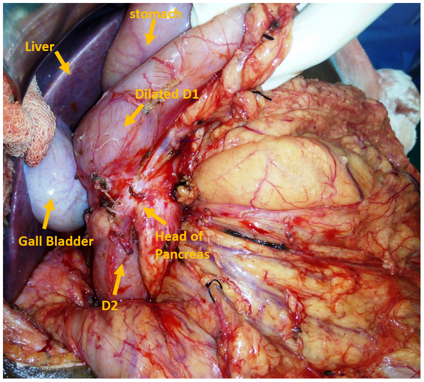
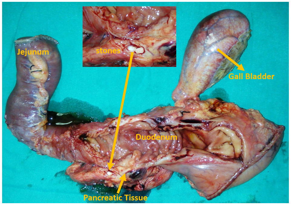
Based on the diagnostic findings, surgical intervention was planned. The patient underwent pylorus-preserving pancreatoduodenectomy under general anesthesia with endotracheal tube intubation. After proper painting and draping, a nasogastric tube was inserted for gastric decompression. An extended right subcostal incision was given to access the abdominal cavity. Intraoperatively, the first part of the duodenum (D1) was found to be dilated Figure 2, with annular pancreatic tissue encircling the second part (D2), causing its narrowing Figure 3) – findings consistent with the preoperative radiological imaging findings. Intra-operatively, a mass-forming lesion was palpated in the HOP. A stapler was fired along the distal part of the stomach, and another was fired approximately 20 cm distal to the ligament of Treitz. A jejunal loop was then brought up retrocolically, and an end-to-side anastomosis was performed, resulting in pancreatojejunostomy, hepaticojejunostomy, and gastrojejunostomy. The specimen was then resected, and the area post-resection was visualized. The macroscopic inspection of the gross specimen by observing the cut section revealed the presence of a stone in the HOP (pancreatolithiasis) Figure 4. The postoperative course was uneventful, with mild abdominal pain and minimal serosanguinous discharge from the drain. The patient was discharged on postoperative day 7 in a stable condition.
Microscopic examination of the resected specimen revealed chronic inflammatory infiltrates with interlobular fibrosis, confirming the diagnosis of chronic calcific pancreatitis. Pancreatic duct dilation and pancreaticolithiasis were noted without evidence of dysplasia or malignancy.
At 6-month postoperative follow-up, the patient remained asymptomatic with complete resolution of vomiting and abdominal pain. No signs of recurrence or complications were observed on CT imaging and labs.
All procedures performed in this study adhered to the ethical standards of the institutional and national research committee and the Helsinki Declaration. Informed written consent was obtained from the patient for the publication of this case report and accompanying images. No identifiable patient information is included in this publication. Ethical approval was waived by the Institutional Review Board (IRB) for the case reports.
Discussion
AP is a rare congenital anomaly resulting from an embryological error during the fifth to seventh weeks of gestation, wherein the ventral pancreatic bud fails to rotate properly, leading to partial or complete encasement of the duodenum. This case illustrates the elusive nature of the condition, consistent with historical autopsy-based prevalence estimations of approximately 1 in 20,000, although more recent radiological studies suggest higher rates [8]. These discrepancies underscore the underrecognized burden of AP, particularly in adults, where incidental diagnoses often mask symptomatic presentations. In our patient, the diagnosis of AP with CCP highlights the clinical challenges posed by its nonspecific symptoms—most commonly abdominal pain, nausea, and vomiting, and less frequently hematemesis. In a large single-institutional cohort study, Nagpal et al. [9] reported that 59.6% of adults with AP were asymptomatic; among symptomatic patients, abdominal pain was the most frequently observed symptom (50%).
To better contextualize our case and understand current trends, we conducted a structured literature review focusing on adult patients with symptomatic annular pancreas who underwent surgical intervention, particularly pancreatoduodenectomy. This review aimed to compare presenting symptoms, diagnostic modalities, and treatment approaches in recently reported cases.
Methodology
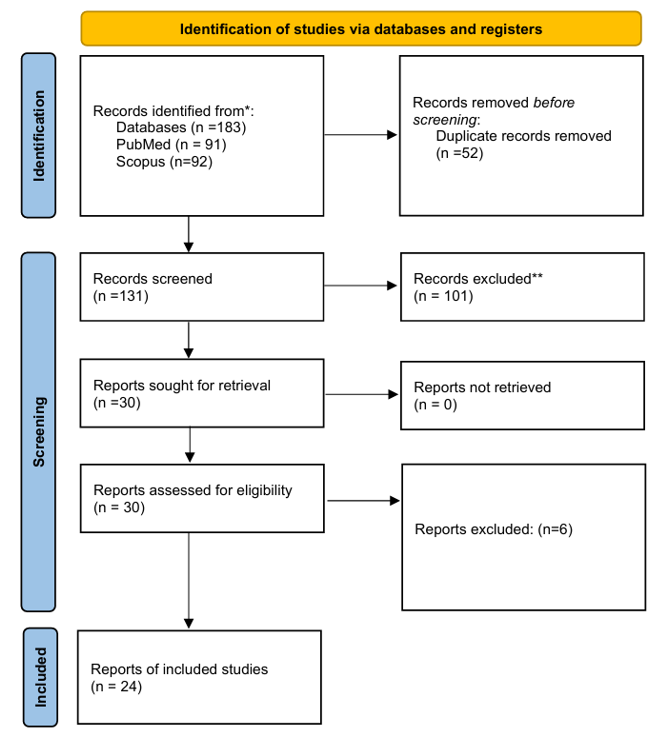
A structured literature review was performed using PubMed and Scopus databases, covering the period from 2018 to 2024. The following Boolean keyword string was applied: (“Annular pancreas” OR “pancreatic ring”) AND (“adult” OR “adult presentation”) AND (“case report” OR “case series”). Filters included: English language, human studies, and adult subjects (aged 18 years or older). The review process was conducted in accordance with the Preferred Reporting Items for Systematic Reviews and Meta-Analyses (PRISMA) guidelines to ensure transparency, reproducibility, and methodological rigor. A PRISMA flow diagram detailing the selection process has been included to summarize the article screening and inclusion process.
Eligibility criteria and study selection
Studies were included if they reported symptomatic adult cases of AP confirmed by imaging or intraoperative findings and provided clinical details including symptoms, diagnostics, and treatment. Exclusion criteria included pediatric-only data, asymptomatic incidental findings with minimal clinical description, and editorials without primary data. Two independent reviewers extracted data including age, gender, presenting symptoms, diagnostic modality, treatment modality, and outcomes. Any discrepancies were resolved through consensus.
Data extraction
Summary statistics were generated using Microsoft Excel. Descriptive percentages were calculated for the following categorical variables: (a) Symptoms, (b) Diagnosing modality, (c) Modality of intervention
Risk of Bias assessment
The methodological quality of the included case reports and case series was assessed using the Joanna Briggs Institute (JBI) Critical Appraisal Checklists, specific to each study design. These standardized tools were used to evaluate the risk of bias and ensure the inclusion of methodologically sound studies in the review.
We reviewed 24 published cases (n = 24) of AP since 2018 and found that patients exhibited abdominal pain (83.3%), vomiting (41.6%), early satiety (16.6), weight loss (16.6%), melena (4.16%), dyspepsia (4.16%), hematochezia (4.16%), and diarrhea (4.16%) – Table 2 Figure 5. These manifestations, often mimicking peptic ulcer disease, pancreatitis, or gastric outlet obstruction, emphasize the need for a high index of suspicion among clinicians.
| No. of patients (n = 24) | ||
|---|---|---|
| Symptoms | No. of patients | Percentage (%) |
| Abdominal pain | 20 | 83.3% |
| Melena | 1 | 4.16% |
| Dyspepsia | 1 | 4.16% |
| Hematochezia | 1 | 4.16% |
| Diarrhea | 1 | 4.16% |
The embryological theories proposed by Leeco and Baldwin [10] offer a foundational framework for understanding the development of AP. Leeco hypothesized that the ventral pancreatic bud adheres to the duodenal wall, while Baldwin suggested that persistent anomalies of the left ventral bud result in the formation of a pancreatic ring—both mechanisms potentially explaining the annular configuration observed in this patient. Imaging studies played a critical role in diagnosis, with CT scan revealing pancreatic tissue posterolateral to the second portion of the duodenum—a finding reported to have 92% sensitivity and 100% specificity for annular pancreas [10]. Complementary modalities such as ERCP and MRCP further improve diagnostic accuracy, although their routine use may be limited by cost and availability.
In our analysis of 24 published case reports comprising 223 patients, CT was the most frequently employed imaging modality for diagnosing annular pancreas. By integrating data from Nagpal et al. [9] with our case series, we summarized the diagnostic tools utilized in Table 3. In the present case, the supra-papillary obstruction corresponded with the literature, which notes involvement of the second portion of the duodenum in approximately 74% of cases, underscoring the anatomical consistency of AP across patient populations [11].
| (No. of patients n = 223) | ||
|---|---|---|
| Modality | No. of patients | Percentage (%) |
| Computed Tomography (CT) scan | 143 | 64.1% |
| Magnetic Resonance Cholangiopancreatography (MRCP) | 52 | 23.3% |
| Intraoperative finding | 15 | 6.72% |
| Endoscopic ultrasound | 8 | 3.58% |
| Endoscopic Retrograde Cholangiopancreatography (ERCP) | 5 | 2.2% |
| Total | 223 | 100% |
Management of the AP remains individualized, as no universal treatment protocol exists. Among patients requiring surgical intervention, gastrojejunostomy is the most frequently performed procedure. By synthesizing data from Nagpal et al [9]with our reviewed case reports, the most common surgical approaches are presented Table 4. In our patient, pancreatoduodenectomy effectively alleviated the obstruction and the symptoms.
| No. of patients (N = 43) | ||
|---|---|---|
| Treatment Modalities | No. of patients | Percentage (%) |
| Gastrojejunostomy | 20 | 46.5% |
| Pancreatoduodenectomy | 10 | 23.2% |
| Duodenoduodenostomy | 5 | 11.6% |
| Duodenojejunostomy | 4 | 9.3% |
| 30 | 2 | 4.6% |
| Annular ring resection | 1 | 2.3% |
| Total pancreatectomy | 1 | 2.3% |
| Total | 43 | 100% |
This case adds to the limited body of literature on adult presentations of AP, where symptomatic manifestations remain less well understood compared to the extensively documented neonatal counterpart. Prevalence estimates vary widely—from 0.0005% to 0.0015% in autopsy series to 3.4 per 100,000 in recent U.S. population-based imaging studies [5]. The true incidence of AP likely lies between these extremes, obscured by asymptomatic cases and variability in diagnostic practices. A slight male predominance has been observed among symptomatic adults, consistent with our patient’s profile.
This report highlights the value of advanced imaging and individualized surgical intervention in managing AP along with CCP, while emphasizing its diagnostic complexity and clinical heterogeneity. As a rare entity, AP continues to present diagnostic and therapeutic challenges, warranting further research to clarify its natural history and optimize treatment strategies, especially in adults, for whom timely diagnosis significantly impacts quality of life.
The case described here is noteworthy for several distinctive features that differentiate it from most reported instances. Unlike the typical neonatal presentation marked by early duodenal obstruction or incidental findings in asymptomatic adults, this 50-year-old male presented with a three-week history of projectile bilious vomiting, postprandial fullness, and abdominal pain. This delayed and atypical symptomatic profile stands out, particularly given the presence of CCP findings that are infrequently emphasized in existing literature. CT Scan confirmed the diagnosis, revealing both annular pancreatic tissue and pancreatic calcification, causing duodenal obstruction—an uncommon constellation. The likely mechanism of obstruction may involve ulceration secondary to compression by the annular tissue in the setting of chronic inflammation.
This combination of a subacute presentation, diagnostic complexity, and the use of an uncommon operative approach —namely, pylorus-preserving pancreatoduodenectomy (Whipple procedure) —distinguishes this case and broadens the current understanding of adult annular pancreas presentations and management.
What sets our review apart from prior studies is its targeted synthesis of 24 recently published case reports (2018–2024), offering a contemporary overview of symptomatic adult AP. We also provide a detailed account of less frequently reported symptoms, such as melena, hematochezia, and dyspepsia, which are often underrepresented in broader reviews. Furthermore, by integrating cohort-level data from Nagpal et al., we enhance the understanding of these symptoms.[9] with our findings, we present a more comprehensive analysis of diagnostic trends and treatment modalities, summarized in Tables 2 and 3. This layered approach enhances both the granularity and relevance of our review compared to earlier, more generalized surveys of the condition.
Conclusion
A review of recent case reports confirms that AP remains a rare condition, often undiagnosed in asymptomatic individuals. Symptomatic patients typically present with abdominal pain and vomiting, though additional symptoms such as weight loss and early satiety may also occur. Abdominal imaging serves as the cornerstone of diagnosis, with CT scans being the most commonly employed modality to confirm the condition. Asymptomatic cases of AP generally require no intervention and are managed conservatively. In contrast, surgical treatment is the primary approach for symptomatic patients, with the specific procedure tailored to the individual’s anatomy and the severity of the pathology. Further timely research is needed to establish standardized protocols for the diagnosis and management of AP.
Conflicts of Interest
GB, SP, RK, and RB declare that they have no financial or non-financial competing interests related to the content of this article. No conflicts of interest, financial ties, or funding sources have influenced the results or interpretations presented in this manuscript.
Funding Source
This research did not receive any specific grant from funding agencies in the public, commercial, or not-for-profit sectors.
Acknowledgments
None
Institutional Review Board (IRB)
This study was conducted in accordance with the Declaration of Helsinki. Ethical approval was waived off by the Institutional Review Board (IRB) for the case reports/case series. Written informed consent was obtained from the patient to participate in the study and publish their clinical information and images. No identifiable patient information is included in this publication.
Large Language Model
None
Authors Contribution
GB and RB supervised, conceptualized, designed methodology, provided resources, investigated, drafted the original manuscript, and reviewed and edited the draft; SP and RK assisted in writing original draft, reviewed and edited the draft. All authors contributed to the manuscript’s text and content, approved the final version, and agreed to be accountable for the work.
Data Availability
All data are included in this published article.
References
- Wani A. A., Maqsood S., Lala P., Wani S.. Annular pancreas in adults: a report of two cases and review of literature. JOP. 2013;14(3):277-9. doi:10.6092/1590-8577/1454 PMID: 23669479
- Pan F. C., Wright C.. Pancreas organogenesis: from bud to plexus to gland. Dev Dyn. 2011;240(3):530-65. doi:10.1002/dvdy.22584 PMID: 21337462
- Newman BM, Lebenthal E. Congenital abnormalities of the exocrine pancreas. The exocrine pancreas: biology, pathobiology and diseases. Raven Press, New York. 1986:773-782.
- Rondelli F., Bugiantella W., Stella P., Boni M., Mariani E., Crusco F., Sanguinetti A., Polistena A., Avenia N.. Symptomatic annular pancreas in adult: Report of two different presentations and treatments and review of the literature. Int J Surg Case Rep. 2016;20S(Suppl):21-4. doi:10.1016/j.ijscr.2016.02.001 PMID: 26867720 PMCID: PMC4883047
- Alkhayyat M., Bachour S., Abou Saleh M., Abureesh M., Almomani A., Simons-Linares C. R., Mansoor E., Sandhu D., Bhatt A., Chahal P.. The Epidemiology of Annular Pancreas in the United States: A Population-based Study. J Clin Gastroenterol. 2022;56(2):186-191. doi:10.1097/MCG.0000000000001531 PMID: 33769394
- Huddleston V. S., Lippuner V., Dyer A. W.. Annular Pancreas in an Adult Presenting with Acute Pancreatitis. J Radiol Case Rep. 2018;12(10):11-16. doi:10.3941/jrcr.v12i10.3461 PMID: 30651906 PMCID: PMC6312117
- Zhou Y., Li X.. Investigation of annular pancreas through multiple detector spiral CT (MDCT) and MRI. J Appl Clin Med Phys. 2022;23(1):e13487. doi:10.1002/acm2.13487 PMID: 34845829 PMCID: PMC8803292
- Paraskevas G., Papaziogas B., Lazaridis C., Gigis P., Papaziogas T.. Annular pancreas in adults: embryological development, morphology and clinical significance. Surg Radiol Anat. 2001;23(6):437-42. doi:10.1007/s00276-001-0437-8 PMID: 11963628
- Nagpal S. J. S., Peeraphatdit T., Sannapaneni S. K., Sharma A., Takahashi N., Kendrick M. L., Majumder S., Vege S. S.. Clinical spectrum of adult patients with annular pancreas: Findings from a large single institution cohort. Pancreatology. 2019;19(2):290-295. doi:10.1016/j.pan.2018.12.009 PMID: 30660391
- Kawaida H., Kono H., Watanabe M., Maki A., Amemiya H., Matsuda M., Fujii H., Fukasawa M., Takahashi E., Sano K., Inoue T.. Pancreaticoduodenectomy for pancreas carcinoma occurring in the annular pancreas: report of a case. Clin J Gastroenterol. 2015;8(4):223-7. doi:10.1007/s12328-015-0579-6 PMID: 26153253 PMCID: PMC4548008
- Sandrasegaran K., Patel A., Fogel E. L., Zyromski N. J., Pitt H. A.. Annular pancreas in adults. AJR Am J Roentgenol. 2009;193(2):455-60. doi:10.2214/AJR.08.1596 PMID: 19620443
- Ahmetgjekaj I., Roy P., Hyseni F., Mamillo K., Syed Z., Parisapogu A., Mian H. S., Pakhrin S., Saha T., Tul Kubra K., Maliha M., Musa J., Ghosh A. S.. Annular pancreas: Beneath the intestinal obstruction-A case report. Radiol Case Rep. 2023;18(3):1364-1367. doi:10.1016/j.radcr.2022.11.083 PMID: 36747589 PMCID: PMC9898573
- Ali I. M., Reddy M. Vs, Shetty V.. Adult-Onset Annular Pancreas: When to Intervene?. Cureus. 2024;16(8):e67728. doi:10.7759/cureus.67728 PMID: 39318953 PMCID: PMC11421865
- Bahja Sarra, Abid Hakima, Abkari Mohammed El, Ahlimine Abdessamad, Bouassria Abdesslam, Bouhaddouti Hicham El. Annular Pancreas in Adults. SAS Journal of Medicine. 2022;8(4):326-329. doi:10.36347/sasjm.2022.v08i04.020
- Castro E., Sanchez-Morales G. E., Hernandez-Acevedo J. D., Sanchez Garcia-Ramos E., Chan C.. Ampullary adenocarcinoma in a patient with portal annular pancreas: Relevance of the surgical approach in the Whipple procedure. Rev Gastroenterol Mex (Engl Ed). 2022;87(1):116-118. doi:10.1016/j.rgmxen.2021.12.007 PMID: 34969645
- Cheng L., Tian F., Zhao T., Pang Y., Luo Z., Ren J.. Annular pancreas concurrent with pancreaticobiliary maljunction presented with symptoms until adult age: case report with comparative data on pediatric cases. BMC Gastroenterol. 2013;13:153. doi:10.1186/1471-230X-13-153 PMID: 24156788 PMCID: PMC4015270
- de la Rosa Rodriguez R., Fogarty A., Israel G. M., Sanchez M. J.. Annular pancreas in a 24-year-old woman with persistent abdominal pain. BMJ Case Rep. 2019;12(6). doi:10.1136/bcr-2019-229223 PMID: 31227569 PMCID: PMC6605914
- Iwao Y., Ban D., Muro S., Kudo A., Tanaka S., Menon K., Tanabe M.. Extraordinary first jejunal arterial variation associated with annular pancreas undergoing pancreaticoduodenectomy for pancreatic cancer: a case report. Surg Radiol Anat. 2021;43(5):805-810. doi:10.1007/s00276-020-02671-9 PMID: 33481131 PMCID: PMC8105220
- Katwal S., Oli R., Bhusal A., Bhattarai H. B.. Partial annular pancreas as an incidental finding in a patient with intermittent bowel obstruction: A case report. Radiol Case Rep. 2023;18(11):3968-3971. doi:10.1016/j.radcr.2023.08.044 PMID: 37680665 PMCID: PMC10480449
- Kryzsko V. M., Tecos M. E., Buesing K. L., Henry R.. Adult presentation of symptomatic annular pancreas treated with gastrojejunostomy. J Surg Case Rep. 2024;2024(10):rjae638. doi:10.1093/jscr/rjae638 PMID: 39411510 PMCID: PMC11475919
- Kweun J. A., Kang H. M., Kim J. E., Park S. J.. Annular Pancreas: A Rare Cause of Upper Gastrointestinal Bleeding in Adults. Korean J Gastroenterol. 2022;79(4):182-186. doi:10.4166/kjg.2022.012 PMID: 35473777
- Nur N. M., Artan A., Omar A. A., Ahmed M. R.. Annular pancreas causing duodenal obstruction in a 23 year old women managed surgically for gastrojejunostomy; a case report. Int J Surg Case Rep. 2022;101:107804. doi:10.1016/j.ijscr.2022.107804 PMID: 36455345 PMCID: PMC9712822
- Patel P., Diko S., Kulkarni S., Persaud C., Sartorato F., Christian D.. Annular pancreas causing pyloric stenosis in a 66-year-old patient treated with gastrojejunostomy: a case report. J Surg Case Rep. 2021;2021(4):rjab125. doi:10.1093/jscr/rjab125 PMID: 33927863 PMCID: PMC8062125
- Qin J., Xu J., Xing J.. Portal annular pancreas: A case report. Asian J Surg. 2019;42(6):708-710. doi:10.1016/j.asjsur.2019.02.001 PMID: 30852073
- Takahashi T., Kakiuchi Y., Kikuch S., Kuroda S., Takeda S., Shigeyasu K., Kondo Y., Teraishi F., Kagawa S., Fujiwara T.. A Case Report of Non-typical Annular Pancreas Diagnosed during Laparoscopic Gastric Surgery. Acta Med Okayama. 2023;77(1):91-95. doi:10.18926/AMO/64368 PMID: 36849152
- Yi D., Ding X. B., Dong S. S., Shao C., Zhao L. J.. Clinical characteristics of adult-type annular pancreas: A case report. World J Clin Cases. 2020;8(22):5722-5728. doi:10.12998/wjcc.v8.i22.5722 PMID: 33344566 PMCID: PMC7716324