Gastrointestinal Non-Hodgkin Lymphoma: A Multifaceted Malady
Mathew Vadukoot LazarⒾ1, George Sarin ZachariaⒾ2, Mudhasir Ahmad3, Syed Mohamed ShimarⒾ4, Ipsita Panda5, Mohammad AliⒾ6, Hardik Patel7
- 1Department of Medical Gastroenterology, Lifecare Hospital, Abu Dhabi, UAE
- 2Department of Internal Medicine & Medical Gastroenterology, Bronx Care Health System, New York, USA
- 3Department of Medical Oncology, Lifecare Hospital, Abu Dhabi, UAE
- 4Department of Anesthesiology, Lifecare Hospital, Abu Dhabi, UAE
- 5Department of Pathology, Lifecare Hospital, Abu Dhabi, UAE
- 6Department of Radiodiagnosis, Lifecare Hospital, Abu Dhabi, UAE
- 7Department of Surgical Gastroenterology, Lifecare Hospital, Abu Dhabi, UAE
Abstract
Lymphomas account for approximately 5% of human malignancies and are classified as Hodgkin or non-Hodgkin variants. Non-Hodgkin lymphomas (NHL) constitute nearly 90% of all lymphomas, with diffuse large B-cell lymphoma (DLBCL) being the most prevalent histological subtype. Although primarily nodal, extranodal involvement is common in NHL, with the gastrointestinal (GI) tract being the most frequently involved extranodal site. Lymphomas constitute only 1–4% of gastrointestinal malignancies, with the appendix being an exceedingly rare site of involvement. Lymphomas are conventionally staged using the Ann Arbor system and risk categorized according to the International Prognostic Index (IPI). Dawson’s criteria differentiate primary versus secondary gastrointestinal lymphoma. Diagnosis of lymphoma relies heavily on conventional and immunostained histopathology. Surgery is often the best therapeutic option for localized diseases, while chemotherapy is preferred for diffuse or non-resectable diseases. We present a unique and exceedingly uncommon combination of recurrent DLBCL with appendicular involvement, gastrointestinal hemorrhage, hepatic metastases, and obstructive jaundice. This case highlights the diagnostic challenges posed by extranodal lymphoma, particularly in rare sites such as the appendix. It highlights the crucial role of multimodal imaging, endoscopic intervention, and immunohistochemical profiling in the diagnosis, staging, and management of intra-abdominal lymphomas.
Keywords: Lymphoma, Appendix, Melena, Liver metastasis, Obstructive jaundice
Article information
Introduction
Lymphomas are neoplasms of lymphocyte origin, accounting for approximately 5% of human malignancies [1]. They are broadly categorized as Hodgkin and non-Hodgkin variants based on the presence of Reed-Sternberg cells in the former and their absence in the latter. Non-Hodgkin lymphomas (NHL) constitute nearly 90% of lymphomas [1]. Overall, more than 70 subcategories of lymphomas have been described in the literature, with the most frequent being diffuse large B-cell lymphoma (DLBCL) [2]. The most frequent sites of involvement are the lymph nodes, most often in the cervical region [3]. Lymphomas can involve tissues other than lymph nodes, referred to as extranodal lymphomas. The most frequent sites are the gastrointestinal (GI) tract and the Waldeyer’s ring. The lymphoma may hematogenously spread to an extranodal site, resulting in secondary extranodal lymphoma, or may originate at an extranodal site, known as primary extranodal lymphoma [4]. Extranodal diseases are most often non-Hodgkin and histologically DLBCL [4]. Advancements in imaging technology and the widespread availability of fluorodeoxyglucose-positron emission tomography (FDG-PET) have led to more accurate detection of extranodal diseases. GI involvement is reported in 5%-20% of extranodal diseases, but is more frequently secondary. Primary GI lymphomas are rare, constituting only 1-4% of gastrointestinal malignancies [5]. The stomach is the most frequent site of GI lymphoproliferative disease, followed by the small intestine and the ileocecal region [5]. Appendicular involvement by lymphoma, whether primary or secondary, is rare, affecting 1%-3% of all GI lymphomas [6]. This report details a case of DLBCL involving the appendix with gastrointestinal bleeding, hepatic metastasis, and diffuse intra-abdominal adenopathy, including porta hepatis nodes complicated by obstructive jaundice due to extrinsic compression.
Case Presentation
The patient, a 44-year-old gentleman, presented with right lower quadrant abdominal pain, unexplained weight loss, and intermittent melena. His history was significant for intra-abdominal lymphadenopathy and a previous diagnosis of non-Hodgkin lymphoma, which had been in remission for four years following chemotherapy. At the time of presentation, he was hemodynamically stable, with an unremarkable physical examination except for pallor, tenderness in the right iliac fossa, and a palpable liver. A digital rectal examination revealed black tarry fecal material. Initial blood sample analysis revealed anemia and leukocytosis, while biochemical parameters were normal. The laboratory reports are summarized in Table 1.
| Parameter | Results | Reference range | |||
|---|---|---|---|---|---|
| Initial presentation | Jaundice presentation | Post ERCP | Post 2-cycle chemo | ||
| Hemoglobin (g/dL) | 10.50 | 9.1 | 8.9 | 11.80 | 14 – 17.5 |
| Leukocyte count (cells/L) | 11900 | 9100 | 6700 | 5500 | 4400 – 11300 |
| Platelet count ( cells/L) | 311 | 292 | 213 | 118 | 150 – 450 |
| Bilirubin Total/Direct (mol/L) | 9.6/5.6 | 32.7/26.2 | 11.8/7.2 | 7.2/2.4 | 0 – 21 / 0 – 5 |
| AST / ALT (U/L) | 27/19 | 83/270 | 55/65 | 23/57 | 5 – 40 |
| ALP (U/L) | 76 | 162 | 126 | 114 | 40 – 129 |
| GGT (U/L) | 71 | 327 | 229 | 208 | 10 – 71 |
| Lipase (U/L) | 33 | 76 | 54 | 57 | 13 – 60 |
| Sodium (mmol/L) | 139 | 138 | 136 | 132 | 135 – 148 |
| Potassium (mmol/L) | 4.95 | 4.1 | 4.2 | 3.8 | 3.5 – 5.5 |
| Albumin (g/L) | 37.10 | 40 | 38 | 34 | 39.7 – 49.4 |
| Prothrombin time (sec) | 11.8 | 11 | 11.2 | 11.1 | 9.1 – 12.1 |
| CRP (mg/L) | 2.3 | 6.4 | 10 | 14 | 0 – 5 |
| Creatinine (mol/L) | 82 | 84 | 86 | 79 | 62 – 106 |
| Lactate dehydrogenase (U/L) | 371 | 392 | 394 | 282 | 135 – 225 |
| Uric acid (mol/L) | 237 | 206 | – | 196 | 202.3 – 416.5 |
AST, Aspartate aminotransferase; ALT, Alanine aminotransferase; ALP, Alkaline phosphatase; GGT, Gamma glutamyl transpeptidase; CRP, C‑reactive protein.
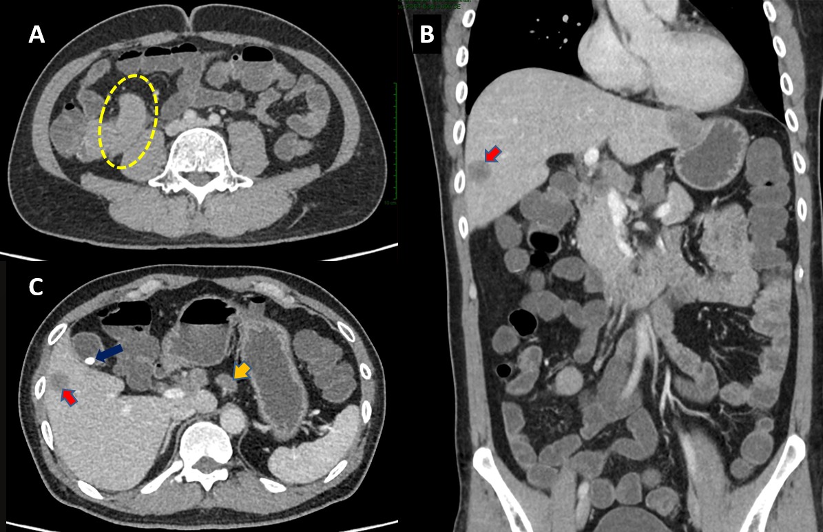
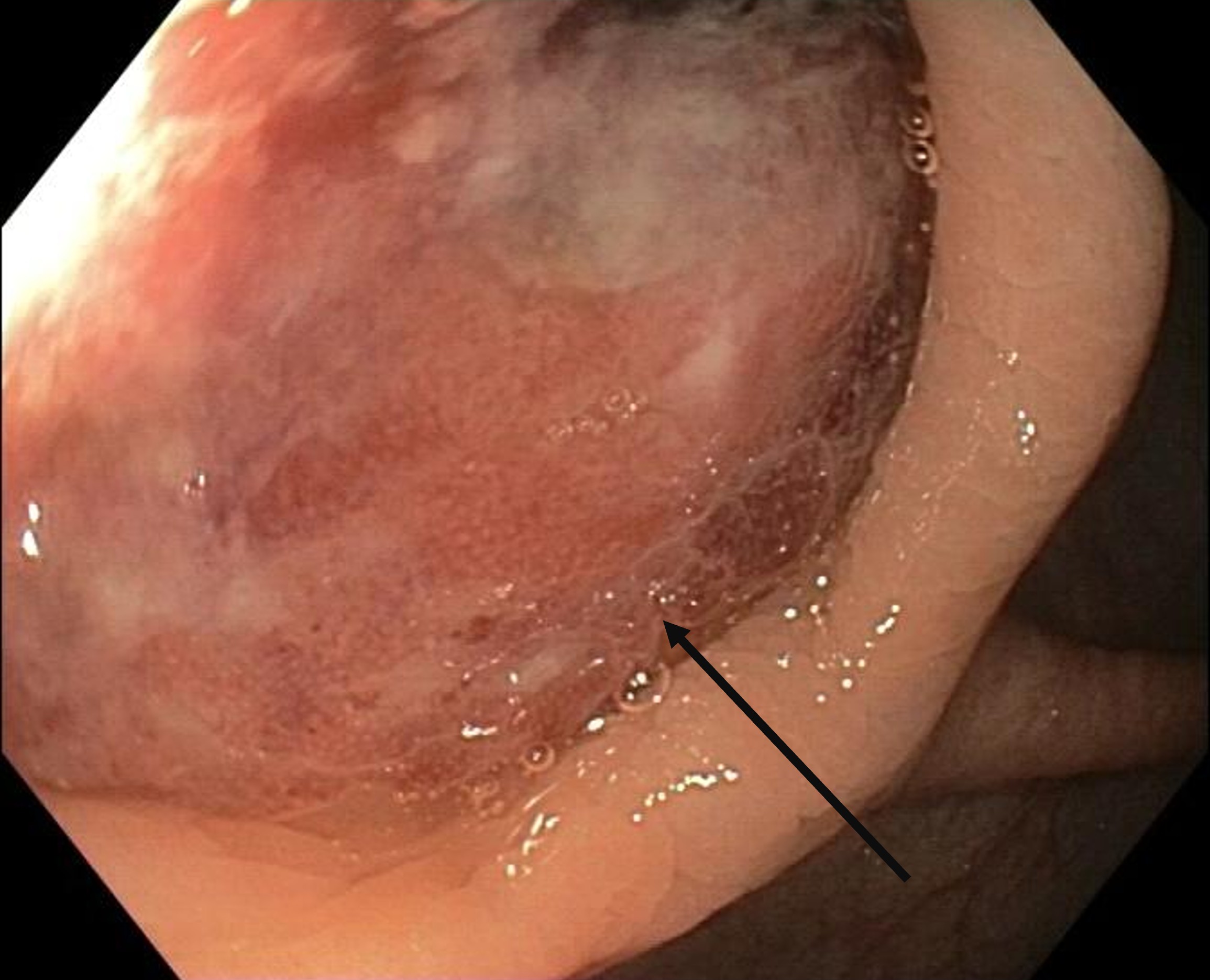
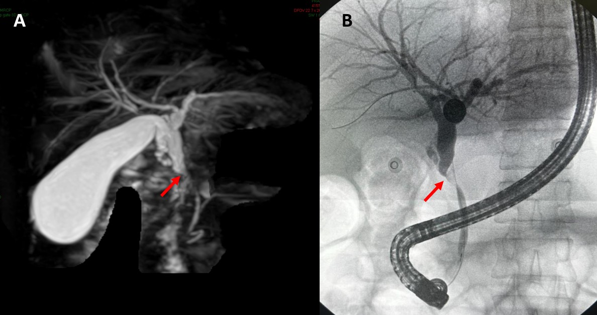
The computed tomography (CT) of the abdomen revealed an enlarged and thickened appendix with a soft tissue mass along its base, with obliteration of the appendicular lumen, measuring 8 mm x 10 mm. Additionally, multiple enlarged lymph nodes were present in the right iliac fossa, porta hepatis, peripancreatic, and left para-aortic regions, with the largest measuring 22 mm x 18 mm. Multiple hypodense areas were imaged in both lobes of the liver, suggestive of hepatic metastasis, the largest in segment six, measuring 26 mm x 20 mm. A solitary 9 mm calculus was seen in the gallbladder, with no evidence of cholecystitis Figure 1. He underwent an image-guided liver biopsy. Owing to the recurrent episodes of melena, he underwent bidirectional endoscopy. An ulcerating mass, bulging from the appendicular lumen, was identified at colonoscopy Figure 2. Biopsies were taken; however, the esophagogastroduodenoscopy was normal. Approximately three weeks after the initial presentation, he noted yellowish discoloration of the body and urine, with biochemical results consistent with cholestatic jaundice Table 1. The magnetic resonance cholangiopancreatography (MRCP) revealed extrahepatic biliary obstruction with porta hepatic lymphadenopathy Figure 3. The endoscopic retrograde cholangiopancreatography (ERCP) suggested bile duct narrowing due to extrinsic compression Figure 3; brush biopsies were taken, and a double pigtail stent was inserted, with subsequent improvement in the icterus Table 1.
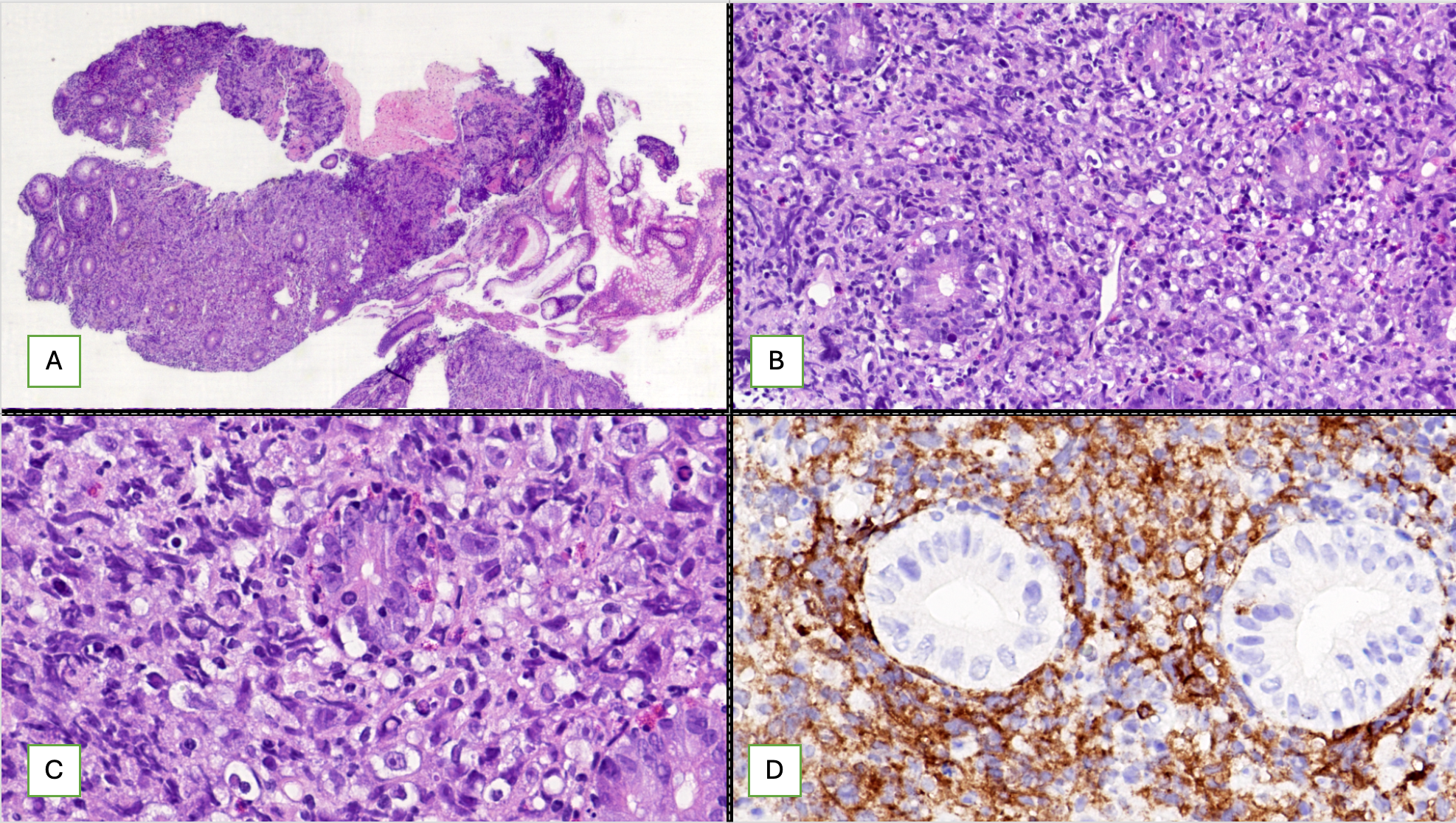
Meanwhile, the hepatic and colonoscopy biopsies were reported as large B-cell lymphoma of a similar immunophenotype. On immunohistochemistry, these tumor cells expressed diffuse positivity for CD45 and were negative for pancytokeratin, confirming their lymphoid lineage. The atypical lymphoid cells expressed CD20, CD19, CD10, CD45, and BCL-6 (>30%), and were immunonegative for CK AE1/AE3, MUM-1 (<30%), BCL-2, CMYC, EBER-ISH, CD117, and CD30; consistent with large B-cell lymphoma, germinal center subtype Figure 4. The Ki-67 labeling index was 70-80%. A diagnosis of recurrent lymphoma, NHL, DLBCL, with appendicular involvement and hepatic metastasis, complicated with appendicular tumor bleed and obstructive jaundice due to extrinsic compression by porta hepatic lymph nodes, was made: Stage IV and high international prognostic index (IPI) risk. The ERCP brush cytology was negative for neoplastic cells. Hematology-oncology consultations were obtained, and the patient underwent a bone marrow biopsy and cerebrospinal fluid analysis, both of which were negative for neoplastic infiltration. The patient’s clinical status, tumor immunohistochemistry, and therapeutic options were discussed at hematology-oncology and multidisciplinary team meetings, as well as the institutional tumor board. The therapeutic options of R-DHAP (Rituximab, Dexamethasone, Cytarabine, Cisplatin), R-ICE (Rituximab, Ifosfamide, Carboplatin, Etoposide), and R-GDP (Rituximab, Gemcitabine, Cisplatin, Dexamethasone) were considered, as these are preferred regimens in patients eligible for transplantation. His Eastern Cooperative Oncology Group Performance Status at presentation was 2; considering this and limited social support, the patient was recommended R-GDP followed by a possible autologous transplant. He was initiated on the regimen every 21 days for 3 to 4 cycles. Following the initial cycle, he felt symptomatically better and is planned for subsequent cycles and an interval positron emission tomography (PET) after two cycles of therapy.
Discussion
The GI tract is the most frequent extranodal site of lymphoproliferative diseases, likely due to the high overall volume of the lymphoid tissue in the tract. GI lymphomas are more frequently secondary and are DLBCL [5]. Additionally, mucosa-associated lymphoid tissue (MALT) lymphomas are commonly reported in the stomach, while mantle cell lymphoma is often reported in the small intestine and colon. Enteropathy-associated T-cell lymphoma (EATL) is widely reported in the jejunum. Except for EATL, most GI lymphomas are of B lymphocyte origin [5]. Beyond the exhaustive battery of genetic and chromosomal aberrations linked with lymphoproliferative diseases, a myriad of infections and diseases have been linked with GI lymphomas: Helicobacter pylori (gastric lymphomas), Campylobacter jejuni (small intestinal lymphoma), celiac disease (EATL), Epstein Barr virus (EBV), human immunodeficiency virus (HIV), human T-cell leukemia virus-1 (HTLV-1), Crohn’s disease, post-transplant immunosuppression, common variable immunodeficiency and many more [7,5]. The stomach accounts for 50%-70% of GI lymphomas, followed by the small intestine (20%-35%) and the colon, primarily the cecum (5%-10%) [8]. The vermiform appendix is an infrequent site for lymphoma, regardless of whether it is a primary tumor or secondary infiltration, accounting for 1%-3% of all gastrointestinal lymphomas [6].
The clinical features of GI lymphoma vary according to the site and severity of involvement, the degree of luminal occlusion, and the presence of metastasis. Gastric lymphoma typically presents with abdominal pain, early satiety, anemia, or GI bleeding; small intestinal lymphoma presents with abdominal pain, GI bleeding, or intestinal obstruction; esophageal lymphoma presents with dysphagia or odynophagia; colorectal lymphomas present with abdominal pain, bleeding, or altered bowel habits. [5]. Patients may report the B symptoms, fever, night sweats, and weight loss. Other, presentations or complications include icterus or incidentally detected abnormal hepatic biochemistry due to hepatic infiltration or biliary outflow obstruction, hollow viscus perforation, intussusception, GI fistula, venous thrombosis, pancreatitis, and peritoneal lymphomatosis [9]. Any bleeding luminal tumor can result in GI bleeding, and lymphomas are no exception, though they are uncommon. Patients with lymphomas may develop jaundice and/or altered liver tests by multiple mechanisms. Lymphomatous involvement in the liver can result in diffuse hepatomegaly or solitary or multiple hepatic mass lesions. Infiltrative liver disease can contribute to hepatic dysfunction and or intrahepatic cholestasis. Enlarged porta hepatis, or peripancreatic nodes, can result in extrinsic compression of the biliary tree and subsequent obstructive jaundice. Rarely, lymphomas may involve the gallbladder, bile ducts, or pancreas, leading to biliary stricture or obstruction. Portal hypertension may result from hepatic parenchymal involvement or portal venous thrombosis [9]. The most frequently reported presentations of appendicular lymphoma are appendicitis and gastrointestinal bleeding [6].
The gold standard for lymphoma diagnosis is a tissue biopsy with both conventional and immunostaining, enabling the histological classification of lymphoproliferative neoplasms. Additional investigations that help in staging include endoscopic ultrasound, cross-sectional imaging (CT or MRI), bone marrow biopsy, and FDG-PET. The typical cross-sectional imaging feature of appendicular lymphoma is a grossly enlarged soft tissue appendix. On CT, a normal appendix rarely exceeds a diameter of 6-7 mm; exceeding this cutoff, especially with a wall thickness of more than 3 mm or peri-appendiceal inflammation/fluid or appendicoliths, suggests appendicitis [10]. In non-tumoral appendicitis, the diameter rarely exceeds 1.5 cm. An appendicular diameter above 1.5cm, or for more specificity, above 2.5 cm, should raise a high suspicion for an appendicular tumor [6]. Peri-appendicular inflammatory changes may result from appendicitis or trans-serosal extension of the neoplasm [6].
Depending on the location and type of lymphoma, patients should be evaluated for associated conditions like Helicobacter pylori, HIV, celiac disease, Crohn’s disease, EBV, immunosuppression, etc. Specific to GI lymphomas, Dawson’s criteria can be used to distinguish between primary and secondary neoplasms. Predominant involvement of the bowel with nodes limited to the immediate vicinity, combined with absent peripheral and mediastinal adenopathy, sparing of liver and spleen, and normal leucocytes on hematology at diagnosis, suggests primary GI lymphoma [11]. The Ann Arbor staging and International Prognostic Index are utilized for staging and prognostication [5]. Managing gastrointestinal conditions, including appendicular lymphoma, depends on the histological type, tumor stage, and presence of active complications. Treatment of the underlying diseases and infections, combined with surgery or chemotherapy, or high-dose chemoradiotherapy coupled with hematopoietic stem cell transplantation, has been utilized. In localized primary disease, surgery offers the best chance of cure. For multifocal, advanced, or non-resectable lesions, multidrug chemotherapy is the preferred treatment. CHOP (cyclophosphamide, doxorubicin, vincristine, prednisone) or hyper-CVAD (hyperfractionated-cyclophosphamide, vincristine, doxorubicin, dexamethasone) regimen with or without Rituximab is utilized for initial chemotherapy [5]. The development of newer chemotherapeutic and targeted therapeutic molecules has further enhanced treatment options and efficacy in lymphomas. Conventional endoscopic hemostasis measures, including thermal therapy, clips, loops, and hemostatic agents, may be employed in bleeding neoplasms, such as lymphoma [12]. When endoscopically inaccessible, life-threatening, or ongoing bleeds occur, interventional radiological procedures or rescue surgeries are alternatives. In patients with obstructive jaundice secondary to extrinsic biliary compression or strictures, ERCP or percutaneous biliary drainage will alleviate the symptoms until the definitive treatment [13].
Our patient, a middle-aged male with a history of NHL–DLBCL, treated with a CHOP regimen, six cycles, with resultant remission, four years before the current presentation, presented with clinical features suggestive of appendicitis and GI bleeding. The abdominal imaging revealed a significantly enlarged appendix with a three-centimeter mass extending to the ceco-appendiceal junction, multiple hepatic mass lesions, and extensive intra-abdominal lymphadenopathy. The endoscopic evaluation for GI bleed revealed an ulcerating mass within the proximal appendicular lumen, immediately beyond a patulous appendicular opening. Image-guided liver biopsy and through-the-scope biopsy from the appendicular mass reported immunophenotypically identical DLBCL. Applying Dawson’s criteria, our patient is most likely a case of secondary GI lymphoma. While awaiting the biopsy reports, the patient developed obstructive jaundice, which was managed with ERCP-guided biliary stenting. Staging was performed using contrast-enhanced CT scans of the neck, thorax, abdomen/pelvis, an MRI of the liver, a bone marrow biopsy, and a CSF analysis. His Eastern Cooperative Oncology Group Performance Status at presentation was 2. The patient was initiated on an R-GDP regimen for relapsed NHL. The oncology team is planning for four cycles of chemotherapy, followed by reassessment, and, in case of complete relapse, to proceed with autologous stem cell therapy: otherwise, chimeric antigen receptor (CAR) T-cell therapy. This case represents an exceedingly rare and complex disease constellation comprising metastatic NHL-DLBCL with appendicular and hepatic involvement, complicated with appendicular tumor bleed and obstructive jaundice due to nodal extrinsic compression.
Conclusions
The gastrointestinal tract is the most frequent extranodal site involved in lymphomas, which can present with diverse and nonspecific clinical features that mimic other gastrointestinal and hepatobiliary disorders. Appendicular mass, gastrointestinal bleed, and obstructive jaundice are a few amongst the exhaustive list of protean manifestations, emphasizing the need for a high index of suspicion and a comprehensive diagnostic approach for lymphoma in patients with atypical and multifocal gastrointestinal manifestations.
Conflicts of Interest
The authors declare that they have no conflicts of interest related to this case report.
Funding Source
This research did not receive any specific grant from funding agencies in the public, commercial, or not-for-profit sectors.
Acknowledgments
None
Institutional Review Board (IRB)
Approval was obtained from the institutional ethics and scientific review board (MOM/COM/ETHICS/100425/007)
Large Language Model
None
Authors Contribution
MVL and GSZ contributed to the study concept. Clinical care and data collection were carried out by MVL, MA, SMS, IP, MA, and HP. The literature review and manuscript preparation were undertaken by GSZ and MVL, while SMS, IP, MA, and HP provided critical review. Manuscript revision was performed by GSZ and MA. Preparation of images and tables was handled by MVL, MA, and IP. All authors read and approved the final version of the manuscript.
Data Availability
All data underlying the findings of this case report are included within the article. Additional information or supporting data are available from the corresponding author upon reasonable request.
References
- Lewis W. D., Lilly S., Jones K. L.. Lymphoma: Diagnosis and Treatment. Am Fam Physician. 2020;101(1):34-41. PMID: 31894937
- Abdalhabib Ezeldine K.. Relative Frequencies and Patterns of Malignant Lymphoma in a Reference Centre in Khartoum, Sudan: A Descriptive Study Based on the WHO Classification of Lymphoid Neoplasms. Asian Pacific Journal of Cancer Care. 2020;5(2):107-112. doi:10.31557/apjcc.2020.5.2.107-112
- Laurent C., Do C., Gourraud P. A., de Paiva G. R., Valmary S., Brousset P.. Prevalence of Common Non-Hodgkin Lymphomas and Subtypes of Hodgkin Lymphoma by Nodal Site of Involvement: A Systematic Retrospective Review of 938 Cases. Medicine (Baltimore). 2015;94(25):e987. doi:10.1097/MD.0000000000000987 PMID: 26107683 PMCID: PMC4504656
- Das J., Ray S., Sen S., Chandy M.. Extranodal involvement in lymphoma - A Pictorial Essay and Retrospective Analysis of 281 PET/CT studies. Asia Ocean J Nucl Med Biol. 2014;2(1):42-56. PMID: 27408858 PMCID: PMC4937711
- Ghimire P., Wu G. Y., Zhu L.. Primary gastrointestinal lymphoma. World J Gastroenterol. 2011;17(6):697-707. doi:10.3748/wjg.v17.i6.697 PMID: 21390139 PMCID: PMC3042647
- Pickhardt P. J., Levy A. D., Rohrmann Jr., C. A., Abbondanzo S. L., Kende A. I.. Non-Hodgkin's lymphoma of the appendix: clinical and CT findings with pathologic correlation. AJR Am J Roentgenol. 2002;178(5):1123-7. doi:10.2214/ajr.178.5.1781123 PMID: 11959713
- Alvarez-Lesmes J., Chapman J. R., Cassidy D., Zhou Y., Garcia-Buitrago M., Montgomery E. A., Lossos I. S., Sussman D., Poveda J.. Gastrointestinal Tract Lymphomas. Arch Pathol Lab Med. 2021;145(12):1585-1596. doi:10.5858/arpa.2020-0661-RA PMID: 33836528
- Engin G., Korman U.. Gastrointestinal lymphoma: a spectrum of fluoroscopic and CT findings. Diagn Interv Radiol. 2011;17(3):255-65. doi:10.4261/1305-3825.DIR.3332-10.3 PMID: 20725903
- Khdhir M., El Annan T., El Amine M. A., Shareef M.. Complications of lymphoma in the abdomen and pelvis: clinical and imaging review. Abdom Radiol (NY). 2022;47(8):2937-2955. doi:10.1007/s00261-022-03567-5 PMID: 35690955 PMCID: PMC10509750
- Vaghela K., Shah B.. Diagnosis of Acute Appendicitis Using Clinical Alvarado Scoring System and Computed Tomography (CT) Criteria in Patients Attending Gujarat Adani Institute of Medical Science - A Retrospective Study. Pol J Radiol. 2017;82:726-730. doi:10.12659/PJR.902246 PMID: 29662595 PMCID: PMC5894019
- Dawson I. M., Cornes J. S., Morson B. C.. Primary malignant lymphoid tumours of the intestinal tract. Report of 37 cases with a study of factors influencing prognosis. Br J Surg. 1961;49:80-9. doi:10.1002/bjs.18004921319 PMID: 13884035
- Ofosu A., Ramai D., Latson W., Adler D. G.. Endoscopic management of bleeding gastrointestinal tumors. Ann Gastroenterol. 2019;32(4):346-351. doi:10.20524/aog.2019.0391 PMID: 31263356 PMCID: PMC6595926
- Sinicrope FA. Biliary Obstruction. 2003.